Mode of Action: Synergistic Interaction of Peppermint - ACG Publications
←
→
Page content transcription
If your browser does not render page correctly, please read the page content below
ORIGINAL ARTICLE

                                  Rec. Nat. Prod. 12:6 (2018) 582-594

           Mode of Action: Synergistic Interaction of Peppermint
    (Mentha x piperita L. Carl) Essential Oil and Meropenem Against
                   Plasmid-Mediated Resistant E. coli

        Shun-Kai Yang 1, Polly Soo-Xi Yap 2, Thiba Krishnan 3, Khatijah
        Yusoff 4, Kok-Gan Chan 4, Wai-Sum Yap 5, Kok-Song Lai 1 and
                            Swee-Hua Erin Lim 6,7*
    1
     Department of Cell and Molecular Biology, Faculty of Biotechnology and Biomolecular Sciences,
                     Universiti Putra Malaysia, 43400 Serdang, Selangor, Malaysia
2
  School of Postgraduate Studies and Research, International Medical University, No. 126, Jalan Jalil
                         Perkasa 19, Bukit Jalil, 57000 Kuala Lumpur, Malaysia
    3
      Division of Genetics and Molecular Biology, Institute of Biological Sciences, Faculty of Science,
                          University of Malaya, 50603 Kuala Lumpur, Malaysia
4
   Department of Microbiology, Faculty of Biotechnology and Biomolecular Sciences, Universiti Putra
                              Malaysia, 43400 Serdang, Selangor, Malaysia
  5
    Department of Biotechnology, Faculty of Applied Sciences, UCSI University, 56000 Cheras, Kuala
                                           Lumpur, Malaysia
6
  Centre for Bioinformatics, School of Data Sciences, Perdana University, MAEPS Building, Serdang,
                                           Selangor, Malaysia
 7
   Health Sciences Division, Abu Dhabi Women’s College, Higher Colleges of Technology, 41012 Abu
                                      Dhabi, United Arab Emirates

         (Received December 22, 2017; Revised February 12, 2018; Accepted February 13, 2018)

Abstract: This study investigated the bactericidal mechanism of peppermint essential oil (PEO) when used
singly and in combination with meropenem against multidrug resistant Escherichia coli. Chemical compositions
of PEO were identified via GC-MS, followed by time-kill analysis which was performed to evaluate the
antibacterial activities of PEO and meropenem. Furthermore, outer membrane permeability test, zeta potential
measurement and scanning electron microscopy were performed to evaluate the ability of PEO in bacterial
membrane disruption. Next, anti-quorum sensing assay was performed to assess the ability of PEO in quorum
sensing inhibition. A complete killing activity was observed within five minutes of treatment with PEO and
meropenem at sub-lethal concentrations. In addition, the outer membrane permeability test and zeta potential
measurement performed indicated increase in the membrane permeability and membrane disruption which can
be observed in the scanning electron micrograph. Furthermore, significant decrease in the light production of E.
coli pSB1075 treated by PEO indicates the presence of quorum sensing inhibitors within PEO. The findings
suggest that PEO has the ability to disrupt the bacterial outer membrane which increases membrane
permeability, in addition to the possible inhibition of bacterial quorum sensing ability in multidrug resistant E.
coli, aiding in the reversal of antibiotic resistance.

Keywords: Escherichia coli; essential oil; membrane permeability; Mentha x piperita L. Carl; quorum sensing;
synergism. © 2018 ACG Publications. All rights reserved.

*
    Corresponding author: E-Mail: erinlim@perdanauniversity.edu.my; Phone: +60389418646 ext. 178
                                     The article was published by ACG Publications
                           www.acgpubs.org/RNP © November-December/ 2018 EISSN:1307-6167
                                     DOI: http://doi.org/10.25135/rnp.59.17.12.078
583 Yang et.al., Rec. Nat. Prod. (2018) 12:6 582-594
1. Introduction
Extensive use of antibiotics in hospitals, communities and agriculture have fueled the ever
increasing threat of antibiotic resistant infections, fast tracking the evolution towards antibiotic
resistance in microorganisms [1]. Escherichia coli and Klebsiella pneumonia expressing genes such as
CMY, IMP, KPC, NDM, OXA and VIM which encode carbapenemases which have been described in
numerous study and are surfacing at an alarming rate [2-5]. Gram negative bacteria often
simultaneously activate two resistant mechanisms whereby the mass production of β-lactamases is
coupled with the decrease in the permeability of their outer membrane (OM) [6-8], making a multi-
targeted drug therapy the ideal treatment method. Preferences for combination therapy by testing
natural products alongside with existing antibiotics have been reported to exert synergistic effects [9].
However, limited information about the exact mechanisms of essential oil functioning as an antibiotic
resistant modifier is known, despite countless reports demonstrating favorable synergistic interactions
[10-13].
In the previous work by Yap et al. 2013, the minimum inhibitory concentration of both
peppermint essential oil (PEO) (8% v/v) and meropenem (4 µg/mL) were determined via resazurin
microplate assay [14]. In subsequent checkerboard assay, it was found that, when combined, PEO and
meropenem reacted synergistically against KPC-3 producing E. coli pMG309, with significantly
lowered minimum inhibitory concentration (MIC) (PEO, 1% v/v; meropenem, 0.5 µg/mL) [14]. This
resulted in a fractional inhibitory concentration (FIC) index of 0.26, indicating a high degree of
synergism. The option for using essential oils in combination with antibiotics hold potential in offering
possibly a novel, multi-targeted method in addressing the issue of antimicrobial resistance (AMR). A
related study demonstrated that PEO and its major component menthol displayed synergistic activity
with oxytetracycline. The finding postulated that the lowered antibiotic effective dosage needed to
achieve total eradication of bacteria is attributed to the ability of PEO in plasmid curing [15].
However, the roles of the components within essential oils have not been explored thoroughly to date.
Therefore, the objective of the current study was carried out to elucidate the possible mechanism of
PEO when used against E. coli pMG309, which harbors plasmid encoding KPC-3 β-lactamase capable
of hydrolyzing a wide range of β-lactam antibiotics such as, penicillins, cephalosporins and
carbapenems [16]
2. Materials and Methods
2.1. Peppermint (Mentha x piperita L. Carl) Essential Oil (PEO) and Meropenem
The peppermint (Mentha x piperita L. Carl) essential oil (Serial number: 7212) purchased from
Aroma Trading Ltd. (Milton Keynes, UK) was used throughout the studies. Both PEO and meropenem
(Sigma Aldrich, St Louis, USA) were dissolved in Mueller-Hinton broth (MHB; oxoid, Cambrigde,
UK) supplemented with Tween80, a solubilizing agent at 1% (v/v) concentration to make desirable
stock solution as described in the Clinical and Laboratory Standards Institute (CLSI) M100-S21
guidelines.
2.2. Gas chromatography-mass Spectrometry (GC-MS) Analysis
The GC-MS analysis was performed with Agilent GC-MS, 7890A GC System with a
triple-axis detector (5975C MSD) and an HP-5MS column (30 m × 250 μm × 0.25 μm)
(Agilent Technologies, California, USA). Helium was used as the carrier gas in the MS. The
sample was injected with an auto-injector heated to 250 °C (Agilent Technologies 7693 Auto-
sampler, California, USA). The oven column, on the other hand, was temperature-
programmed from 40 °C (2 min) to 175 °C at a rate of 5 °C min-1 within 10 min. The
temperature then rose to 250 °C at a rate of 90 °C min-1 within the next 5 min. The flow rate
of the column was 1 mL min-1 with a split ratio of 40:1. EI mode with scan range 30-450 m/z
was used to analyze the MS. The temperature of the MS source was set at 250 °C whereas the
MS quad at 150 °C. Identification of compounds was solely based on the comparison of theSynergistic interaction of peppermint essential oil and meropenem 584
mass spectra with those in National Institute of Standards and Technology libraries. The
relative percentage of the identified compounds was computed from their GC peak area and
only components with total abundance exceeding 0.1 % were presented.
2.3. Bacterial Strains and Growth Conditions
Escherichia coli pMG309 harboring a plasmid encoding β-lactamase, KPC-3, a kind gift from
George A. Jacoby (Lahey Clinic, MA, USA) was cultured in Mueller-Hinton broth (MHB; Oxoid,
Cambridge, UK) and all test conditions were carried out according to the CLSI M07-A8 guidelines.
Biosensor strains E. coli pSB1075 and E. coli pSB401 used for the anti-quorum sensing assay were
detailed in Table 1. Both biosensor strains were cultured in Luria Bertani (LB; Oxoid, Cambridge,
UK) broth supplemented with the antibiotics tetracycline and ampicillin (Thermo Fisher Scientific,
Massachusetts, USA) at 37 °C with shaking at 200 rpm.
Table 1. Biosensor E. coli strains and their plasmid used
Biosensor Description Source
Escherichia lasR lasI’ (Pseudomonas aeruginosa PAO1) : luxCDABE [17]
coli pSB1075 (Photorhabdus luminescens ATCC 29999) fusion in pUC18
AmpR, AHL biosensor producing bioluminescence in
response to long chain AHL.
Escherichia luxR luxI’ (Photobacterium fischeri ATCC 7744) [17]
coli pSB401 :luxCDABE (Photorhabdus luminescens ATCC 29999)
fusion; pACYC184-derived, TetR, AHL biosensor producing
bioluminescence in response to short chain AHL.
2.4. Time-kill Assay
The spread-plate method was used for viable counts of bacteria in the time kill assay. The sub-
lethal concentrations of PEO and meropenem used in the time-kill assay were determined in previous
work [14], control (without treatment); PEO (1 % v/v); meropenem (0.5 µg/mL); PEO (1 % v/v) in
combination with meropenem (0.5 µg/mL). Directly after addition of the inoculum and after each
interval of incubation (every 4 h), 100 µL of samples was removed for viable counting as a
preliminary screening (data not shown). Sampling time was then shortened to 5, 10, 30, 60, 120, 180,
240 min in the events of rapid killing rate. Samples were serially diluted with 0.85% (w/v) sodium
chloride, then plated onto Mueller-Hinton (MH) agar followed by an overnight incubation at 37°C.
The minimum time required to inhibit the growth of bacteria undergoing combinatory treatment was
used in subsequent assays as the optimal treatment time. The experiment was performed in triplicates.
2.5. Outer Membrane (OM) Permeability Test
OM permeability test was performed using sub-lethal concentration of PEO (1% v/v) and
meropenem (0.5 µg/mL), as detailed previously [14], using 0.1 % (w/v) of anionic detergent, sodium
dodecyl sulfate (SDS; Thermo Fisher Scientific, Massachusetts, USA) as a permeabilizing probe [18].
The absorbance at 625 nm were measured and compared amongst treatment group with and without
SDS via a UV-visible and visible spectrophotometer (Thermo Fisher Scientific, Massachusetts, USA).
The experiment was performed in 3 biological replicate in order to minimize the variance between
sample of the same treatment group.
2.6. Bacterial Surface Charge: Zeta Potential Measurement
The zeta potential of all treatment groups was quantified using Nano Zetasizer (Malvern
Instruments, Malvern, UK). After treatment with sub-lethal concentration of PEO (1% v/v) and
meropenem (0.5 µg/mL) alone, and in combination for 5 minutes, cells were washed thoroughly with585 Yang et.al., Rec. Nat. Prod. (2018) 12:6 582-594
phosphate buffered saline (PBS) (pH 7.4) for in preparation for measurements. The non-treated cell
suspension in PBS was used as a control. The experiment was performed in 3 biological replicate in
order to minimize the variance between sample of the same treatment group.
2.7. Scanning Electron Microscopy
SEM were performed on non-treated cells, cell treated with PEO (1% v/v) alone and in
combination with meropenem (0.5 µg/mL) for 5 min, as determined in the time kill assay. After the 5
minute treatment, cells were harvested and washed with PBS (pH 7.4) for at least three times to
remove the treatment residue completely. Then, the samples were processed according to the reported
protocol with slight modification [19]. Samples were fixed with 4% glutaraldehyde and 1% osmium
tetroxide at 4°C followed by dehydration via sequential exposure to graded acetone (35-100%). The
samples were then subjected to critical point drying and finally sputter-coated with gold, followed by
SEM observations via JEOL JSM-6400 (JEOL Ltd., Tokyo, Japan). All treatment groups were
compared visually to the untreated group based on the criteria of modification towards the cell surface
(blebbing/corrugation) and cell to cell contact (adhesion).
2.8. Anti-quorum Sensing Assay: Quantification of Light Production from E. coli pSB1075
and E. coli pSB401
Light produced by both biosensor strains of E. coli was measured using an Infinite M200 Pro
Tecan Microplate Reader (Tecan, Männedorf, Switzerland). A concentration of 0.5 μg/mL 3-oxo-C6-
HSL and 10 μg/mL 3-oxo-C12-HSL (Sigma-Aldrich, Missouri, USA) were prepared in acetonitrile
(Sigma-Aldrich, Missouri, USA) and stored at −20 °C. An overnight culture of E. coli biosensor cells
were diluted to 0.5 McFarland Standard. Then, 200 μL of E. coli biosensor cells with essential oil
(0.01%, 0.025% and 0.05%) was added into the well of Greiner 96-well microtitre plate (Greiner Bio-
One, Kremsmünster, Austria). For comparison, E. coli pSB401 and E. coli pSB1075 were
supplemented with 2 μL of 3-oxo-C6-HSL (0.005 μg/mL) and 2 μL of 3-oxo-C12-HSL (0.1 μg/mL),
respectively. The light signal produced was determined every 30 min for 24 h at 495 nm by the
microplate reader. Production of light was measured as relative light units (RLU), whereby the total
light production is directly proportional to the growth rate as well as the quorum sensing ability of the
biosensor strains [20]. A decrease of light production in E. coli pSB1075 and E. coli pSB401 indicates
anti-quorum sensing properties of the PEO.
2.9. Statistical Analysis
All results represent the average of three independent experiments. The data were presented as
mean ± standard deviation (SD) and analyzed by one-way analysis of variance (ANOVA) and
Student’s t-test. The P value was calculated via GraphPad Prism 5 statistical software with P < 0.05
considered as significant.
3. Results and Discussion
The chemical composition of peppermint (Piperita x mentha) essential oil is tabulated in Table
2. The major compounds (>1.0%) found in PEO are menthol (50.1435%), followed by menthone
(24.4276%), menthyl acetate (6.1701%), eucalyptol (5.7894%), cis-β-Terpineol (1.8929%), o-cymene
(1.0384%) and caryophyllene oxide (1.0148%).Synergistic interaction of peppermint essential oil and meropenem 586
Table 2. Chemical composition of peppermint (Mentha x piperita) essential oil
Peak Library/ID RIa RIb Composition %
1 Menthol 1177 1177 41.36
2 p-menthone 1156 1150 25.72
3 l-menthone 1166 1150 17.78
4 Isomenthol acetate 1295 1295 4.59
5 Isopulegol 1147 1145 1.60
6 Piperitone 1256 1253 1.49
7 Caryophyllene 1423 1420 1.15
8 (±)-pulegone 1241 1234 1.07
9 Neoisomenthol 1185 1173 0.96
10 Terpineol 1193 1189 0.52
11 (-)-β-bourbonene 1388 1384 0.46
12 (-)-lavandulol 1169 1168 0.34
13 p-methan-1-ol 1191 1110 0.25
14 Germacrene D 1458 1480 0.22
15 Elixene 1339 1337 0.19
16 D-limonene 1028 1029 0.17
17 γ-muurolene 1502 1476 0.14
18 Caryophyllene oxide 1589 1580 0.13
19 Linalool 1101 1099 0.12
20 (-)-elemene 1395 1390 0.10
21 1-decanol 1274 1272 0.05
Time-kill assay indicated synergism between PEO and meropenem whereby the combination
treatment results in a reduction of ≥ 2-log10 CFU/mL compared to the growth curve of cells treated
singly with essential oil and antibiotic and the control [20]. The PEO and meropenem combination
showed rapid and complete killing profile within 5 mins after the addition of PEO and meropenem.
However, when applied singly, PEO or meropenem did not eradicate the cells completely or as rapidly
as the combinatory counterpart (Figure 1).
Figure 1. Growth curves of E. coli pMG309
Untreated E. coli pMG309 (♦), E. coli pMG309 treated with PEO (●) and meropenem (*) alone and in
combination (▲).587 Yang et.al., Rec. Nat. Prod. (2018) 12:6 582-594
Sodium dodecyl sulfate (SDS) was the permeabilizing probe used to detect a disruption in the
bacterial outer membrane in this experiment. Differences in absorbance between pre- and post-
treatment of the control, essential oil or/and meropenem with or without 0.1% SDS was presented in
Table 3. Sample treated with PEO alone and the combination of PEO and meropenem reduced the cell
viability significantly (P < 0.05) when compared to the non-treated group after 5 minutes of treatment
time (denoted by superscript a in Table 3). Absorbance of cells treated with meropenem alone (with
and without 0.1% SDS) remained constant throughout the study and was not significant (P > 0.05).
Table 3. Outer membrane permeabilization of E. coli pMG309 treated with PEO and/or meropenem
Treatment OD625 ± SD (n=3)
Time (min) 0 5 10 30 60
Control
with 0.1% SDS 0.33±0.001 0.32±0.005 0.31±0.006 0.30±0.001 0.30±0.002
without 0.1% SDS 0.32±0.003 0.33±0.003 0.33±0.004 0.33±0.005 0.33±0.006
Peppermint (1% v/v)
with 0.1% SDS 0.25±0.005a 0.23±0.007b 0.22±0.007b 0.21±0.001b 0.22±0.003b
without 0.1% SDS 0.26±0.001a 0.26±0.004b 0.25±0.002b 0.25±0.003b 0.26±0.011b
Meropenem (0.5µg/mL)
with 0.1% SDS 0.33±0.002 0.33±0.005 0.32±0.007 0.33±0.010 0.33±0.003
without 0.1% SDS 0.33±0.002 0.32±0.010 0.33±0.009 0.32±0.001 0.32±0.005
Peppermint (1% v/v) + Meropenem (0.5µg/mL)
with 0.1% SDS 0.26±0.004a 0.23±0.006b 0.21±0.002b 0.21±0.006b 0.20±0.008b
without 0.1% SDS 0.26±0.004a 0.25±0.004b 0.25±0.005b 0.26±0.004b 0.25±0.003b
a
Significant difference among treatment groups when compared to the corresponding control groups (with or
without 0.1% SDS). (P < 0.05)
b
Significant difference between samples treated and non-treated with 0.1% SDS at the corresponding time
points. (P < 0.05)
Values are mean OD625 ± S.D of three replicates.
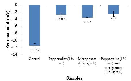
The surface charge of bacterial cells is determined based on their zeta potential value, which
can be measured based on the mobility of cells exposed to an electrophoretic force under defined pH
and salt concentrations. Under normal condition, E. coli pMG309 had an original negative surface
charge of -11.52 mV. Singular treatment of PEO and meropenem further increases the surface charge
to -2.82 mV and -3.67 mV while combinatory treatment yielded the further increases the surface
charge to -2.56. Thus, all the treatments applied significantly reduced the negative charges on the cell
surface with the cells treated with the combination of PEO and meropenem being the least negative (P
< 0.05) (Figure 2).Synergistic interaction of peppermint essential oil and meropenem 588
Figure 2. Zeta potential values (mV) of E. coli pMG309 when treated with PEO or/and meropenem
Figure 3. Scanning electron micrographs of E. coli pMG309
(a) Untreated E. coli pMG309, (b) cells treated with PEO (1% v/v), (c) cells treated with meropenem (0.5µg/mL)
and (d) cells treated with the combination of peppermint (1% v/v) and meropenem (0.5µg/mL) for five minutes.
The outer membrane of the treated cells appeared to be corrugated (shown in white arrows).589 Yang et.al., Rec. Nat. Prod. (2018) 12:6 582-594
The scanning electron micrographs further depict the morphological changes of E. coli
pMG309 treated with PEO and meropenem, alone and in combination (Figure 3). In the non-treated
controls, a rod-shaped morphology which is characteristic of E. coli was observed (Figure 3a). It was
observed that samples treated with PEO alone (Figure 3b) and in combination with meropenem
(Figure 3d) resulted in severe corrugation of the bacterial cell membrane when compared with
untreated sample (Figure 3a) and sample treated with meropenem alone (Figure 3c).
An anti-quorum sensing assay was also performed to investigate the ability of PEO as a
quorum sensing inhibitor by using biosensor E. coli. In the presence of a quorum sensing inhibitor,
light production by the biosensor cells will decrease. Prior to the assay, resazurin microplate assay
were used to determine the concentration of PEO that can be used for the anti-QS assay without
harming the biosensor cell (data not shown). As observed from Figure 4, increment in the
concentration of PEO from 0.01%, 0.025% to 0.05% significantly inhibited light production activity in
E. coli pSB1075 (Figure 4a) but not in E. coli pSB401 (Figure 4b) when compared to the controls
(untreated cells and cells treated with Tween80).
Figure 4. Light production by biosensor E. coli pSB1075 and E. coli pSB401
Both biosensor cells were treated with PEO at concentration 0.01% (▲), 0.025% (■), and 0.05% (●). Tween 80
(♦) and E. coli pSB401 and pSB1075 supplemented with 3-oxo-C6-HSL and 3-oxo-C12-HSL, were also
included respectively, serving as controls (▼). The data were presented as RLU/OD to account for any
differences in growth. Data were analyzed via one-way analysis of variance, with p < 0.05 being significant
(indicated by * in figure).Synergistic interaction of peppermint essential oil and meropenem 590
In previous publication [14], five essential oils (cinnamon bark, tea tree, peppermint,
marjoram and lavender essential oil) and seven antibiotics (ampicillin, carbenicillin, cefazolin,
cefuroxime, piperacillin, ceftazidime and meropenem) had been screened for their minimum inhibitory
concentration individually against 3 different strains of E. coli harboring different plasmid which
encodes different β-lactamases, via resazurin microplate assay [14]. Following that, checkerboard
assay had also been performed on all possible combinations of essential oil and antibiotics. Of all
pairs, only 4 combinations of EO and antibiotic exhibited synergism with one another in nature and
one of it was the combination of PEO and meropenem against E. coli pMG309 harboring KPC-3 β-
lactamase. The MIC of PEO and meropenem were 8 % and 4 µg/mL respectively when used alone.
However, when applied in combination, the MIC of PEO and meropenem were reduced to 1 % and 0.5
µg/mL respectively, yielding a high degree of synergism with fractional inhibitory concentration index
of 0.26. Thus, this particular combination was chosen to be further studied in the current work.
GC-MS had been performed in order to understand and postulate the possible compounds
which might be associated with the mechanism of PEO in combinatory treatment. According to the
GC-MS analysis, PEO consisted of a total of 21 compounds (≥0.1%) as presented in Table 2. The
major compounds found within PEO were menthol (41.36 %), p-menthone (25.72%), l-menthone
(17.78%) and isomenthol acetate (4.59%). For comparison, a previous study in Poland reported that
menthol, menthone, and menthyl acetate were the major components in PEO [22]. In addition, similar
studies carried out by researchers in Germany and Africa also obtained a similar result whereby
menthol and menthone are the major components of PEO [23, 24]. According to the study conducted
by [24], it was found that menthol disturbs the plasma membrane of bacteria, resulting in the alteration
of membrane permeability and intracellular leakage [25]. However, there is no reported mechanism to
date regarding the second most abundant component in PEO, menthone.
In the time-kill analysis, combinatory treatment with PEO and meropenem at their sub-lethal
concentrations showed a complete and rapid killing profile when compared to the other three treatment
groups. Therefore the bactericidal activity of PEO was not affected by the β-lactamase produced.
Action of PEO against the E. coli might be due to the general toxicity effects of the essential oil itself
on the bacterial outer membrane [25]. A similar study carried out on one of the major components of
peppermint, menthol also caused disruption of outer membrane, leading to an increase in the
membrane permeability and leakage of intracellular content [26]. To further validate this theory, the
OM permeability test and zeta potential measurement were performed to assess the ability of PEOs in
altering the OM permeability. In OM permeability test, anionic detergent, SDS was used as a
permeabilizing probe whereby it was excluded extracellularly by the OM under normal condition [27].
However, a disruption in the membrane enabled the influx of SDS which eventually led to cell death
when the accumulated concentration of SDS reached its threshold. Thus, SDS functions as a
permeabilizing probe whereby it initiated cell lysis after the bacterial membrane had reached a critical
extent of membrane disordering [28,29]. As shown in Table 3, PEO alone and PEO-meropenem
combination altered the OM permeability of E. coli pMG309 significantly when compared to the other
two treatment groups. Sub-lethal concentration of meropenem alone did not increase outer membrane
permeability, thus there were no influx of SDS into the cells observed wherein the OD readings were
not decreased significantly (P > 0.05) throughout the entire course of this experiment. This is due to
the hydrophilic nature of meropenem, a β-lactam antibiotic which did not cause any
lipopolysaccharide destabilisation, and thus did not potentiate the influx of SDS into the cells [29].
These results suggest that the outer membrane was disrupted solely by PEO, increasing the bacterial
membrane permeability, subsequently decreasing the membrane’s ability to osmoregulate the cell
adequately, thus, promoting the influx of SDS, eventually lysing the cell [30]. In the current study, the
possible mode of action of PEO is through the disruption of the bacterial membrane, either directly or
indirectly, both at lethal and sub-lethal concentrations which increases the non-specific penetration of
the antibiotic into the bacteria [31,32]. As discussed before [33], alteration in the OM of the bacteria
would facilitate the diffusion of antibiotics through the cell barrier eventually elevating the
intracellular concentration of antibiotics which subsequently leads to cell death [33]. This explains the
synergism observed which caused rapid killing of multidrug resistant E. coli as observed in the time-
kill assay. Zeta potential measurement was then carried out to further clarify the effect of PEOs on the
bacterial membrane. The zeta potential of bacterial cell membrane provides information of the
membrane potential which reflects the metabolic state of the bacteria, whereby the zeta potential591 Yang et.al., Rec. Nat. Prod. (2018) 12:6 582-594
values were inversely proportional to the growth rate of the bacteria [34,35]. Bacterial cell surfaces are
normally negatively charged under physiological conditions due to the presence of anionic groups
such as carboxyl and phosphate in their membranes but the value becomes less negative when the
bacteria is under stressful conditions. In this study, the zeta potential of E. coli pMG309 treated with
PEO alone and in combination with meropenem was significantly higher than the non-treated cells,
indicating disruption in the membrane thus leading to slow metabolic rate. This might be due to the
acidification of OM by the PEO which increases the zeta potential of the bacteria, leading to
membrane damage [36].
The scanning electron micrograph (Figure 3) revealed that the overall cell surface of E. coli
pMG309 treated with PEO alone and in combination with meropenem were structurally different from
the control, validating the ability of PEO in bacterial OM disruption. Observed morphological changes
were due to the disruption of outer membrane as evidenced by the OM permeability test and zeta
potential measurement as well as the previous findings by other researchers [37-39]. As shown in
Figure 3c, cells treated with meropenem alone had minor corrugation on their outer membrane.
Similar observations have been reported on bacterial cells treated with imipenem [40]. The minor
corrugation was attributed to the nature of β-lactam antibiotic which interferes with the peptidoglycan
linkage, eventually altering the integrity of the cell towards osmotic pressure and thus disrupting the
outer membrane [41]. As shown in Figure 3b and 3d, a severe alteration on the cellular surface of E.
coli pMG309 was observed in the presence of PEO alone and in combination with meropenem.
Therefore, OM permeability test, zeta potential measurement and scanning electron microscopy used
in this study has provided strong evidence that PEO possess the capability to disrupt the outer
membrane of E. coli pMG309, leading to an influx of meropenem into the cell which eventually
eradicates the cell synergistically.
In this study, the anti-quorum sensing assay had also been carried out to detect the quorum
sensing inhibitor compound which might be present within PEO. Quorum sensing is the language used
between bacteria to communicate with each other solely through chemical signalling. QS regulates a
vast number of bacterial functions including virulence and pathogenic potential of bacteria [41]. This
communicative capability is also one of the major causes of antibiotic resistance nowadays. Under the
stress of antimicrobials, a bacterial community produces signals which enhance the secretion of
extracellular polymeric substances, leading to the formation of biofilm which serves as a protective
shield to the bacterial community from antibiotics [42]. Meropenem is a broad-spectrum carbapenem
antibiotic with a known mode of action that penetrates bacterial cells readily and interferes with the
synthesis of the vital cell wall components [43]. In this current study, PEO was tested individually to
investigate its anti-quorum sensing potential as an important anti-infective target that does not rely on
antibiotics. Hence, the combination of meropenem/PEO aims to address gaps in the development
carbapenem resistance such as increased expression of the efflux system, porin deficiency and
increased chromosomal carbapenemases activity [44]. Results shown in Figure 4 indicates that
increment in the concentration of peppermint oil significantly decreases the light production in
biosensor E. coli pSB1075, subsequently increasing in quorum sensing inhibition. However, quorum
sensing inhibition was not observed in control biosensor E. coli pSB401. Hence, PEO was shown to
inhibit quorum sensing activity in E. coli pSB1075 carrying the lasR receptor gene but not in E. coli
pSB401 carrying the luxR. lasR gene is commonly found in opportunistic pathongens such as P.
aeruginosa while luxR gene is found in harmless bacteria such as Vibrio harveyi [45].
One of the most prominent mechanisms postulated in recent studies for the synergistic
interactions between essential oil and antibiotics is via the disruption of the bacterial cytoplasmic
membrane. Further investigation by researchers also showed the specific mechanism of essential oil
components responsible for the leakage of potassium ions, loss of 260-nm-absorbing material and salt
tolerance as well as up/down-regulation of protein found in bacterial membrane [37,38,40,46]. A
standardized method for the evaluation of membrane permeability is yet to be developed and
limitations exist whereby the activity of the compounds tested in unrelated studies cannot be compared
to each other directly.
In the current study, PEO was found to act synergistically with meropenem showing
disruption of the bacterial membrane as evidenced by findings from the OM permeability test, zeta
potential measurement and scanning electron microscopy (SEM) observation. The findings implied
that PEO has the potential to increase the bacterial susceptibility towards β-lactam antibiotics; in thisSynergistic interaction of peppermint essential oil and meropenem 592
case, meropenem may operate via two mechanisms: generalized bacterial outer membrane disruption
which potentiates antibiotics influx into the cytoplasm, eventually leading to cell death and anti-
quorum sensing ability. It should be pointed out that this study serves as preliminary work on the
mode of action of PEO in its synergistic interaction with meropenem. It has been discussed and
hypothesized that the actions of PEO are broad and may provide promising cell targets other than the
membrane effects. This possibility deserves further investigation. Additionally, since the membrane
permeabilizing effects of PEO are demonstrated, their possible effect on up/down-regulation of the
membrane proteins opens an interesting avenue for further study using high-throughput screening such
as microarray. Further work such as better understanding of the mechanisms at the molecular level are
required before the actual applications of combinatory therapy using essential oils and antibiotics as a
potential drug treatment strategy in the clinical setting can be fully employed.
Acknowledgments
This study was funded by the Fundamental Research Grant Scheme (FRGS) from the Ministry
of Higher Education (MOHE), Malaysia [FRGS/1/2011/SKK/IMU/03/3] and UCSI University
Internal Grant [Proj-In-FAS-044]. The author (Kok Gan Chan) acknowledges Universiti Malaya for
the High Impact Research Grant [HIR/MOHE Grant A000001-50001]. Authors would also like to
thank Dr. George A Jacoby for providing the bacterial strain used in this study and fellow members of
IMU Research Laboratory and Floral Biotechnology Laboratory, Universiti Putra Malaysia. SKY and
PSXY drafted the manuscript. KSL and KY edited the manuscript whereas SHEL significantly refined
the manuscript. All authors read and approved the final manuscript.
ORCID
Shun Kai Yang: 0000-0002-9275-3653
Polly Soo Xi Yap: 0000-0002-1615-3138
Thiba Krishnan: 0000-0003-1791-7627
Khatijah Yusoff: 0000-0002-7683-4760
Kok Gan Chan: 0000-0002-1883-1115
Wai Sum Yap: 0000-0003-1589-0176
Kok Song Lai: 0000-0002-1887-2232
Swee Hua Erin Lim: 0000-0001-5177-0257
References
[1] S.K. Yang, L.Y. Low, P.S.X. Yap, K. Yusoff, C.W. Mai, K.S. Lai and S.H.E. Lim (2018). Plant-derived
antimicrobials: insights into mitigation of antimicrobial resistance, Rec. Nat. Prod. 12(4), 295-316.
[2] P. Nordmann, G. Cuzon and T. Naas (2009). The real threat of Klebsiella pneumoniae carbapenemase-
producing bacteria, The Lancet Infect. Dis. 9(4), 228-236.
[3] W.H. Goessens, A.K. van der Bij, R. van Boxtel, J.D. Pitout, P. van Ulsen and D.C. Melles (2013).
Antibiotic trapping by plasmid-encoded CMY-2 beta-lactamase combined with reduced outer membrane
permeability as a mechanism of carbapenem resistance in Escherichia coli, Antimicrob. Agents.
Chemother. 57(8), 3941-3949.
[4] H. Yigit, A.M. Queenan, G.J. Anderson, A. Domenech-Sanchez, J.W. Biddle and C.D. Steward (2001).
Novel carbapenem-hydrolyzing beta-lactamase, KPC-1, from a carbapenem-resistant strain of Klebsiella
pneumoniae, Antimicrob. Agents Chemother. 45(4), 1151-1161.
[5] D. Yong, M.A. Toleman, C.G. Giske, H.S. Cho, K. Sundman and K. Lee (2009). Characterization of a new
metallo-beta-lactamase gene, blaNDM-1, and a novel erythromycin esterase gene carried on a unique
genetic structure in Klebsiella pneumoniae sequence type 14 from India, Antimicrob. Agents. Chemother.
53(12), 5046-5054.
[6] M. Doumith, M.J. Ellington, D.M. Livermore and N. Woodford (2009). Molecular mechanisms disrupting
porin expression in ertapenem-resistant Klebsiella and Enterobacter spp. clinical isolates from the UK, J.
Antimicrob. Chemother. 63(4), 659-667.593 Yang et.al., Rec. Nat. Prod. (2018) 12:6 582-594
[7] Y.F. Liu, J.J. Yan, W.C. Ko, S.H. Tsai and J.J. Wu (2008). Characterization of carbapenem-non-susceptible
Escherichia coli isolates from a university hospital in Taiwan, J. Antimicrob. Chemother. 61(5), 1020-
1023.
[8] Q. Yang, H. Wang, H. Sun, H. Chen, Y. Xu and M. Chen (2010). Phenotypic and genotypic
characterization of Enterobacteriaceae with decreased susceptibility to carbapenems: results from large
hospital-based surveillance studies in China, Antimicrob. Agents Chemother. 54(1), 573-577.
[9] W.T. Langeveld, E.J. Veldhuizen and S.A. Burt (2013). Synergy between essential oil components and
antibiotics: a review, Crit. Rev. Microbiol. 40(1), 76-94.
[10] H. Si, J. Hu, Z. Liu and Z. Zeng (2008). Antibacterial effect of oregano essential oil alone and in
combination with antibiotics against extended-spectrum b-lactamase-producing Escherichia coli, FEMS
Immunol. Med. Microbiol. 53, 190-194.
[11] A. Rosato, C. Vitali, N. De Laurentis, D. Armenise and M. Antonietta Milillo (2007). Antibacterial effect
of some essential oils administered alone or in combination with Norfloxacin, Phytomedicine 14(11), 727-
732.
[12] N. Radulovic, M. Misic, J. Aleksic, D. Dokovic, R. Palic and G. Stojanovic (2007). Antimicrobial
synergism and antagonism of salicylaldehyde in Filipendula vulgaris essential oil, Fitoterapia 78(7-8),
565-570.
[13] K. Palaniappan and R.A. Holley (2010). Use of natural antimicrobials to increase antibiotic susceptibility
of drug resistant bacteria, Int. J. Food Microbiol. 140(2-3), 164-168.
[14] P.S. Yap, S.H. Lim, C.P. Hu and B.C. Yiap (2013). Combination of essential oils and antibiotics reduce
antibiotic resistance in plasmid-conferred multidrug resistant bacteria, Phytomedicine 20(8-9), 710-713.
[15] G. Spengler, A. Molnar, Z. Schelz, L. Amaral, D. Sharples and J. Molnar (2006). The mechanism of
plasmid curing in bacteria, Curr. Drug Target. 7(7), 823-841.
[16] J. Alba, Y. Ishii, K. Thomson, E.S. Moland and K. Yamaguchi (2005). Kinetics study of KPC-3, a plasmid-
encoded class A carbapenem-hydrolyzing beta-lactamase, Antimicrob. Agents Chemother. 49(11), 4760-
4762.
[17] M.K. Winson, S. Swift, L. Fish, J.P. Throup, F. Jorgensen, S.R. Chhabra, B.W. Bycroft, P. Williams and
G.S.A.B. Stewart (1998). Construction and analysis of luxCDABE-based plasmid sensors for investigating
N-acyl homoserine lactone-mediated quorum sensing, FEMS. Microbiol. Lett. 163(2), 185-192.
[18] P.S. Yap, T. Krishnan, K.G. Chan and S.H. Lim (2015). Antibacterial mode of action of Cinnamomum
verum bark essential oil, alone and in combination with piperacillin, against a multi-drug-resistant
Escherichia coli strain, J. Microbiol. Biotechnol. 25(8), 1299-1306.
[19] K.S. Lai, K. Yusoff and M. Maziah (2011). Extracellular matrix as the early structural marker for Centella
asiatica embryogenic tissues, Biol. Plant. 55, 549-553.
[20] K. Winzer, C. Falconer, N.C. Garber, S.P. Diggle, M. Camara and P. Williams (2000). The Pseudomonas
aeruginosa lectins PA-IL and PA-IIL are controlled by quorum sensing and by RpoS, J. Bacteriol.
182(22), 6401-6411.
[21] V. Lorian (2005). Antibiotics in laboratory medicine, 5th edition. Lippincott Williams & Wilkins,
Philadelphia.
[22] A. Bertoli, M. Leonardi, J. Krzyzanowska, W. Oleszek and L. Pistelli (2012). In vitro production of M. x
piperita not containing pulegone and menthofuran, Acta Biochim. Pol. 59(3), 417-423.
[23] I.H. Bassole, A. Lamien-Meda, B. Bayala, S. Tirogo, C. Franz and J. Novak (2010). Composition and
antimicrobial activities of Lippia multiflora Moldenke, Mentha x piperita L. and Ocimum basilicum L.
essential oils and their major monoterpene alcohols alone and in combination, Molecules 15(11), 7825-
7839.
[24] E. Schmidt, S. Bail, G. Buchbauer, I. Stoilova, T. Atanasova and A. Stoyanova (2009). Chemical
composition, olfactory evaluation and antioxidant effects of essential oil from Mentha x piperita, Nat.
Prod. Commun. 4(8), 1107-1112.
[25] D.G. Barceloux (2008). Peppermint Oil (Mentha x piperita L.), in medical toxicology of natural
substances: foods, fungi, medicinal herbs, plants, and venomous animals. John Wiley & Sons, Inc, New
Jersey.
[26] D. Trombetta, F. Castelli, M.G. Sarpietro, V. Venuti, M. Cristani and C. Daniele (2005). Mechanisms of
antibacterial action of three monoterpenes, Antimicrob. Agents Chemother. 49(6), 2474-2478.
[27] S.K. Yang, K. Yusoff, C.W. Mai, W.M. Lim, W.S. Yap, S.H.E. Lim and K.S. Lai (2017). Additivity vs
synergism: Investigation of the additive interaction of cinnamon bark oil and meropenem in combinatory
therapy, Molecules 22(11), 1733.
[28] M. Nazari, M. Kurdi and H. Heerklotz (2012). Classifying surfactants with respect to their effect on lipid
membrane order, Biophys J. 102(3), 498-506.
[29] D.M. Livermore (1996). Are all beta-lactams created equal?, Scand. J. Infect. Dis. 101, 33-43.Synergistic interaction of peppermint essential oil and meropenem 594
[30] P. Gilbert, D.J. Evans, E. Evans, I.G. Duguid and M.R. Brown (1991). Surface characteristics and adhesion
of Escherichia coli and Staphylococcus epidermidis, J. Appl. Bacteriol. 71(1), 72-77.
[31] G. Eumkeb, S. Siriwong, S. Phitaktim, N. Rojtinnakorn and S. Sakdarat (2012). Synergistic activity and
mode of action of flavonoids isolated from smaller galangal and amoxicillin combinations against
amoxicillin-resistant Escherichia coli, J. Appl. Microbiol. 112(1), 55-64.
[32] G. Eumkeb, S. Siriwong and K. Thumanu (2012). Synergistic activity of luteolin and amoxicillin
combination against amoxicillin-resistant Escherichia coli and mode of action, J. Photochem. Photobiol. B
Biology. 117, 247-253.
[33] J.M. Bolla, S. Alibert-Franco, J. Handzlik, J. Chevalier, A. Mahamoud and G. Boyer (2011). Strategies for
bypassing the membrane barrier in multidrug resistant Gram-negative bacteria, FEBS Lett. 585(11), 1682-
1690.
[34] E.E. Tymczyszyn, M. del Rosario Diaz, A. Gomez-Zavaglia and E.A. Disalvo (2007). Volume recovery,
surface properties and membrane integrity of Lactobacillus delbrueckii subsp. bulgaricus dehydrated in
the presence of trehalose or sucrose, J. Appl. Microbiol. 103(6), 2410-2419.
[35] H.C. van der Mei, J. de Vries and H.J. Busscher (1993). Hydrophobic and electrostatic cell surface
properties of thermophilic dairy streptococci, Appl. Environ. Microbiol. 59(12), 4305-4312.
[36] A. Borges, C. Ferreira, M.J. Saavedra and M. Simoes (2013). Antibacterial activity and mode of action of
ferulic and gallic acids against pathogenic bacteria, Microb. Drug. Resist. 19(4), 256-265.
[37] V.K. Bajpai, A. Sharma and K.H. Baek (2013). Antibacterial mode of action of Cudrania tricuspidata fruit
essential oil, affecting membrane permeability and surface characteristics of food-borne pathogens, Food
Control. 32(2), 582-590.
[38] A. Sharma, V.K. Bajpai and K.H. Baek (2013). Determination of antibacterial mode of action of Allium
sativum essential oil against foodborne pathogens using membrane permeability and surface characteristic
parameters, J. Food. Safety. 33(2), 197-208.
[39] P. Souren, R.C. Dubey, D.K. Maheswari and K. S.C (2011). Trachyspermum ammi (L.) fruit essential oil
influencing on membrane permeability and surface characteristics in inhibiting food-borne pathogens,
Food Control. 22, 725-731.
[40] Y.S. Cho, J.J. Oh and K.H. Oh (2011). Synergistic anti-bacterial and proteomic effects of epigallocatechin
gallate on clinical isolates of imipenem-resistant Klebsiella pneumoniae, Phytomedicine 18(11), 941-946.
[41] C. Fuqua and E.P. Greenberg (2002). Listening in on bacteria: Acyl-homoserine lactone signalling, Nat.
Rev. Mol. Cell. Biol. 3, 685–695.
[42] S.J. Hagen (2016). The physical basis of bacterial quorum communication. Springer New York, New York.
[43] J.R. Edwards and M.J. Betts (2000). Carbapenems: the pinnacle of the β-lactam antibiotics or room for
improvement?, J. Antimicrob. Chemother. 45(1), 1-4.
[44] G. Meletis, M. Exindari, N. Vavatsi, D. Sofianou and E. Diza (2012). Mechanisms responsible for the
emergence of carbapenem resistance in Pseudomonas aeruginosa, Hippokratia 16(4), 303-307.
[45] Y.H. Li and X. Tian (2012). Quorum sensing and bacterial social interactions in biofilms, Sensors (Basel).
12(3), 2519-2538.
[46] C.F. Carson, B.J. Mee and T.V. Riley (2002). Mechanism of action of Melaleuca alternifolia (tea tree) oil
on Staphylococcus aureus determined by time-kill, lysis, leakage, and salt tolerance assays and electron
microscopy, Antimicrob. Agents. Chemother. 46(6), 1914-1920.
© 2018 ACG PublicationsYou can also read