Heterotrophic Carbon Fixation in a Salamander-Alga Symbiosis
←
→
Page content transcription
If your browser does not render page correctly, please read the page content below
ORIGINAL RESEARCH
                                                                                                                                           published: 04 August 2020
                                                                                                                                      doi: 10.3389/fmicb.2020.01815

                                             Heterotrophic Carbon Fixation in a
                                             Salamander-Alga Symbiosis
                                             John A. Burns 1,2* , Ryan Kerney 3 and Solange Duhamel 1,4
                                             1
                                              Division of Biology and Paleo Environment, Lamont-Doherty Earth Observatory of Columbia University, Palisades, NY,
                                             United States, 2 Bigelow Laboratory for Ocean Sciences, East Boothbay, ME, United States, 3 Department of Biology,
                                             Gettysburg College, Gettysburg, PA, United States, 4 Department of Molecular and Cellular Biology, The University
                                             of Arizona, Tucson, AZ, United States

                                             The unique symbiosis between a vertebrate salamander, Ambystoma maculatum, and
                                             unicellular green alga, Oophila amblystomatis, involves multiple modes of interaction.
                                             These include an ectosymbiotic interaction where the alga colonizes the egg capsule,
                                             and an intracellular interaction where the alga enters tissues and cells of the salamander.
                                             One common interaction in mutualist photosymbioses is the transfer of photosynthate
                                             from the algal symbiont to the host animal. In the A. maculatum–O. amblystomatis
                                             interaction, there is conflicting evidence regarding whether the algae in the egg capsule
                                             transfer chemical energy captured during photosynthesis to the developing salamander
                           Edited by:        embryo. In experiments where we took care to separate the carbon fixation contributions
                Alfonso Benítez-Páez,
                                             of the salamander embryo and algal symbionts, we show that inorganic carbon fixed by
      Principe Felipe Research Center
                         (CIPF), Spain       A. maculatum embryos reaches 2% of the inorganic carbon fixed by O. amblystomatis
                        Reviewed by:         algae within an egg capsule after 2 h in the light. After 2 h in the dark, inorganic carbon
                       Kathleen Scott,       fixed by A. maculatum embryos is 800% of the carbon fixed by O. amblystomatis algae
           University of South Florida,
                         United States
                                             within an egg capsule. Using photosynthesis inhibitors, we show that A. maculatum
               Seana Kelyn Davidson,         embryos and O. amblystomatis algae compete for available inorganic carbon within
     University of Washington Bothell,
                                             the egg capsule environment. Our results confirm earlier studies suggesting a role of
                         United States
                                             heterotrophic carbon fixation during vertebrate embryonic development. Our results also
                  *Correspondence:
                        John A. Burns        show that the considerable capacity of developing A. maculatum embryos for inorganic
                   jburns@bigelow.org        carbon fixation precludes our ability to distinguish any minor role of photosynthetically
                                             transferred carbon from algal symbionts to host salamanders using bicarbonate
                   Specialty section:
         This article was submitted to       introduced to the egg system as a marker.
                 Microbial Symbioses,
               a section of the journal      Keywords: symbiosis, carbon fixation, photosymbiosis, mutualism, heterotroph, alga, salamander, embryo
             Frontiers in Microbiology
        Received: 23 February 2020
           Accepted: 10 July 2020
                                            INTRODUCTION
         Published: 04 August 2020
                                            During embryonic development, egg capsules of multiple amphibian species found in the Northern
                            Citation:
                                            Hemisphere are colonized by a green alga, Oophila amblystomatis. Particular attention has been
             Burns JA, Kerney R and
    Duhamel S (2020) Heterotrophic
                                            given to the conspicuous association between the common spotted salamander of North America
Carbon Fixation in a Salamander-Alga        (Ambystoma maculatum) and its O. amblystomatis symbiont (Orr, 1888; Gilbert, 1942). While
Symbiosis. Front. Microbiol. 11:1815.       recent studies have focused on the unique facultative endosymbiotic association of algal cells
     doi: 10.3389/fmicb.2020.01815          inside the embryo host (Kerney et al., 2011, 2019; Burns et al., 2017), there is a long history

Frontiers in Microbiology | www.frontiersin.org                                    1                                           August 2020 | Volume 11 | Article 1815
Burns et al. Animal Carbon Fixation During Photosymbiosis
of research into the ecto-symbiotic association between free- in TCA intermediates (Jitrapakdee et al., 2006); and fatty acid
living Oophila inside the embryonic egg capsule (Kerney, 2011). biosynthesis where the first committed step involves adding
This intracapsular Oophila has a role in oxygenating the egg bicarbonate to acetyl-CoA to produce malonyl-CoA, the fatty
capsule microenvironment (Gilbert, 1942, 1944; Bachmann et al., acid building block precursor (Blanchard and Waldrop, 1998).
1986; Pinder and Friet, 1994; Mills and Barnhart, 1999; Bianchini Heterotrophic carbon fixation was first described by Harlan
et al., 2012) and potentially in removal of nitrogenous waste Wood in his studies of propionibacteria (Wood et al., 1941) and
from the host (Goff and Stein, 1978; Bianchini et al., 2012; later in animals through his work on pigeon liver physiology
Small et al., 2014). Other modes of interaction between alga (Wood et al., 1945; Kresge et al., 2005). The discovery of
and embryo during the ecto-symbiotic stage of this association heterotrophic carbon fixation was at first met with skepticism,
are relatively unexplored. One intriguing possibility is that the but later revealed in multiple bacterial and animal systems
intracapsular Oophila fixes carbon from the atmosphere, uses (Kresge et al., 2005). While there are six canonical autotrophic
energy from the sun to build fixed carbon into energy storage pathways to carbon fixation (Berg, 2011), many additional
molecules like carbohydrates, and transfers that chemical energy carboxylases have been characterized in heterotrophic cellular
to the salamander by exporting metabolically active compounds physiology (Erb, 2011). The scale of these “dark carbon fixation”
(Hammen and Hutchison, 1962; Goff and Stein, 1978; Graham pathways are often overlooked and may have considerable
et al., 2013, 2014). Such a mechanism is at play in other bearing on models of global carbon cycling (Baltar and Herndl,
animal-alga photosymbioses, such as the coral-dinoflagellate 2019). Coincidentally, many of the animal systems that revealed
mutualism, where the photosymbiont captures and transfers metazoan carbon fixation utilized amphibian embryos (Biggers
energy to the animal in the form of sugars and sugar alcohols and Bellve, 1974). Several definitive studies on carbon fixation
like glucose and glycerol in nutrient-poor waters (Venn et al., by frog embryos were performed by Nobel laureate Stanley
2008; Tremblay et al., 2012; Raven, 2017). However, none of these Cohen (1954, 1963), who later went on to discover nerve and
parallel naturally occurring animal-algal photosymbioses include epidermal growth factors (Shampo, 1999). Interestingly, the study
a vertebrate host. of heterotrophic carbon fixation in amphibian embryos has
There is an under-appreciated controversy in the published received little research attention since the 1970’s despite decades
literature on metabolite transfer within the Oophila – of biochemical and molecular research into the mechanisms
A. maculatum symbiosis. Recent studies have found evidence of development (Elinson and del Pino, 2011) and parallel
of 14 C-labeled photosynthate transfer from Oophila to their research attention into the mechanisms of carbon fixation
embryonic spotted-salamander hosts (Graham et al., 2013, (Gong et al., 2016).
2014). However, an earlier study (Hammen and Hutchison, In amphibians, carbon fixation was noted during early
1962), which also used a 14 C label to detect intracapsular development of embryos of several frog species in the genus Rana
Oophila photosynthate transfer, came to the opposite conclusion: (Cohen, 1954, 1963; Flickinger, 1954) and in the European newt
“The results of these experiments indicate that the facultative Triton (Tiedemann and Tiedemann, 1954). Those early studies
mutualism of Ambystoma embryos and the alga Oophila is generally concluded that in early development, carbon fixation
not simply one of photosynthetic carbon dioxide fixation by proceeded via the action of carbamoyl phosphate synthetase,
the alga with subsequent transport of labeled carbohydrate to feeding into both de novo pyrimidine biosynthesis and nascent
the embryo” (Hammen and Hutchison, 1962). Instead, this mRNA production, as well as directly into the urea cycle
earlier report found that the embryos themselves were fixing (Biggers and Bellve, 1974). Carbon fixation has been noted in
a considerable amount of inorganic carbon, in the form of other vertebrates as well, including fish (Mounib and Eisan,
either carbon dioxide or bicarbonate ions. The possibility of 1969) and mammals (Wales et al., 1969). In whole animals and
heterotrophic carbon fixation was not explored by either recent embryos, the bulk of carbon fixation was observed to be tissue-
paper and may be a confounding variable in their analysis and specific reflecting the metabolic demands of different cell types
subsequent conclusions. (Biggers and Bellve, 1974).
In aquatic systems, carbon dioxide (CO2 ) and bicarbonate Here, we revisit carbon fixation and translocation experiments
(HCO− 3 ) are critical for energy storage and central metabolism taking care to separate algal and salamander contributions.
(Brinson et al., 1981; Boston et al., 1989). Heterotrophs primarily We found that heterotrophic carbon fixation by salamander
get the carbon they need from organic molecules like sugars and embryos is considerable and precludes our ability to distinguish
lipids while plants and algae collect carbon from the air or water metabolite transfer of algal-fixed carbon from heterotrophic
and build it into the requisite organic compounds (Allen et al., carbon fixation by the salamander using bulk measurements of
2005). There are, however, several metabolic processes common whole embryos or egg capsules. Our results are incompatible
to both plants and animals that require inorganic carbon in with the notion that a measurable quantity of photosynthetically
the form of carbon dioxide or bicarbonate as substrates. Those fixed carbon is transferred from alga to salamander in the
include the urea cycle where the initial step of ammonia removal egg capsule and are in agreement with the results from
involves combining ammonium ions with bicarbonate and ATP Hammen and Hutchison, 1962. These results further indicate
by the enzyme carbamoyl phosphate synthetase to eventually that more precise imaging of labeled carbon is required to
remove the nitrogen in urea (Holden et al., 1999); the citric definitively reveal whether algal metabolites are transferred to
acid cycle where the enzyme pyruvate carboxylase combines the host during the extracellular and intracellular portions of
carbon dioxide with pyruvate to produce oxaloacetate to fill this symbiosis.
Frontiers in Microbiology | www.frontiersin.org 2 August 2020 | Volume 11 | Article 1815Burns et al. Animal Carbon Fixation During Photosymbiosis
MATERIALS AND METHODS reported as disintegrations per minute (DPM) as a measure of
14 C incorporation.
Egg Mass Collection and Maintenance
Ambystoma maculatum egg clutches were collected from Decapsulated Embryo Experiments
Michaux State Forest in central Pennsylvania (PA Fish and Boat For experiments where embryos were removed from eggs prior to
Commission permit PA-727 type A) and Castamine Maine in incubation with 14 C bicarbonate, individual eggs were removed
March/April, 2019 (ME Department of Inland Fisheries and from the jelly mass by gentle manipulation with gloved hands.
Wildlife permit 2020-590). A. maculatum egg clutches were Eggs were pierced with jeweler’s forceps and embryos were
maintained in modified Holtfreter’s solution (15 mM NaCl, transferred to clean Holtfreter’s solution without bicarbonate;
0.6 mM NaHCO3 , 0.2 mM KCl, 0.2 mM CaCl2 , 0.2 mM algae were discarded. Embryos were transferred through clean
MgSO4 ·7H2 0) at 4◦ C to control the rate of development of Holtfreter’s solution without bicarbonate three times to remove
the salamander embryos and with constant light to maintain exogenous algae. Individual decapsulated and washed embryos
intracapsular algae. Prior to carbon fixation experiments, egg were transferred to scintillation vials containing 2.5 ml modified
clutches were transferred to 18◦ C with a 12 h/12 h light/dark Holtfreter’s solution without bicarbonate. Controls consisting
cycle for several days to facilitate development (Hammen and of killed embryos were completed by transferring decapsulated,
Hutchison, 1962; Graham et al., 2013, 2014). Development was washed embryos to a solution of 0.6× PBS containing 2.5%
monitored by visual inspection using a binocular microscope. (w/v) glutaraldehyde and incubating the embryos in that solution
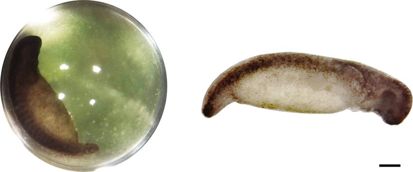
When embryos reached Harrison stage 32–34 (Harrison, 1969; for 15 min at room temperature. Following incubation in
Figure 1), individual eggs were removed from the jelly glutaraldehyde, embryos were washed three times in Holtfreter’s
mass. Algal presence was confirmed by the green color of solution without bicarbonate.
individual eggs. Cultured algae were labeled by adding 300 µL of 14 C
bicarbonate (at 37 MBq/mL) to 100 mL of Oophila algae in
14 C exponential growth in AF6 medium (at around 200,000 cells
Bicarbonate Incubation per mL) and incubating algae with 14 C bicarbonate for 4 h
Basic experimental design and methods for 14 C bicarbonate at 16◦ C under 260 µmol photons/m2 /s. Following incubation,
incubation experiments are visualized in Supplementary algae were transferred to 50 mL falcon tubes and centrifuged at
Figure S1. 300 × g for 8 min. The supernatant was discarded and algae were
resuspended in 20 mL complete Holtfreter’s solution. Algae were
Whole Eggs pelleted a second time at 300 × g for 8 min, and then resuspended
For experiments with whole eggs, individual eggs were removed in 1 mL complete Holtfreter’s solution. In parallel, unlabeled algae
from the jelly mass by gentle manipulation with gloved hands, were subjected to the same centrifugation and wash procedure.
and transferred to individual scintillation vials in 2.5 ml modified Unlabeled algae from the resuspended pellet were counted on
Holtfreter’s solution without sodium bicarbonate per egg. To a hemocytometer.
each egg, 1.5 µl of 14 C sodium bicarbonate [Perkin Elmer, For experiments with pre-labeled algae, a volume of 14 C
37 MBq/mL] was added. Adapting the protocol from Graham labeled algae approximately equivalent to 150,000 algal cells
et al. (2014), five individual eggs per experiment were incubated was added to live and glutaraldehyde killed decapsulated and
in the light or dark for 2 h. Following the initial incubation with washed embryos. Live and dead embryos were incubated
14 C bicarbonate, eggs were removed from the solution and rinsed with 14 C bicarbonate (1.5 µL at 37 MBq/mL per embryo in
with ultra-pure water to remove unincorporated 14 C bicarbonate 2.5 mL Holtfreter’s without bicarbonate) or 14 C labeled algae
from outside the egg. Following the rinse, eggs were transferred for 2 h in the dark at 16◦ C. Following incubation with 14 C
to 2.5 ml modified Holtfreter’s solution (not radioactive) per sodium bicarbonate or 14 C labeled algae, embryos were washed
egg and incubated in the dark for 2 additional hours. After three times with unlabeled Holtfreter’s solution, transferred to
the dark incubation, eggs were pierced with jeweler’s forceps, individual scintillation vials, crushed in 500 µL of 2M HCl,
and the embryo was removed. Intracapsular fluid, including all incubated for 24+ hours in open vials in a chemical fume
algae, and the egg membrane were retained in one scintillation hood to remove unincorporated bicarbonate, mixed with 10 mL
vial. Embryos were washed three times with Holtfreter’s solution scintillation cocktail, and subjected to scintillation counting as
without sodium bicarbonate to remove carry-over algae and described above.
were placed into a separate scintillation vial. Embryos were
homogenized in 500 µl of 2M HCl and incubated for 24 h in Photosynthesis Inhibitor Experiments
an open vial in a chemical fume hood to remove unincorporated Individual eggs were removed from the jelly mass by gentle
sodium bicarbonate as CO2 . 500 µl of 2M HCl was added to manipulation with gloved hands. Six eggs per experiment were
the algal fraction, which was also incubated for 24 h in an open placed together in a 50 mL falcon tube in 15 mL modified
vial in a chemical fume hood to remove unincorporated sodium Holtfreter’s solution without sodium bicarbonate. Eggs were pre-
bicarbonate. After 24 h incubation, 10 mL of scintillation cocktail incubated in the light at 16◦ C with two photosynthesis inhibitors,
(Perkin Elmer, Ultima Gold LLT) was added to all scintillation DCMU (3-(3,4-dichlorophenyl)-1,1-dimethylurea) and DBMIB
vials before vortexing, and radioactivity was assayed using a (2,5-dibromo-3-methyl-6-isopropylbenzoquinone) at 20 µM
Perkin Elmer Tri-Carb 3110TR scintillation counter. Results are final concentration for 1 h. Following pre-incubation, 9 µl of
Frontiers in Microbiology | www.frontiersin.org 3 August 2020 | Volume 11 | Article 1815Burns et al. Animal Carbon Fixation During Photosymbiosis
FIGURE 1 | Typical stage 33 A. maculatum embryo from these experiments. (A) Single egg capsule containing stage 33 embryo. Green hue is from intracapsular
algae. (B) Decapsulated stage 33 embryo. Scale bar: 1 mm.
14 C sodium bicarbonate, was added to each 50 mL Falcon glutaraldehyde killed embryos (p < 0.05). There was no
tube containing six eggs. Eggs were incubated in the light or significant difference between live or dead embryos when
dark for 2 h. Following incubation, eggs were removed from incubated with pre-labeled algae (p = 0.9) (Figure 2B). The
the Falcon tubes, washed with ultra-pure water, opened with result suggests that some algae persist through the washes but
jeweler’s forceps, and separated into individual scintillation vials. living A. maculatum embryos do not actively accrue additional
Intracapsular fluid with algae and the egg membrane from measurable photosynthate from lab-cultured algae in these
one egg were collected in one vial, each embryo was washed conditions. We note that although cultured algae are in a
three times with Holtfreter’s solution and placed in a separate different transcriptional state from intracapsular algae (Kerney
scintillation vial. Algae and embryos were processed as described et al., 2019), lab-cultured algae are able to interact with and
above for scintillation counting. invade A. maculatum embryos ex situ, similar to algae in the
intracapsular environment (Kerney et al., 2019). In this study,
Statistical Analyses decapsulated embryos incubated in the dark accumulated
Statistical analyses were performed in the R programming significantly more fixed carbon (Figure 2B) compared to
language (R Core Team, 2017). Plots were generated using the embryos incubated with pre-labeled algae (p < 0.001) or
ggplot2 package (Wickham, 2016). One-way ANOVA followed embryos killed with glutaraldehyde prior to incubation with
by the non-parametric Games-Howell post hoc test (used due to bicarbonate (p < 0.001).
violation of homogeneity of variance in the data) was performed
using the “userfriendlyscience” package (Peters, 2018). A. maculatum Embryos Compete With
O. amblystomatis Algae for Bicarbonate
Embryos from whole eggs incubated in the dark fixed
RESULTS significantly more carbon than embryos from whole eggs
incubated in the light (Figure 3A, p < 0.01). To test whether this
A. maculatum Embryos Fix Carbon was due to greater inorganic carbon availability when algae are
Decapsulated A. maculatum embryos (Figure 1B) incubated in not actively photosynthesizing (i.e., in the dark), photosynthesis
the dark incorporated inorganic carbon into their tissues as acid inhibitors were added to the eggs in the light and the dark.
stable forms (Figure 2A) while control embryos had no such Chemical inhibition of algal photosynthesis in the light decreased
signal (p < 0.01). Decapsulated Embryos incubated in the dark carbon fixation in the algae (Figure 3B, p < 0.01), and resulted in
exhibited significantly more 14 C-bicarbonate incorporation than elevated fixed carbon levels in salamander embryos, reproducing
total algae from whole eggs (as in Figure 1A) incubated in the the effect of placing the egg in the dark (Figure 3C).
dark (p < 0.01) indicating that the elevated radioactive signal in
the embryos could not have come from residual algae on or near
the dark incubated embryos (Figure 2A). DISCUSSION
To explicitly test whether residual algae that persisted
through wash steps could account for the fixed carbon signal In plants and algae, carbon dioxide is seen as a building block,
from embryos, 14 C-bicarbonate or pre-labeled O. amblystomatis a molecule captured from the air or surrounding fluid and
cultures were added to live or glutaraldehyde killed A. maculatum fixed into organic matter as part of photosynthesis. In animals,
embryos. When incubated with 14 C-bicarbonate, live embryos carbon dioxide is largely a by-product of respiration that is
exhibited an elevated fixed carbon signal compared to also co-opted as an important biological buffer in body fluids.
Frontiers in Microbiology | www.frontiersin.org 4 August 2020 | Volume 11 | Article 1815Burns et al. Animal Carbon Fixation During Photosymbiosis FIGURE 2 | A. maculatum embryos fix carbon. Box and whisker plots showing raw data (dark circles) range (thin lines) averages (thick horizontal lines) and upper and lower quartiles (box). In each plot, the y-axis represents radioactivity assayed per sample (DPM, disintegrations per minute); the x-axis represents the treatments: (A) embryo or algae incubated in the dark without (no label) or with 14 C-bicarbonate (14 C-bic). (B) Glutaraldehyde-killed or live embryo (embryo-killed and embryo-live, respectively) incubated in the light with 14 C-labeled alga (14 C-alg) or with 14 C-bicarbonate. n.s. = not significant, ∗ p < 0.05, ∗∗ p < 0.01, ∗∗∗ p < 0.001. Significance levels were determined by ANOVA followed by the Games-Howell post-hoc test. In addition to these roles, carbon dioxide and its hydration 2012). These changes are inferred to have a direct effect on product, bicarbonate, participate in several biosynthetic reactions algal cellular physiology, which interferes with the net benefit outside of photosynthesis. All animals can presumably re- Oophila confers to the host embryos. The balance between algal capture respiratory carbon for use in biosynthetic pathways and embryo manipulation of local pH through the action of (Windmueller and Spaeth, 1980; Marini, 2016). Aquatic animals, excreted carboxylases (Shiraiwa et al., 1993) and photosynthetic and their eggs and embryos in particular, however, have access and heterotrophic carbon fixation may additionally influence the to environmental carbon dioxide dissolved in water and can rate and quantity of carbon fixation at different embryonic stages import that exogenous carbon for use in biosynthetic processes (Cohen, 1954). (Flickinger, 1954; Mounib and Eisan, 1973). During the salamander-alga symbiosis, the carbon fixation In addition to biosynthetic roles, fixation of intracapsular processes observed in other animals are likely active in carbon dioxide by amphibian embryos may be important for A. maculatum embryos. The results presented here are consistent localized pH regulation. Amphibian embryos vary in their pH with heterotrophic fixation of exogenous bicarbonate by tolerances, and A. maculatum are particularly sensitive (Pierce, A. maculatum embryos (Table 1). As demonstrated in 1985). Their mortality can increase from 60% by other systems, such carbon fixation is necessary for proper lowering the pH from 7.0 to 6.0 and approaches 100% by pH 4.0 development, and here we provide evidence that the algae and (Pough, 1976). Previous research in the A. maculatum-Oophila salamander compete for exogenously supplied bicarbonate symbiosis found a pH of 4.5 decreases the partial pressure of (Table 1). Our results are consistent with the findings of oxygen in the egg capsule and increases intracapsular ammonia Hammen and Hutchison, 1962, suggesting that there is no as well as embryonic ammonia and lactate (Bianchini et al., measurable exchange of photosynthate from ecto-symbiotic Frontiers in Microbiology | www.frontiersin.org 5 August 2020 | Volume 11 | Article 1815
Burns et al.                                                                                                                Animal Carbon Fixation During Photosymbiosis

  FIGURE 3 | A. maculatum embryos compete with O. amblystomatis algae for bicarbonate. In each image, the y-axis represents radioactivity assayed per sample
  (DPM, disintegrations per minute); the x-axis represents the treatments. (A) Algae from whole eggs incubated in the light (L) or dark (D). (B) Cultured algae incubated
  in the light or dark with and without photosynthesis inhibitors (PI). (C) Carbon fixation by embryos in the whole egg environment, in the light or dark with and without
  photosynthesis inhibitors. n.s. = not significant, ∗∗ p < 0.01. Significance levels were determined by ANOVA followed by the Games-Howell post-hoc test.

TABLE 1 | Results summary.                                                               formation of glycerol, formate, or acetate (Catalanotti et al.,
                                                                                         2013), which are used in other photosynthetic endosymbioses
Experiment                                           Embryo                  Algae
                                                                                         (glycerol; Léon and Galván, 1995) or ectosymbiotic bacterial
Decapsulated embryo–light                              +++                     na        associations (formate and acetate; Den Besten et al., 2013;
Decapsulated embryo–dark                               +++                     na        Karasov and Douglas, 2013). While there is no evidence that
Decapsulated embryo–killed                              −                      na        endosymbiotic Oophila enable A. maculatum embryos to utilize
Decapsulated embryo+labeled algae                       −                      na        photosynthesis as a direct energy source, the chemical dialogue
Whole egg–light                                         ++                   ++++        between this intracellular mutualist and its vertebrate host is a
Whole egg–dark                                         +++                      +        fascinating research topic for subsequent studies.
Whole egg–light–PI                                     +++                      +
Whole egg–dark–PI                                      +++                      +
                                                                                         AUTHOR’S NOTE
The number of pluses (+) indicates the relative amount of inorganic carbon
assimilated under the indicated conditions: (+) low, (++) moderate, (+++) high,
(++++) max observed. A dash (−) indicates that no carbon assimilation was                This manuscript has been released as a pre-print at BioRxiv
observed above background. An na indicates “not applicable” in experiments               (Burns et al., 2020).
where algae were not present or were pre-labeled. “PI” indicates the addition of
photosynthesis inhibitors in those experiments.
                                                                                         DATA AVAILABILITY STATEMENT
algae to salamander embryos. The studies suggesting such
                                                                                         The datasets generated for this study are available on request to
an exchange (Graham et al., 2013, 2014) were likely only
                                                                                         the corresponding author.
measuring variability in heterotrophic carbon fixation between
individual embryos and did not control for the possibility that
the salamander embryos themselves were fixing significant                                ETHICS STATEMENT
quantities of exogenously supplied bicarbonate.
   Further studies on endosymbiotic algae are needed to reveal                           Ethical review and approval was not required for the animal
whether intracellular algal metabolites which are produced by                            study because research on pre-hatchling archosaur and ectotherm
the subset of endosymbiotic Oophila inside A. maculatum                                  embryos does not require Institutional Animal Care and Use
host cells are assimilated by their embryonic hosts. Our                                 Committee protocol approval according to Public Health Service
previous transcriptomic analysis has revealed the metabolic                              policy (NIH 2015; National Research Council 2011). Embryos
shift of intracellular algae from oxidative phosphorylation to                           were treated with the same ethical standards as free-living larvae
fermentation (Burns et al., 2017), which may coincide with the                           and care was taken to minimize stress and the number of embryos

Frontiers in Microbiology | www.frontiersin.org                                      6                                              August 2020 | Volume 11 | Article 1815
Burns et al. Animal Carbon Fixation During Photosymbiosis
used in this study. National Research Council (2011) Guide collection and analysis, decision to publish, or preparation of
for the Care and Use of Laboratory Animals, Eighth Edition. the manuscript.
That National Academies Press. Washington D.C. https://doi.
org/10.17226/12910. NIH Office of Laboratory Animal Welfare
(2015) Public Health Service Policy on the Humane Care ACKNOWLEDGMENTS
and Use of Laboratory Animals. United States Department of
Health and Human Services. https://olaw.nih.gov/policies-laws/ The authors thank the Challenge Yourself to Change (CYTC)
phs-policy.htm. community group in East Stroudsburg, PA, especially Rocky
Sayles, Marquise Long, Dominic Kaps, Julius Patterson,
and others who put on waders when collecting spotted
AUTHOR CONTRIBUTIONS salamander egg masses that contributed to this research. The
authors also thank Darryl Speicher, Roger Spotts, and Kettle
JB and SD conceived, planned, and conducted the experiments.
Creek Environmental Education Center for aid in egg mass
JB performed the data analysis. JB, SD, and RK discussed the
collection and Hui Yang for thoughtful comments on drafts of
results and implications and co-wrote the manuscript. All authors
the manuscript.
contributed to the article and approved the submitted version.
FUNDING SUPPLEMENTARY MATERIAL
This work was supported by the Gordon and Betty Moore The Supplementary Material for this article can be found
Foundation Grant #GBMF5604 (https://doi.org/10.37807/ online at: https://www.frontiersin.org/articles/10.3389/fmicb.
GBMF5604). The funders had no role in study design, data 2020.01815/full#supplementary-material
REFERENCES Cohen S. (1954). The metabolism of 14 CO2 during amphibian development. J. Biol.
Chem. 211, 337–54.
Allen, A. P., Gillooly, J. F., and Brown, J. H. (2005). Linking the global carbon cycle Cohen, S. (1963). 14CO2 fixation and the accumulation of malonic acid in
to individual metabolism. Funct. Ecol. 19, 202–213. doi: 10.1111/j.1365-2435. amphibian hybrids (R. pipiens - female x R. sylvatica - male). Exp. Cell Res.
2005.00952.x 29, 207–211. doi: 10.1016/0014-4827(63)90376-8
Bachmann, M. D., Carlton, R. G., Burkholder, J. M., and Wetzel, R. G. Den Besten, G., van Eunen, K., Groen, A. K., Venema, K., Reijngoud, D. J., and
(1986). Symbiosis between salamander eggs and green algae: microelectrode Bakker, B. M. (2013). The role of short-chain fatty acids in the interplay between
measurements inside eggs demonstrate effect of photosynthesis on oxygen diet, gut microbiota, and host energy metabolism. J. Lipid Res. 54, 2325–2340.
concentration. Can. J. Zool. 64, 1586–1588. doi: 10.1139/z86-239 doi: 10.1194/jlr.r036012
Baltar, F., and Herndl, G. J. (2019). Ideas and perspectives: is dark carbon fixation Elinson, R. P., and del Pino, E. M. (2011). Developmental diversity of amphibians.
relevant for oceanic primary production estimates? Biogeosciences 16, 3793– Wiley Interdiscip. Rev. Dev. Biol. 1, 345–369. doi: 10.1002/wdev.23
3799. doi: 10.5194/bg-16-3793-2019 Erb, T. J. (2011). Carboxylases in natural and synthetic microbial pathways. Appl.
Berg, I. A. (2011). Ecological aspects of the distribution of different autotrophic Environ. Microbiol. 77, 8466–8477. doi: 10.1128/AEM.05702-11
CO2 fixation pathways. Appl. Environ. Microbiol. 77, 1925–1936. doi: 10.1128/ Flickinger, R. A. (1954). Utilization of 14CO2 by developing amphibian embryos,
AEM.02473-10 with special reference to regional incorporation into individual embryos. Exp.
Bianchini, K., Tattersall, G. J., Sashaw, J., Porteus, C. S., and Wright, P. A. (2012). Cell Res. 6, 172–180. doi: 10.1016/0014-4827(54)90159-7
Acid water interferes with salamander-green algae symbiosis during early Gilbert, P. W. (1942). Observations on the eggs of Ambystoma maculatum with
embryonic development. Physiol. Biochem. Zool. 85, 470–480. doi: 10.1086/ especial reference to the green algae found within the egg envelopes. Ecology 23,
667407 215–227. doi: 10.2307/1931088
Biggers, J., and Bellve, A. (1974). “Carbon dioxide in developmental systems,” Gilbert, P. W. (1944). The alga-egg relationship in Ambystoma maculatum, a case
in Proceedings of the Carbon Dioxide and Metabolic Regulations Satellite of symbiosis. Ecology 25, 366–369. doi: 10.2307/1931284
SYmposium of the XXV Internaitonal Congress of Physiology, Monaco. Goff, L. J., and Stein, J. R. (1978). Ammonia: basis for algal symbiosis in salamander
Blanchard, C. Z., and Waldrop, G. L. (1998). Overexpression and kinetic egg masses. Life Sci. 22, 1463–1468. doi: 10.1016/0024-3205(78)90641-0
characterization of the carboxyltransferase component of acetyl-CoA Gong, F., Cai, Z., and Li, Y. (2016). Synthetic biology for CO2 fixation. Sci. China
carboxylase. J. Biol. Chem. 273, 19140–19145. doi: 10.1074/jbc.273.30.19140 Life Sci. 59, 1106–1114. doi: 10.1007/s11427-016-0304-2
Boston, H. L., Adams, M. S., and Madsen, J. D. (1989). Photosynthetic strategies Graham, E. R., Fay, S. A., Davey, A., and Sanders, R. W. (2013). Intracapsular algae
and productivity in aquatic systems. Aquat. Bot. 34, 27–57. doi: 10.1016/0304- provide fixed carbon to developing embryos of the salamander Ambystoma
3770(89)90049-1 maculatum. J. Exp. Biol. 216, 452–459. doi: 10.1242/jeb.076711
Brinson, M. M., Lugo, A. E., and Brown, S. (1981). Primary productivity, Graham, E. R., McKie-Krisberg, Z. M., and Sanders, R. W. (2014). Photosynthetic
decomposition and consumer activity in freshwater wetlands. Annu. Rev. Ecol. carbon from algal symbionts peaks during the latter stages of embryonic
Evol. Sci. 12, 123–161. doi: 10.1146/annurev.es.12.110181.001011 development in the salamander Ambystoma maculatum. BMC Res. Notes 7:764.
Burns, J. A., Kerney, R., and Duhamel, S. (2020). Heterotrophic carbon fixation in a doi: 10.1186/1756-0500-7-764
salamander-alga symbiosis. bioRxiv [Preprint]. doi: 10.1101/2020.02.14.948299 Hammen, C. S., and Hutchison, V. H. (1962). Carbon dioxide assimilation in
Burns, J. A., Zhang, H., Hill, E., Kim, E., and Kerney, R. (2017). Transcriptome the symbiosis of the salamander Ambystoma maculatum and the alga Oophila
analysis illuminates the nature of the intracellular interaction in a vertebrate- amblystomatis. Life Sci. 1, 527–532. doi: 10.1016/0024-3205(62)90113-3
algal symbiosis. eLife 6:e22054. doi: 10.7554/eLife.22054.049 Harrison, R. G. (1969). “Harrison stages and description of the normal
Catalanotti, C., Yang, W., Posewitz, M. C., and Grossman, A. R. (2013). development of the spotted salamander, Amblystoma punctatum,” in
Fermentation metabolism and its evolution in algae. Front. Plant Sci. 4:150. Organization and Development of the Embryo, ed. S. Wilens (London:
doi: 10.3389/fpls.2013.00150 Yale University Press), 44–66.
Frontiers in Microbiology | www.frontiersin.org 7 August 2020 | Volume 11 | Article 1815Burns et al. Animal Carbon Fixation During Photosymbiosis
Holden, H. M., Thoden, J. B., and Raushel, F. M. (1999). Carbamoyl phosphate R Core Team (2017). R: A Language And Environment For Statistical Computing.
synthetase: an amazing biochemical odyssey from substrate to product. Cell. Vienna: R Foundation for Statistical Computing.
Mol. Life. Sci. 56, 507–522. doi: 10.1007/s000180050448 Raven, J. A. (2017). “Symbiosis involving photosynthetic organisms,” in Algal and
Jitrapakdee, S., Vidal-Puig, A., and Wallace, J. C. (2006). Anaplerotic roles of Cyanobacteria Symbioses, eds M. Grube, J. Seckbach, and A. Muggia (Singapore:
pyruvate carboxylase in mammalian tissues. Cell. Mol. Life. Sci. 63, 843–854. World Scientific), 3–41. doi: 10.1142/9781786340580_0001
doi: 10.1007/s00018-005-5410-y Shampo, M. A. (1999). Stamp vignette on medical science: Stanley Cohen–Nobel
Karasov, W. H., and Douglas, A. E. (2013). Comparative digestive physiology. Laureate for growth factor. Mayo Clin. Proc. Rochester 74:600. doi: 10.4065/74.
Compr. Physiol. 3, 741–783. doi: 10.1002/cphy.c110054 6.600
Kerney, R. (2011). Symbioses between salamander embryos and green algae. Shiraiwa, Y., Goyal, A., and Tolbert, N. E. (1993). Alkalization of the medium by
Symbiosis 54, 107–119. doi: 10.1007/s13199-011-0134-2 unicellular green algae during uptake of dissolved inorganic carbon. Plant Cell
Kerney, R., Kim, E., Hangarter, R. P., Heiss, A. A., Bishop, C. D., and Hall, B. K. Physiol. 34, 649–657. doi: 10.1093/oxfordjournals.pcp.a078467
(2011). Intracellular invasion of green algae in a salamander host. Proc. Natl. Small, D. P., Bennett, R. S., and Bishop, C. D. (2014). The roles of oxygen
Acad. Sci. U.S.A. 108, 6497–6502. doi: 10.1073/pnas.1018259108 and ammonia in the symbiotic relationship between the spotted salamander
Kerney, R., Leavitt, J., Hill, E., Zhang, H., Kim, E., and Burns, J. (2019). Co-cultures Ambystoma maculatum and the green alga Oophila amblystomatis during
of Oophila amblystomatis between Ambystoma maculatum and Ambystoma embryonic development. Symbiosis 64, 1–10. doi: 10.1007/s13199-014-0297-8
gracile hosts show host-symbiont fidelity. Symbiosis 78, 73–85. doi: 10.1007/ Tiedemann, H., and Tiedemann, H. (1954). Einbau von14CO2 in gefurchte und
s13199-018-00591-2 ungefurchte Eihälften und in verschiedene entwicklungsstadien von Triton.
Kresge, N., Simoni, R., and Hill, R. (2005). The discovery of heterotrophic carbon Naturwissenschaften 41, 535–535. doi: 10.1007/BF00623059
dioxide fixation by Harland G. Wood. J. Biol. Chem. 280:e15. Tremblay, P., Grover, R., Maguer, J. F., Legendre, L., and Ferrier-Pagès, C.
Léon, R., and Galván, F. (1995). Glycerol photoproduction by free and Ca-alginate (2012). Autotrophic carbon budget in coral tissue: a new 13C-based model
entrapped cells of Chlamydomonas reinhardtii. J. Biotechnol. 42, 61–67. doi: of photosynthate translocation. J. Exp. Biol. 215, 1384–1393. doi: 10.1242/jeb.
10.1016/0168-1656(95)00069-3 065201
Marini, J. C. (2016). Interrelationships between glutamine and citrulline Venn, A. A., Loram, J. E., and Douglas, A. E. (2008). Photosynthetic symbioses in
metabolism. Curr. Opin. Clin. Nutr. 19, 62–66. doi: 10.1097/mco.00000000 animals. J. Exp. Bot. 59, 1069–1080. doi: 10.1093/jxb/erm328
00000233 Wales, R. G., Quinn, P., and Murdoch, R. N. (1969). The fixation of carbon dioxide
Mills, N. E., and Barnhart, M. C. (1999). Effects of hypoxia on embryonic by the eight-cell mouse embryo. J. Reprod. Fertil. 20, 541–543. doi: 10.1530/jrf.
development in two Ambystoma and two Rana species. Physiol. Biochem. Zool. 0.0200541
72, 179–188. doi: 10.1086/316657 Wickham, H. (2016). Ggplot2: Elegant Graphics For Data Analysis. Berlin: Springer.
Mounib, M. S., and Eisan, J. S. (1969). Metabolism of pyruvate and glyoxylate Windmueller, H. G., and Spaeth, A. E. (1980). Respiratory fuels and
by eggs of salmon (Salmo salar). Comp. Biochem. Physiol. 29, 259–264. doi: nitrogen metabolism in vivo in small intestine of fed rats. Quantitative
10.1016/0010-406x(69)91742-3 importance of glutamine, glutamate, and aspartate. J. Biol. Chem. 255,
Mounib, M. S., and Eisan, J. S. (1973). Fixation of carbon dioxide and some of the 107–112.
enzymes involved in cod eggs. Int. J. Biochem. 4, 207–212. doi: 10.1016/0020- Wood, H. G., Vennesland, B., and Evans, E. A. (1945). The mechanism of carbon
711x(73)90014-1 dioxide fixation by cell-free extracts of pigeon liver: distribution of labeled
Orr, H. (1888). Memoirs: note on the development of amphibians, chiefly carbon dioxide in the products. J. Biol. Chem. 159, 153–158.
concerning the central nervous system; with additional observations on the Wood, H. G., Werkman, C. H., Hemingway, A., and Nier, A. O. (1941). Heavy
hypophysis, mouth, and the appendages and skeleton of the head. J. Cell Sci. carbon as a tracer in heterotrophic carbon dioxide assimilation. J. Biol. Chem.
2, 295–324. 139, 365–376.
Peters, G. (2018). Userfriendlyscience: Quantitative Analysis Made Accessible.
R package version 0.7.2. Available online at: https://userfriendlyscience.com Conflict of Interest: The authors declare that the research was conducted in the
(accessed July 22, 2020). absence of any commercial or financial relationships that could be construed as a
Pierce, B. A. (1985). Acid tolerance in amphibians. BioScience. 35, 239–243. doi: potential conflict of interest.
10.2307/1310132
Pinder, A., and Friet, S. (1994). Oxygen transport in egg masses of the amphibians Copyright © 2020 Burns, Kerney and Duhamel. This is an open-access article
Rana sylvatica and Ambystoma maculatum: convection, diffusion and oxygen distributed under the terms of the Creative Commons Attribution License (CC BY).
production by algae. J. Exp. Biol. 197, 17–30. The use, distribution or reproduction in other forums is permitted, provided the
Pough, F. H. (1976). Acid precipitation and embryonic mortality of spotted original author(s) and the copyright owner(s) are credited and that the original
salamanders, Ambystoma maculatum. Science 192, 68–70. doi: 10.1126/science. publication in this journal is cited, in accordance with accepted academic practice. No
3852 use, distribution or reproduction is permitted which does not comply with these terms.
Frontiers in Microbiology | www.frontiersin.org 8 August 2020 | Volume 11 | Article 1815You can also read