CEFTIZOXIME LOADED ZNO/L CYSTEINE BASED AN ADVANCED NANOCARRIER DRUG FOR GROWTH INHIBITION OF SALMONELLA TYPHIMURIUM - NATURE
←
→
Page content transcription
If your browser does not render page correctly, please read the page content below
www.nature.com/scientificreports
OPEN Ceftizoxime loaded ZnO/l‑cysteine
based an advanced nanocarrier
drug for growth inhibition
of Salmonella typhimurium
M. S. Bacchu1,2, M. R. Ali1,2, M. A. A. Setu3, S. Akter3 & M. Z. H. Khan1,2*
l-Cysteine coated zinc oxide (ZnO) nano hollow spheres were prepared as a potent drug delivery agent
to eradicate Salmonella enterica serovar Typhimurium (S. typhimurium). The ZnO nano hollow spheres
were synthesized by following the environmentally-friendly trisodium citrate assisted method and
l-cysteine (L-Cys) conjugate with its surface. ZnO/L-Cys@CFX nanocarrier drug has been fabricated
by incorporating ceftizoxime with L-Cys coated ZnO nano hollow spheres and characterized using
different techniques such as scanning electron microscope (SEM), attenuated total reflection Fourier
transform infrared (ATR-FTIR), and X-ray diffraction (XRD) etc. Furthermore, the drug-loading
and encapsulation efficiency at different pH levels was measured using UV–vis spectrometer and
optimized. A control and gradual manner of pH-sensitive release profile was found after investigating
the release profile of CFX from the carrier drug. The antibacterial activity of ZnO/L-Cys@CFX and CFX
were evaluated through the agar disc diffusion method and the broth dilution method, which indicate
the antibacterial properties of antibiotics enhance after conjugating. Surprisingly, the ZnO/L-Cys@
CFX exhibits a minimum inhibitory concentration (MIC) of 5 µg/ml against S. typhimurium is lower than
CFX (20 µg/ml) itself. These results indicate the nanocarrier can reduce the amount of CFX dosed to
eradicate S. typhimurium.
Antibiotic is the greatest invention at last century used to eradicate pathogens by reducing pathogen growth
and destroying t hem1,2. Conventional antibiotics have drawbacks, such as multidrug-resistance (MDR) bacteria,
minimum bioavailability, less diffusion capacity to the outer membrane, etc.3. World health organization (WHO)
defines these MDR bacteria as microorganisms that can grow in the presence of one or more a ntibiotics4. The
recovery from these MDR strains caused the healthcare-associated infection to become a more significant chal-
lenge day by day. High doses of antibiotics are administrated to overcome this challenge, which may generate
toxic and adverse effects. A promising strategy to manage MDR bacteria-caused infections is applying nano-
particles (NPs) to restrain the MDR m icroorganism5–7. NPs can be the carrier of antibiotics and enhance the
antibacterial activity of the a ntibiotics8. This nanocarrier can enter cell’s intracellular endocytic pathways and
deliver the drug at a targeted position9. The NP-based carrier for antibiotics significantly reduces the antibiotic
doses required to eradicate pathogen several times than the dose of the free antibiotics.
Salmonella typhimurium is a highly gram-negative pathogenic bacteria which causes salmonellosis for human
and animals10. The global concern about the treatment of S. typhimurium poses infection is day by day increas-
ing due to its antibiotic resistance behavior. The antibiotic susceptibility properties of S. typhimurium were
decreased, and this pathogen is resistant to chloramphenicol, tetracycline, streptomycin, ampicillin, etc.11. For
this reason, we choose ceftizoxime (CFX) (member of cephalosporin antibiotic group) to treat S. typhimurium
caused the infection. CFX is a third-generation antibiotic which is effective for both gram-positive and gram-
negative bacteria. The minimum amount of CFX requires to inhibit pathogenic growth for human body tissue
causes different adverse e ffects12. It is crucial to improve the therapeutic efficiency of CFX, which can be achieved
by developing CFX loaded nano-drug carrier (NDC) with the minimum side e ffect13. This NDC increases the
therapeutic efficiency and reduces the drug consumption rate by the sustainable release of antibiotics. Different
inorganic nanoparticles are widely used as carriers for antibiotics such as gold nanoparticles, silver nanoparticles,
1
Department of Chemical Engineering, Jashore University of Science and Technology, Jashore 7408,
Bangladesh. 2Laboratory of Nano‑Bio and Advanced Materials Engineering (NAME), Jashore University of Science
and Technology, Jashore 7408, Bangladesh. 3Department of Microbiology, Jashore University of Science and
Technology, Jashore 7408, Bangladesh. *email: zaved.khan@just.edu.bd
Scientific Reports | (2021) 11:15565 | https://doi.org/10.1038/s41598-021-95195-0 1
Vol.:(0123456789)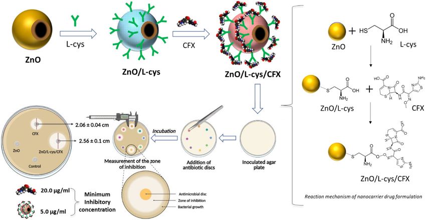
www.nature.com/scientificreports/
iron oxide nanoparticles, silica nanoparticles, zinc oxide nanoparticles, etc. Different biocompatible polymers
are generally used in NDC for coating with nanoparticles to achieve specific targeted and sustainable r elease14.
We choose a noble NDC based on l-cysteine (L-Cys) coated hollow Zinc oxide nanosphere to reduce antibiotic
consumption. ZnO is a safe metal oxide approved by the US food and drug administration (FDA), suggesting the
micro to larger size ZnO are generally safe, but nano shape might be toxic. It is accepted that discrete polymer
coating on the surface of ZnO reduces its toxicity and makes it more efficient for drug d elivery15. Nano-shaped
ZnO exhibited antibacterial properties over different pathogenic bacteria such as Escherichia coli, Staphylococcus
aureus, etc.16–22. A variety of nanostructured ZnO is generally used in the drug delivery system, such as quantum
dots, nanospheres, nanoshells, nanosheets, nanorods, nano hollow spheres, nanobelts, and nanobelts, etc. Among
them, nano hollow spheres take greater attention due to substantial internal space in their core. This core helps
in drug transport, specific targeted drug delivery to the particular regime, and maintains release p rofile23,24.
l-Cysteine (L-Cys) is a member of amino acid groups that are widely used in targeted drug delivery because
of its active groups (carboxylic –COOH, amines –NH2, and thiol -SH). The thiol group present in L-Cys gives
some exciting properties such as metal conjugating, damage skin repairing, and cross-linking25. The three func-
tional groups of L-Cys may interact with the ZnO sphere surface where the thiol group of L-Cys reacts with
Zn2+ of ZnO and form a complex26. L-Cys works as a scavenger free radical that create oxidative stress to cause
cellular damage27.
In this work, we present CFX loaded NDC named ZnO/L-Cys@CFX for controlled drug delivery for the
growth inhibition of S. typhimurium. Here, the carrier matrix is L-Cys coated ZnO nano hollow sphere synthe-
sized in simple and environmentally-friendly trisodium citrate assisted method. Finally, the antimicrobial test,
such as the zone inhibition analysis and minimum inhibitory concentration (MIC) analysis, is carried out over
S. typhimurium to characterize the NDC.
Materials and methods
Materials and reagent. All chemicals and reagents used in this study were in high analytical grade. Triso-
dium citrate (Na3C6H5O7), Zinc nitrate hexahydrate (Zn(NO3)2‧6H2O), sodium chloride (NaCl) were purchased
from Merck (Germany). Hexamine (C6H12N4) was brought from PT. SMART LAB (Indonesia). CFX and L-Cys
were purchased from Aladdin Reagent Ltd. (China). Salmonella Shigella (SS) agar and Mueller Hinton agar were
purchased from Oxoid(UK). Ultrapure water from Evoqua (Germany, resistivity > 18 Ω) was used for all types
of preparations. Phosphate buffer saline (PBS) of 0.01 M used for the release study was purchased from Aladdin
Reagent (China).
Instruments. Scanning electron microscope (SEM) of different nanoparticles and drug carriers was per-
formed using ZEISS GeminiSEM 500 field emission scanning electron microscope (FE-SEM). A NICOLERT
iS20 ATR-FTIR, Bruker D8 Advance PXRD, and Shimadzu lab solution UV–VIS (UV-1900i) were also used
to investigate the different physicochemical characterization and drug loading efficiency. An electrochemical
workstation of Corrtest CS300 was used to conduct in vitro released behavior of the carrier drug.
Synthesis of hollow ZnO nanosphere. The ZnO nano hollow spheres were synthesized by following a
slightly modified procedure, as previously r eported28. In short, a 200 ml aqueous solution of 15 mM zinc nitrate
hexahydrate, 15 mM hexamine, and 4.08 mM trisodium citrate were transferred into 250 ml round bottom flux
made of bomex glass. Then, the flux was sealed using a sealing plug and placed in the oven at 90 °C for two
hours. After completing the reaction period, the white color residue was found, and the precipitate was collected
by centrifuging at 5000 rpm after naturally cooling. The resulting ZnO was washed with ultrapure water several
times to remove the residue and dried at 60 °C for 24 h. The pure hollow shape ZnO was found after annealing
at 400 °C for 2 h and finely grinding.
Preparation of ZnO/L‑Cys and ZnO/L‑Cys@CFX. To prepare ZnO/L-Cys, 100 mg of hollow ZnO
nanosphere, 50 mg of L-Cys were dispersed in 10 ml normal saline (0.85% NaCl) separately. Each solution was
ultrasonicated for 5 min and stirred magnetically by using a magnetic stirred for 1 h. After formulating the
homogenous solution, the required amount of ZnO and L-Cys solutions were taken in 40 ml normal saline to
get a suspension of 1 mg ZnO and 0.5 mg L-Cys. Then, the resulting solution was stirred using a magnetic stirrer
for 1 h to prepare L-Cys coated hollow ZnO nanosphere (ZnO/L-Cys).
To prepare ZnO/L-Cys@CFX, 1 mg of CFX was immersed in 10 ml normal saline to prepare 100 µg/ml CFX
stock solution and stirred magnetically to make a homogenous solution. An aliquot of required volume CFX
was added to the ZnO/L-Cys solution to prepare ZnO/L-Cys@CFX where the concentrations of CFX were 1,
5, 10, 20 µg/ml and stirred in a magnetic stirrer for 1 h. The ZnO/L-Cys@CFX of different concentrations (1,
5, 10, 20 µg/ml based on CFX) were denoted as C 1, C2, C3, C4. Then the prepared solutions were stored at 4 °C
and used for different antibacterial tests. Scheme 1 represents the preparation steps of the ZnO/L-Cys@CFX
nanocarrier and its reaction mechanism.
Drug loading and encapsulation efficiency. The amount of drug loading was determined by using a
UV–Vis spectrometer to assess the effect of pH on drug adsorption. For this study, ZnO/L-Cys@CFX ( C3) was
prepared in ultrapure water of different pH values ranging from 1.5 to 7.5 according to a similar procedure as
previously stated. Then, the resulting solutions were centrifuged at 6000 rpm, and the supernatant was collected.
To determine the amount of drug loading and encapsulation efficiency, the supernatants were analyzed with
UV–Vis spectrometer at the wavelength of 298 nm29 using a calibration curve of CFX in the concentration of
1–15 µg/ml (y = 0.0152x + 0.0302 and R2 = 0.985) shown in Fig. 1A. The amount of drug loading was calculated
Scientific Reports | (2021) 11:15565 | https://doi.org/10.1038/s41598-021-95195-0 2
Vol:.(1234567890)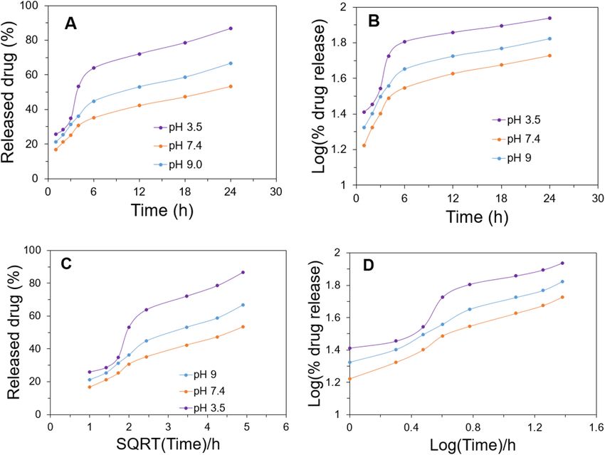
www.nature.com/scientificreports/

                                  Scheme 1.  Step-by-step preparation technique of proposed nanocarrier drug and its working principle.

                                  Figure 1.  Calibration curve for measuring drug loading efficiency by using UV–Vis spectroscopy (A).
                                  Calibration curve for evaluating drug release percentage by using the electrochemical sensor at pH 3.5 (B), pH
                                  7.4 (C), and pH 9.0 (D).

                                  by subtracting the total amount of drug that remains in supernatants from the total amount of drug initially
                                  used. The encapsulation efficiency was calculated by dividing the total amount of drug loading by the total
                                  amount of drug loading drug initially used.

                                  In vitro drug release. For the drug release study, a certain amount of ZnO/L-Cys@CFX was sequentially
                                  immersed into 50 ml of PBS solution of (pH 3.5, 7.4 and 9.0) in a water bath at 37 °C with continuous shaking. At
                                  specific time intervals, 4 ml of the resulting solutions were withdrawn and the amount of release was calculated
                                  by using an electrochemical sensor where MWCNT/P-Cys@MIP modified glassy carbon electrode (GCE) was
                                  used as a working electrode, silver/silver chloride (Ag/AgCl) electrode and platinum wire were used as refer-
                                  ence and counter electrode. The amount of drug released was evaluated according to our previously reported

Scientific Reports |   (2021) 11:15565 |              https://doi.org/10.1038/s41598-021-95195-0                                                   3

                                                                                                                                            Vol.:(0123456789)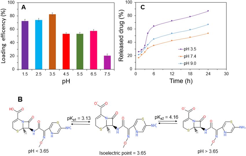
www.nature.com/scientificreports/
work30. Calibration curves of different pH were also done to determine the amount of CFX released, as shown
in Fig. 1B–D.
Antibacterial tests. Bacterial culture. For the antimicrobial assay, we have used CFX sensitive S. typhimu-
rium (ATCC 19585) strain. We resuscitated the lyophilized cells on nutrient broth and culture on MacConkey
agar medium. Pure discrete typical colonies from the plate were subcultured on Mullier Hinton agar and used
further for the antimicrobial susceptibility tests.
Antibiotic susceptibility assay. Agar disc diffusion method. We followed the National Committee for Clini-
cal Laboratory Standards (NCCLS) guideline31,32 and performed the antibiotic susceptibility assay by modified
Kirby–Bauer’s disc diffusion m ethod33 on Mueller–Hinton Agar media. We have used CFX antibiotic at differ-
ent concentrations (1.0. 5.0, 10.0 and 20.0 μg/ml) alone named D 1, D5, D10, D20 and in combination with 1 mg/
ml ZnO nano hollow sphere and 0.5 mg/ml l-cysteine cross-linked nanoparticle named C1, C2,C3, and C4. The
bacterial suspension was prepared using overnight grown pure culture on Muller Hinton agar media into nor-
mal physiological saline (0.85% NaCl) and adjusted the turbidity comparing with 0.5% MacFarland standard. A
non-absorbent cotton swab was dipped in the suspension and spread over a Muller Hinton agar plate to make
a bacterial lawn. Blank antimicrobial susceptibility discs (ThermoScientific Oxoid) of 0.5 cm are placed at the
center, and measured antibiotic solutions (with or without nanoparticles) are poured on the disc. The Petri
dishes are incubated upside up the position at 37 °C for 18 h. The zone of bacterial growth inhibition around the
discs is measured manually using a cm-scale.
Broth dilution method to compare the effect of nanoparticles conjugated with antibiotic on bacterial suscepti-
bility. We compare the bactericidal effect of ZnO/L-Cys@CFX and CFX by broth dilution method according
to EUCAST, 2003 guidelines of European Society of Clinical Microbiology & Infectious Disease (ESCMID).
Ceftizoxime antibiotic concentrations were set at 1.0 μg/ml, 5.0 μg/ml, 10.0 μg/ml and 20.0 μg/ml named D1, D2,
D3, D4. Tubes with antibiotic alone and, in combination with 1 mg/ml ZnO nano hollow sphere and 0.5 mg/ml
l-cystine cross-linked nanoparticle named C 1, C2, C3, and C
4 were prepared in normal saline. All the tubes were
inoculated with a measured amount of antibiotic-sensitive S. typhimurium strain. All the tubes were incubated
at 37 °C, and after 6 h of an interval, viable bacterial count (CFU/ml) was measured by the drop plate method on
MacConkey agar plates. The plates were incubated overnight (17–20 h) at 37 °C. The colony-forming unit (CFU)
of the three drops in the countable dilution range were averaged, and the total count was scaled up34. The same
procedure has been adopted for determining the antimicrobial activity of ZnO nano hollow spheres, L-Cys, and
ZnO/L-Cys. In this case, five different concentrations of ZnO nano hollow sphere, L-Cys, and ZnO/L-Cys of
three were used as test suspensions and followed the above-mention protocol to detect its effect on the bacterial
strain.
Results and discussions
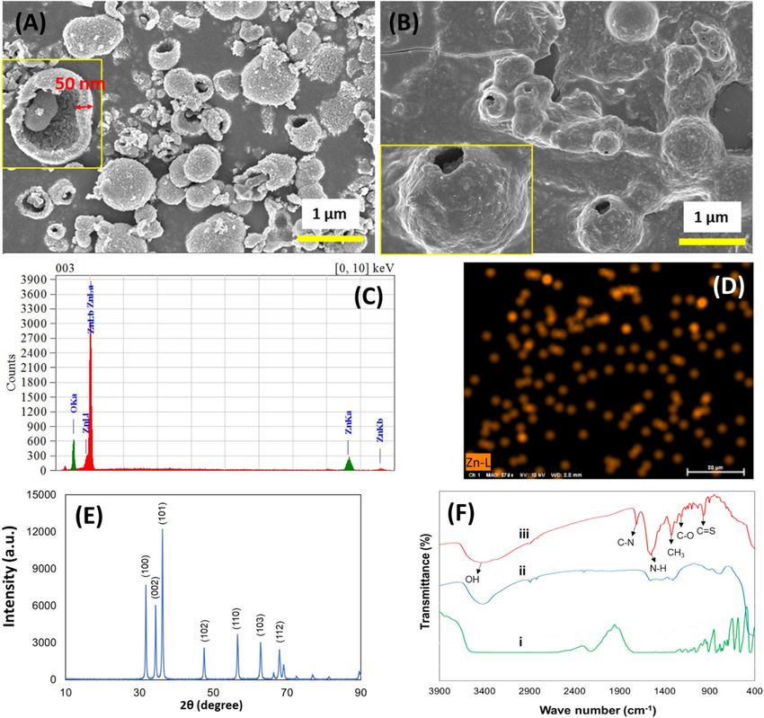
Physio‑chemical properties of Carrier matrix. The morphology of the obtained nanocarrier was char-
acterized using field emission scanning electron microscopy as shown in Fig. 2. Figure 2A shows the low and
high magnification SEM images of ZnO hollow spheres. The average diameter of individual particles ranged
from 95 to 350 nm with sphere-shaped nanocrystal structures and the thickness of the walls is about 50 nm. The
SEM image of the CFX loaded ZnO/L-Cys nanocarrier was shown in Fig. 2B. It can be observed that the overall
morphology of the nanospheres remained unchanged even after the incorporation of CFX which suggests strong
binding of drug molecules with nanocarrier through cross-linking of L-Cys. The EDX spectra (Fig. 2C) and ele-
mental mapping of the same (Fig. 2D) confirm the presence and uniform distribution of Zn in the carrier drug.
The changes of phase structure and crystallite size of the as-prepared ZnO hollow sphere were investigated
by XRD measurement and represented in Fig. 2E. The observed diffraction peaks are in good agreement with
the pure ZnO hollow microsphere structure (JCPDS card 36-1451)35, which confirm the hollow structure of
the sample with P63mc symmetry. No other peaks of impurities and different crystalline phases existed, which
ensures the purity of synthesized materials, which is in good agreement with SEM results. The earlier researcher
reported the higher entrapment capacity of hollow nanocarriers for greater concentrated release of drugs14.
ATR-FTIR analyses were performed to evaluate the functional groups of the nanocarrier surface and the
following conjugation of the target drug (Fig. 2F). The presence of a Zn–O stretching mode is confirmed by the
intense broadband at 430–500 cm−1 in the spectrum of ZnO hollow spheres. The spectrum of L-Cys (Fig. 2Fi) can
be labeled as (–NH) bending vibration at 1140 cm−1 and while peaks around 900 cm−1 prove the –COOH group.
In addition, a weak band attributed to the (–SH) group of cysteine molecule is observed near 2350 cm−1 which
completely disappears (Fig. 2Fii) due to the cleavage of S–H bonds and bonding with ZnO hollow spheres via
the thiol group. The bifunctional ligand like L-Cys can afford thiol groups to form a chemical bond on the NPs
surface36. FTIR spectrums of ceftizoxime loaded ZnO/L-Cys are displayed in Fig. 2Fiii. The spectra revealed the
peaks at 3440 c m−1 that confirmed the presence of an OH group. The observed other peaks showed changes in
recorded phases as the drug was entrapped in the ZnO/L-Cys structure.
Effect of pH on drug loading and efficiency of encapsulation. To investigate the effect of pH on the
amount of drug loading, a series of drug encapsulation efficiency analyses were carried out under different pH at
room temperature. Figure 3A shows that the percentage of drug loading at different pH and maximum efficiency
was found at pH 3.5, which means the maximum amount of drug adsorbed at this pH. This fact may be con-
firmed by using the bipolar structure of CFX. The isoelectric point of CFX is 3.65 that means the positive part of
the CFX molecule ( NH3+) will be activated at pH lower than 3.65 and turn into action. The positive charge of the
Scientific Reports | (2021) 11:15565 | https://doi.org/10.1038/s41598-021-95195-0 4
Vol:.(1234567890)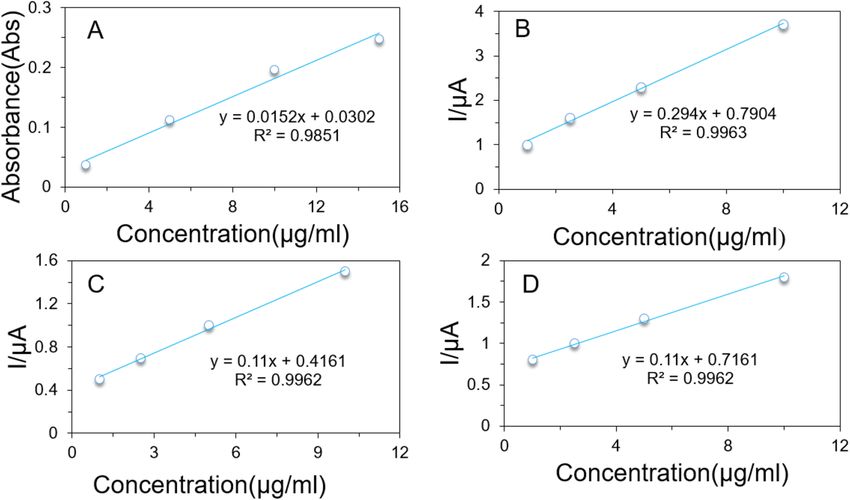
www.nature.com/scientificreports/
Figure 2. SEM images of as-synthesized ZnO nanosphere (A) and ZnO/L-Cys@CFX (B). The inset
shows a high magnification image of the same. EDX spectra (C) and elemental mapping (D) of ZnO in the
nanocomposite. (E) XRD peaks of synthesized pure ZnO sphere and (F) ATR-FTIR spectra of (i) L-Cys powder;
(ii) ZnO/L-Cys, and (iii) ZnO/L-Cys@CFX.
molecule increases with decreasing pH, as shown in Fig. 3B. The negative part of CFX (COO−) is activated after
a rising pH greater than 3.65, and the negative charge increase with an increasing pH value of the molecule. The
maximum number of positive and negative charges were found at pH 3.13 and 4.16, respectively. The probable
cause of maximum adsorption found at pH 3.5 is the better interaction between the amine group of CFX and the
carboxylic group of L-Cys due to a maximum number of charge differences between them. The pH value greater
than 3.5, the electrostatic repulsion between CFX and L-Cys becomes higher, so the drug loading efficiency get-
ting lower. The maximum amount of CFX loaded per grams of nanocarrier is 36 µg/ml which is briefly discussed
in the supplementary information section (Fig S2).
In vitro drug release profile analysis. To access the usability of ZnO/L-Cys as an effective nano-drug
carrier, a released profile analysis of the carrier using CFX was recorded. Figure 3C shows the drug release profile
from ZnO/L-Cys@CFX under different pH at 37 °C. Rapid release behavior of the carrier was found at the initial
stage. Almost 30–55 percent of CFX was released in the first 4 h at different reservoir pH, but a constant release
profile was achieved from 4 to 24 h. The pattern of drug release is acceptable in cases where a sufficient amount
of drug is required to improve the therapeutic efficiency of antibiotics37. The release pattern of CFX revealed the
concentration of released CFX was higher at acidic and basic concentrations than the pH of 7.4, suggesting pH-
dependent drug release behavior. In acidic conditions, the drug-releasing rate was higher than that of another
Scientific Reports | (2021) 11:15565 | https://doi.org/10.1038/s41598-021-95195-0 5
Vol.:(0123456789)www.nature.com/scientificreports/
Figure 3. Percentage of drug loading at different pH (A), CFX ionic state (B) drug release profile of CFX under
different pH at 37 ± 0.5 °C (C).
pH and almost 86.7% of CFX was released over 24 h releasing periods. The probable reason for this burst release
behavior will be the loosening of the grafted L-Cys @CFX from ZnO bonding. The grafted composite of L-Cys@
CFX became positively charged because the sulfur atom of the thiol group of L-Cys may be protonated at pH 3.5
(pKa value –SH group of L-Cys was 8.33) but the thiol group is neutral at normal blood p H38. So, a pH-depend-
ent release profile of CFX from the carrier drug is found, which can be justified by previously reported w ork39.
To investigate the release kinetics of CFX from ZnO/L-Cys@CFX, the drug release data were fitted to a suitable
model such as zero-order (percentage of drug release versus time), first-order (log percentage of drug release
versus time), Korsmeyer–Peppas (log percentage of drug release versus log time), and Higuchi (percentage of
drug release vs square root of time) shown in Fig. 4. To calculate the release rate constant (K) and the regression
coefficient (R2), excel 2019 (Microsoft Corporation) was used. The best-fitted model is the Korsmeyer–Peppas
model which is shown in Table 1.
Antibacterial activity Of ZnO/L‑Cys@CFX. Zone of inhibition analysis. In the disc diffusion test, we
found a growth inhibition zone as low as 1.0 μg/ml ZnO/L-Cys@CFX, where no zone was found around the disc
of 1.0 μg/ml ceftizoxime alone. At higher concentrations, nanoparticle conjugated antibiotics produced a more
significant zone than antibiotics alone at similar concentrations (Fig. 5). The details result of zone of inhibition
analysis by using disk diffusion test were interpreted in supplementary information (Table S1, Fig. S1). The na-
noparticle itself did not produce any zone of bacterial inhibition. Thus, the nanoparticle conjugated antibiotic
showed greater efficiency. The Ceftizoxime solution was in a non-buffered medium; when assayed using the disc
agar-diffusion method, it loses potency quicker37. The nanoparticle conjugation might also help the antibiotic
retain its potency.
Minimum inhibitory concentration. We found the minimum inhibitory concentration (MIC) of ceftizoxime as
20.0 μg/ml and nanoparticle conjugated ceftizoxime as 5.0 μg/ml in bacterial suspension (3.67 ± 0.47 × 10⁶ CFU/
ml). We use the broth dilution method and measure the viable bacterial count by drop plate method at 6 h and
12 h of the antibiotic treatment. Surprisingly we found a higher viable count after 12 h of treatment than 6 h
which can be seen in Table 2. The sensitivity of the ceftizoxime assay was reported to decrease by increasing the
incubation temperature40. In this study, we have used the incubation temperature at 37 °C, which might also
cause loss of antibiotic potency with time and thus explains the bacterial count to increase slightly with treat-
ment. Table 3 represents different third-generation cephalosporin grouped antibiotics conjugated nanocarrier
drug targets to inhibit the growth of different pathogenic bacteria. Compared to previously reported work, the
ZnO and L-Cys-based nanocarrier drugs are highly effective to kill S. typhimurium29,36,39–42.
Effect of nanocarriers on MIC. Each component of the nanoparticle was also evaluated for the antibac-
terial effect in the broth dilution method. The 1.0 mg/ml ZnO nano hollow sphere, 0.5 mg/ml l-cysteine and
ZnO/L-Cys nanocarrier made by nanocomposite of 1 mg/ml ZnO nano hollow sphere and 0.5 mg/ml l-cystine
were tested. ZnO nano hollow sphere was found to have some bacteriostatic effect at 1.0 mg/ml and 0.2 mg/ml
Scientific Reports | (2021) 11:15565 | https://doi.org/10.1038/s41598-021-95195-0 6
Vol:.(1234567890)www.nature.com/scientificreports/
Figure 4. Kinetics model for release of CFX from ZnO/L-Cys@CFX (A) zero-order (B) first order (C) Higuchi
and (D) Korsmeyer–Peppas model of kinetics.
Korsmeyer–
Zero-order First-order Higuchi Peppas
Formulation pH K0 R2 K1 R2 KH R2 K R2
9.0 1.86 0.90 0.02 0.81 11.54 0.97 0.37 0.99
ZnO/L-Cys@ CFX 7.4 1.48 0.91 0.02 0.80 9.13 0.98 0.36 0.99
3.5 2.54 0.82 0.02 0.72 15.53 0.91 0.41 0.93
Table 1. The release rate constant (K) and regression coefficient (R2) values of the different models.
Figure 5. Zone diameter (cm) of S. typhimurium growth inhibition zone at different concentrations of CFX and
CFX conjugated nanocarrier in disc diffusion method (S/N = 3).
and not below that level which can be seen in Table 4. The major reason for this finding is the ZnO produces
reactive oxygen species to damage the cell membrane mechanically38. Withdrawing or diluting the ZnO nano
hollow sphere retain the viability of the bacterium, hence prove the non-bactericidal effect. Table 4 represents,
the ZnO/L-Cys nanocarrier had some bacteriostatic effect as well but far lesser efficiency than ZnO spheres.
The observation also refers that the lesser MIC and increasing zone of inhibition for nanoparticle conjugated
Scientific Reports | (2021) 11:15565 | https://doi.org/10.1038/s41598-021-95195-0 7
Vol.:(0123456789)www.nature.com/scientificreports/
Name Bacterial count (CFU/ml) at 0 h Bacterial count (CFU/ml) at 6 h Bacterial count (CFU/ml) at 12 h
C1 3.67 ± 0.47 × 10⁶ 2.33 ± 0.47 × 10⁴ 2.5 ± 0.82 × 10⁵
6
C2 3.67 ± 0.47 × 10 TFTC TFTC
C3 3.67 ± 0.47 × 10⁶ TFTC TFTC
C4 3.67 ± 0.47 × 10⁶ TFTC TFTC
D1 3.67 ± 0.47 × 10⁶ 1.67 ± 0.47 × 10⁶ 1.90 ± 0.82 × 10⁶
6 6
D2 3.67 ± 0.47 × 10 1.17 ± 0.12 × 10 2.4 ± 0.82 × 10⁶
6 5
D3 3.67 ± 0.47 × 10 5.50 ± 0.50 × 10 7.50 ± 0.50 × 10⁵
D4 3.67 ± 0.47 × 10⁶ TFTC TFTC
Nanocarrier 3.67 ± 0.47 × 10⁶ 6.67 ± 0.94 × 10⁵ 3.67 ± 0.47 × 10⁵
Control 3.67 ± 0.47 × 10⁶ TNTC TNTC
Table 2. Effect of nanocarrier drug compare to free drug on bacterial susceptibility (mean count ± SD) in
broth dilution method. TNTC too numerous to count, TFTC too few to count, C1, C2, C3, C4 nano carrier with
1, 5, 10, and 20 µg/ml CFX respectively, D1, D2, D3, D4 1, 5, 10, and 20 µg/ml CFX, respectively.
Antibacterial activity
Carrier matrix Antibiotics Targeted bacteria MIC (µg/ml) ZOI compared to the free drug (Za/Zb) References
GO@CoFe2O4@Ag Ciprofloxacin E. coli 2.5 – 37
ZnO Ciprofloxacin S. aureus 20 – 40
AuNPs Ciprofloxacin S. epidermidis 3.9–15.62 1.05–1.09 41
FAuNPs Cefotaxime K. pneumoniae 0.562 – 42
Pectin CFX B. cereus – 1.14 29
Fe3O4@BSM Cephalexin S. typhimurium – 1.14 43
ZnO/L-Cys CFX S. typhimurium 5 1.5 This work
Table 3. Comparison of antibacterial activities of some Cephalosporin grouped antibiotic-loaded nanocarrier
for different targeted bacteria as previously reported. Za ZOI of nanocarrier conjugated antibiotics, Zb ZOI of
the reference drug.
Compound Bacterial count (CFU/ml)
Dilution (times) of nano carriers 0 5 25 125
ZnO 1.70 ± 0.16 × 10³ 2.67 ± 0.34 × 10³ TNTC TNTC
L-Cys TNTC TNTC TNTC TNTC
ZnO/L-Cys 6.03 ± 0.12 × 10³ 4.77 ± 0.17 × 10⁴ TNTC TNTC
Control TNTC
Table 4. Comparison of antibacterial activity of different nanocarriers on bacterial growth inhibition after 6 h
incubation. TNTC too numerous to count.
antibiotics were not contributed by the nanoparticle, but the antibiotic itself. Nanoparticle enhances the potency
or stability of the antibiotics.
Conclusion
In the present study, the ZnO nano hollow sphere was successfully synthesized from an environmentally friendly
method and coated with L-Cys, confirmed by SEM, FTIR. The ZnO/L-Cys was conjugated with CFX and the
resulting ZnO/L-Cys@CFX was also confirmed by the different investigations. The maximum amount of CFX
conjugate with the carrier matrix was found at pH 3.5 due to electrostatic interaction between L-Cys and CFX.
A pH-sensitive release profile of CFX shows around 60% of CFX released after 24 h released period at blood
pH, which will be advantageous for time-consuming treatments. The growth inhibition investigation against
S. typhimurium confirmed that the ZnO/L-Cys@CFX had been better antimicrobial properties and the ability
to eradicate S. typhimurium more effectively than the reference drug. The MIC of CFX and ZnO/L-Cys@CFX
expressed the carrier drug decrease amount CFX required to eradicate S. typhimurium. Therefore, ZnO/L-Cys@
CFX nanocomposite could be an effective tool to restrain S. typhimurium type MDR pathogen.
Scientific Reports | (2021) 11:15565 | https://doi.org/10.1038/s41598-021-95195-0 8
Vol:.(1234567890)www.nature.com/scientificreports/
Received: 2 June 2021; Accepted: 21 July 2021
References
1. Fischbach, M. A. & Walsh, C. T. Antibiotics for emerging pathogens. Science 325(5944), 1089–1093. https://doi.org/10.1126/scien
ce.1176667 (2009).
2. Linares, J. F., Gustafsson, I., Baquero, F. & Martinez, J. L. Antibiotics as antimicrobial signaling agents instead of weapons. Proc.
Natl. Acad. Sci. 103(51), 19484–19489. https://doi.org/10.1073/pnas.0608949103 (2006).
3. Pizzolato-Cezar, L. R., Okuda-Shinagawa, N. M. & Machini, M. T. Combinatory therapy antimicrobial peptide-antibiotic to
minimize the ongoing rise of resistance. Front. Microbiol. 10, 1703. https://doi.org/10.3389/fmicb.2019.01703 (2019).
4. Balaban, N. et al. A problem of persistence: Still more questions than answers?. Nat. Rev. Microbiol. 11, 587–591. https://doi.org/
10.1038/nrmicro3076 (2013).
5. Baptista, P. V. et al. Nano-strategies to fight multidrug resistant bacteria—” a battle of the titans”. Front. Microbiol. 9, 1441. https://
doi.org/10.3389/fmicb.2018.01441 (2018).
6. Muzammil, S. et al. Nanoantibiotics: Future nanotechnologies to combat antibiotic resistance. Front. Biosci. (Elite Ed) 10, 352–374.
https://doi.org/10.2741/e827 (2018).
7. Natan, M. & Banin, E. From nano to micro: Using nanotechnology to combat microorganisms and their multidrug resistance.
FEMS Microbiol. Rev. 41, 302–322. https://doi.org/10.1093/femsre/fux003 (2017).
8. Burygin, G. et al. On the enhanced antibacterial activity of antibiotics mixed with gold nanoparticles. Nanosc. Res. Lett. 4, 794–801.
https://doi.org/10.1007/s11671-009-9316-8 (2009).
9. Huang, X., Zheng, X., Xu, Z. & Yi, C. ZnO-based nanocarriers for drug delivery application: From passive to smart strategies. Int.
J. Pharm. 534, 190–194. https://doi.org/10.1016/j.ijpharm.2017.10.008 (2017).
10. Lotha, R., Sundaramoorthy, N. S., Shamprasad, B. R., Nagarajan, S. & Sivasubramanian, A. Plant nutraceuticals (Quercetrin and
Afzelin) capped silver nanoparticles exert potent antibiofilm effect against food borne pathogen Salmonella enterica serovar
typhimurium and curtail planktonic growth in zebrafish infection model. Microb. Pathog. 120, 109–118. https://doi.org/10.1016/j.
micpath.2018.04.044 (2018).
11. Wang, X. et al. Antibiotic resistance in Salmonella typhimurium isolates recovered from the food chain through national antimi-
crobial resistance monitoring system between 1996 and 2016. Front. Microbiol. 10, 985. https://doi.org/10.3389/fmicb.2019.00985
(2019).
12. Richards, D. M. & Heel, R. C. Ceftizoxime. A review of its antibacterial activity, pharmacokinetic properties and therapeutic use.
Drugs 29, 281–329. https://doi.org/10.2165/00003495-198529040-00001 (1985).
13. Singh, R. & Lillard, J. W. Jr. Nanoparticle-based targeted drug delivery. Exp. Mol. Pathol. 86(3), 215–223. https://doi.org/10.1016/j.
yexmp.2008.12.004 (2009).
14. Puvvada, N. et al. Novel ZnO hollow-nanocarriers containing paclitaxel targeting folate-receptors in a malignant pH-microenvi-
ronment for effective monitoring and promoting breast tumor regression. Sci. Rep. 5, 11760. https://doi.org/10.1038/srep11760
(2015).
15. Hafeli, U. O. et al. Cell uptake and in vitro toxicity of magnetic nanoparticles suitable for drug delivery. Mol. Pharm. 6, 1417–1428.
https://doi.org/10.1021/mp900083m (2009).
16. Brayner, R. & Ferrari-Iliou, R. Toxicological impact studies based on Escherichia coli bacteria in ultrafine ZnO nanoparticles col-
loidal medium. Nano Lett. 6(4), 866–870. https://doi.org/10.1021/nl052326h (2006).
17. Jones, N., Ray, B., Ranjit, K. T. & Manna, A. C. Antibacterial activity of ZnO nanoparticle suspensions on a broad spectrum of
microorganisms. FEMS Microbiol. Lett. 279(1), 71–76. https://doi.org/10.1111/j.1574-6968.2007.01012.x (2008).
18. Jalal, R. et al. ZnO nanofluids: Green synthesis, characterization, and antibacterial activity. Mater. Chem. Phys. 121(1), 198–201.
https://doi.org/10.1016/j.matchemphys.2010.01.020 (2010).
19. Seil, J. T. & Webster, T. J. Antimicrobial applications of nanotechnology: Methods and literature. Int. J. Nanomed. 7, 2767–2781.
https://doi.org/10.2147/IJN.S24805 (2012).
20. Emami-Karvani, Z. & Chehrazi, P. Antibacterial activity of ZnO nanoparticle on gram-positive and gram-negative bacteria. Afr.
J. Microbiol. Res. 5(12), 1368–1373. https://doi.org/10.5897/AJMR10.159 (2011).
21. Padmavathy, N. & Vijayaraghavan, R. Enhanced bioactivity of ZnO nanoparticles—an antimicrobial study. Sci. Technol. Adv. Mater.
9(3), 035004. https://doi.org/10.1088/1468-6996/9/3/035004 (2008).
22. Raghupathi, K. R., Koodali, R. T. & Manna, A. C. Size-dependent bacterial growth inhibition and mechanism of antibacterial
activity of zinc oxide nanoparticles. Langmuir 27(7), 4020–4028. https://doi.org/10.1371/journal.pone.0085981 (2011).
23. Goethals, E. C., Elbaz, A., Lopata, A. L., Bhargava, S. K. & Bansal, V. Decoupling the effects of the size, wall thickness, and porosity
of purcumin-loaded chitosan nanocapsules on their anticancer efficacy: Size is the winner. Langmuir 29, 658–666. https://doi.org/
10.1371/10.1021/la3033836 (2012).
24. Yuan, Q. et al. Responsive core−shell latex particles as colloidosome microcapsule membranes. Langmuir 26, 18408–18414. https://
doi.org/10.1021/la1033564 (2010).
25. Bak, D. W., Bechtel, T. J., Falco, J. A. & Weerapana, E. Cysteine reactivity across the subcellular universe. Curr. Opin. Chem. Biol.
48, 96–105. https://doi.org/10.1016/j.cbpa.2018.11.002 (2019).
26. Mocanu, A. et al. Self-assembly characteristics of gold nanoparticles in the presence of cysteine. Col. Surf. A Physicochem. Eng.
Asp. 338, 93–101. https://doi.org/10.1016/j.colsurfa.2008.12.041 (2009).
27. McCarty, M. F. & DiNicolantonio, J. J. An increased need for dietary cysteine in support of glutathione synthesis may underlie the
increased risk for mortality associated with low protein intake in the elderly. Age 37, 96. https://d
oi.o
rg/1 0.1 007/s 11357-0 15-9 823-8
(2015).
28. Ge, L. et al. Trisodium citrate assisted synthesis of ZnO hollow spheres via a facile precipitation route and their application as gas
sensor. J. Mater. Chem. 21, 10750–10754. https://doi.org/10.1039/C0JM04405A (2011).
29. Kumar, P., Kumar, V., Kumar, R. & Pruncu, C. I. Fabrication and characterization of ceftizoxime-loaded pectin nanocarriers.
Nanomaterials 10, 1452. https://doi.org/10.3390/nano10081452 (2020).
30. Ali, M. R. et al. Sensitive MWCNT/P-Cys@MIP sensor for selective electrochemical detection of ceftizoxime. J. Mater. Sci. 56,
12803–12813. https://doi.org/10.1007/s10853-021-06115-6 (2021).
31. NCCLS. Performance Standards for Antimicrobial Susceptibility Testing. Nineteenth Informational Supplement (C. a. L. S. I. (CLSI),
2009).
32. CLSI, C. a. L. S. I. Performance Standards for Antimicrobial Susceptibility Testing. 22nd Informational Supplement M100-S22, 2012.
33. Bauer, A. W. et al. Antibiotic susceptibility testing by a standardized single disk method. Am. J. Clin. Pathol. 45(4), 493–496 (1966).
34. Herigstad, B. et al. How to optimize the drop plate method for enumerating bacteria. J. Microbio. Methods 44(2), 121–129. https://
doi.org/10.1016/S0167-7012(00)00241-4 (2001).
35. Li, X. et al. Template-free microwave-assisted synthesis of ZnO hollow microspheres and their application in gas sensing. Cryst-
EngComm 15, 2949–2955. https://doi.org/10.1039/C2CE26539G (2013).
36. Li, L. et al. Application of l-cysteine capped core-shell CdTe/ZnS nanoparticles as a fluorescence probe for cephalexin. Anal.
Methods 6, 2715–2721. https://doi.org/10.1039/C4AY00094C (2014).
Scientific Reports | (2021) 11:15565 | https://doi.org/10.1038/s41598-021-95195-0 9
Vol.:(0123456789)www.nature.com/scientificreports/
37. Wang, J. X., Wang, Z. H., Chen, J. F. & Yun, J. Direct encapsulation of water-soluble drug into silica microcapsules for sustained
release applications. Mater. Res. Bull. 43(12), 3374–3381. https://doi.org/10.1016/j.materresbull.2008.02.011 (2008).
38. Dourado, A. H. B., Batista, A. P. L., Oliveira-Filho, A. G. S., Sumodjo, P. T. A. & Torresi, S. I. C. l-Cysteine electrooxidation in
alkaline and acidic media: A combined spectroelectrochemical and computational study. RSC Adv. 7, 7492–7501. https://doi.org/
10.1039/C6RA26576F (2017).
39. Kooti, M. et al. Magnetic graphene oxide inlaid with silver nanoparticles as antibacterial and drug delivery composite. Appl.
Microbiol. Biotechnol. 102, 3607–3621. https://doi.org/10.1007/s00253-018-8880-1 (2018).
40. Uri, J. V. Stability of ceftizoxime in aqueous solution. Experientia 39, 1085–1086. https://doi.org/10.1007/BF01943119 (1983).
41. Jang, Y. J., Simer, C. & Ohm, T. Comparison of zinc oxide nanoparticles and its nano-crystalline particles on the photocatalytic
degradation of methylene blue. Mater. Res. Bull. 41(1), 67–77. https://doi.org/10.1016/j.materresbull.2005.07.038 (2006).
42. Patra, P. et al. Ciprofloxacin conjugated zinc oxide nanoparticle: A camouflage towards multidrug resistant bacteria. Bull. Mater.
Sci. 37, 199–206. https://doi.org/10.1007/s12034-014-0637-6 (2014).
43. Roshmi, T. et al. Effect of biofabricated gold nanoparticle-based antibiotic conjugates on minimum inhibitory concentration of
bacterial isolates of clinical origin. Gold Bull. 48, 63–71. https://doi.org/10.1007/s13404-015-0162-4 (2015).
Author contributions
S.B., M.R.A., M.A.A.S. performed the lab work. M.Z.H.K. designed and supervised the whole work. S.A. super-
vised the Microbiological part of the experiment. The Scheme 1 has been drawn by M.Z.H.K. with the help of
M.A.A.S. All the authors contributed in the manuscript writing.
Competing interests
The authors declare no competing interests.
Additional information
Supplementary Information The online version contains supplementary material available at https://doi.org/
10.1038/s41598-021-95195-0.
Correspondence and requests for materials should be addressed to M.Z.H.K.
Reprints and permissions information is available at www.nature.com/reprints.
Publisher’s note Springer Nature remains neutral with regard to jurisdictional claims in published maps and
institutional affiliations.
Open Access This article is licensed under a Creative Commons Attribution 4.0 International
License, which permits use, sharing, adaptation, distribution and reproduction in any medium or
format, as long as you give appropriate credit to the original author(s) and the source, provide a link to the
Creative Commons licence, and indicate if changes were made. The images or other third party material in this
article are included in the article’s Creative Commons licence, unless indicated otherwise in a credit line to the
material. If material is not included in the article’s Creative Commons licence and your intended use is not
permitted by statutory regulation or exceeds the permitted use, you will need to obtain permission directly from
the copyright holder. To view a copy of this licence, visit http://creativecommons.org/licenses/by/4.0/.
© The Author(s) 2021
Scientific Reports | (2021) 11:15565 | https://doi.org/10.1038/s41598-021-95195-0 10
Vol:.(1234567890)You can also read