Structure of the PH domain and Btk motif from Bruton's tyrosine kinase: molecular explanations for X-linked agammaglobulinaemia
←
→
Page content transcription
If your browser does not render page correctly, please read the page content below
The EMBO Journal Vol.16 No.12 pp.3396–3404, 1997

Structure of the PH domain and Btk motif from
Bruton’s tyrosine kinase: molecular explanations for
X-linked agammaglobulinaemia

Marko Hyvönen and Matti Saraste1                             pathways involving Btk and arrest development of B cells
                                                              at the pre-B cell stage. Interestingly, a similar but milder
European Molecular Biology Laboratory, Postfach 10.2209,      disease in mice, X-linked immunodeficiency (xid), is also
69012 Heidelberg, Germany
                                                              caused by a mutation within the Btk PH domain (Rawlings
1Corresponding  author                                        et al., 1993; Thomas et al., 1993).
e-mail: saraste@embl-heidelberg.de
                                                                 Btk is a cytoplasmic protein tyrosine kinase (PTK)
                                                              which belongs to the Tec family (Bolen, 1995). In addition
Bruton’s tyrosine kinase (Btk) is an enzyme which is          to the Src homology 2 and 3 (SH2 and SH3) domains
involved in maturation of B cells. It is a target for         and the catalytic kinase domain, which are found in
mutations causing X-linked agammaglobulinaemia                proteins belonging to the Src superfamily of PTKs, the
(XLA) in man. We have determined the structure of             members of the Tec family contain an N-terminal pleck-
the N-terminal part of Btk by X-ray crystallography           strin homology (PH) domain and a so-called Tec homology
at 1.6 Å resolution. This part of the kinase contains a       domain that includes a Btk motif and a proline-rich
pleckstrin homology (PH) domain and a Btk motif.              region. They occupy the place of the unique domain and
The structure of the PH domain is similar to those
                                                              myristylation site of Src. The Btk motif comprises only
published previously: a seven-stranded bent β-sheet
                                                              26 residues which characteristically include three fully
with a C-terminal α-helix. Individual point mutations
                                                              conserved cysteines and a histidine (Vihinen et al., 1994).
within the Btk PH domain which cause XLA can be
                                                              This motif is found in only a few proteins outside the Tec
classified as either structural or functional in the light
                                                              family of PTKs and is always adjacent to a PH domain.
of the three-dimensional structure and biochemical
                                                                 Btk can be activated by different extracellular signals
data. All functional mutations cluster into the positively
                                                              such as cross-linking of the B-cell antigen receptor or
charged end of the molecule around the predicted
                                                              binding of interleukin 5 (IL-5) (de Weers et al., 1994;
binding site for phosphatidylinositol lipids. It is likely
                                                              Sato et al., 1994). Upon activation of the B-cell receptor,
that these mutations inactivate the Btk pathway in
                                                              Btk is phosphorylated on Tyr551 by Src family tyrosine
cell signalling by reducing its affinity for inositol
phosphates, which causes a failure in translocation of        kinases and subsequently localized to the plasma mem-
the kinase to the cell membrane. A small number of            brane (Rawlings et al., 1996). This transphosphorylation
signalling proteins contain a Btk motif that always           of Btk leads to autophosphorylation of Tyr223 in the
follows a PH domain in the sequence. This small               Btk SH3 domain (Park et al., 1996). Conversely, cells
module has a novel fold which is held together by a           simulated with IL-5 do not show increased tyrosine
zinc ion bound by three conserved cysteines and a             phosphorylation but rather an enhanced activity of Btk
histidine. The Btk motif packs against the second half        (Sato et al., 1994).
of the β-sheet of the PH domain, forming a close                 The PH domains are small signal transduction domains
contact with it. Our structure opens up new ways to           which have been found in close to 100 different eukaryotic
study the role of the PH domain and Btk motif in the          proteins up to now (Gibson et al., 1994). Although
cellular function of Btk and the molecular basis of its       sequence conservation between different domains is very
dysfunction in XLA patients.                                  low, their three-dimensional structures appear to share a
Keywords: Bruton’s tyrosine kinase/Btk motif/inositol         common fold and a remarkable electrostatic polarization.
phosphates/PH domain/ X-linked agammaglobulinaemia            The positively charged end of several PH domains bind
                                                              inositol phosphates in vitro with Kds ranging from 210 nM
                                                              to 1.23 mM for phospholipase Cδ1 (PLCδ1) and dynamin
                                                              domains, respectively (Harlan et al., 1994; Lemmon et al.,
Introduction                                                  1995; Salim et al., 1996). This binding enhances recruit-
X-linked agammaglobulinaemia (XLA) is a hereditary            ment of PLCδ1 to the cell membrane in vivo (Paterson
immune disease which is estimated to affect one in every      et al., 1995). Two three-dimensional structures of PH
150 000 human males (Smith et al., 1994). XLA patients        domains have been determined in complex with inositol(1,
lack circulating B cells and subsequently have very low       4,5)trisphosphate [Ins(1,4,5)P3, herein we use the inositol
levels of immunoglobulins in their serum. They are very       phosphate nomenclature of Divecha and Irvine (1995)]
vulnerable to infectious diseases and require continuous      (Ferguson et al., 1995; Hyvönen et al., 1995).
medical treatment. The cause of this disease has recently        In most cases, the PH domains bind preferentially to
been mapped to the gene encoding for Bruton’s tyrosine        Ins(1,4,5)P3. However, a recent study by Salim et al.
kinase (Btk) (Tsukada et al., 1993; Vetrie et al., 1993). A   (1996) showed that the Btk PH domain specifically binds
large number of mutations in Btk have since been found        PtdIns(3,4,5)P3 rather than PtdIns(4,5)P2. Interestingly,
in patients suffering from XLA (Vihinen et al., 1996).        this specificity for PtdIns(3,4,5)P3 appears to be regulated
The XLA-causing mutations inactivate signal transduction      by divalent ions, as it was only observed in the presence

3396                                                                                            © Oxford University Press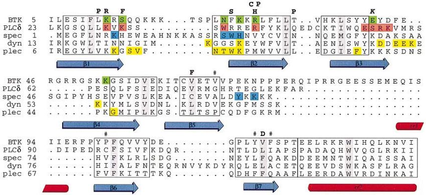
PH domain of Bruton’s tyrosine kinase

Fig. 1. Solubility of the mutants. The figure shows the SDS–PAGE of the wild-type and mutant proteins expressed in E.coli as described in
Materials and methods. Total cell lysates (T) and high-speed supernatants (S) were mixed with Laemmli sample buffer, boiled and run in a 15%
polyacrylamide gel. The wild-type protein is shown as control (WT), and the mutants are marked on top of the gel (F25S, R28H, R28C, T33P,
V64F, V113D). The position of the expressed proteins is marked with an arrow. Molecular weight markers (in kDa) are indicated on the side
of the gel.

of 2 mM Mg2⫹ or Ca2⫹. Similar results have also been                        groups: (i) folding mutations, which prevent formation of
obtained with soluble inositol phosphates (Fukuda et al.,                   a stable native-like structure, and (ii) functional mutations
1996). The Btk PH domain showed tightest binding to                         which do not affect the overall fold, but which can be
Ins(1,3,4,5)P4 compared with the other tested compounds,                    expected to disrupt ligand-binding site(s) on the domain.
with 40 nM affinity.                                                        As shown in Figure 1, wild-type protein and mutants
   Most of the structural and functional studies on the                     F25S, R28C and R28H are soluble in E.coli, whereas
interaction of PH domains with their ligands have been                      mutants T33P, V64F and V113D fail to yield soluble
carried out using soluble inositol phosphates, but they are                 protein under the same conditions. Structural reasons for
thought to be relevant for binding of phosphatidylinositol                  this behaviour are discussed later. The far-UV circular
lipids to the same sites. Consequently, in the following,                   dichroism (CD) spectra of the purified wild-type protein
we shall regard the ligand as a (soluble) phospholipid                      and soluble mutants are virtually identical (data not
head group.                                                                 shown).
   Several XLA-causing point mutations in Btk are located                      The wild-type protein and all the soluble mutants were
in the PH domain and one in the Btk motif. This is the                      used for crystallization trials, but only the mouse xid
only case in which mutations in a PH domain are known                       mutant R28C gave high quality crystals. Crystals belong
to cause a human disease. We have studied the roles of                      to space group P21 (unit cell a ⫽ 49.15, b ⫽ 59.87, c ⫽
individual mutations in the PH domain of human Btk and                      55.94 Å, β ⫽ 98.2°), with two molecules in the asymmetric
determined the structure of the R28C mutant corresponding                   unit. Crystals diffracted beyond 1.5 Å on a synchrotron
to the xid mutation in mouse by X-ray crystallography at                    source. The structure was solved with a single heavy
1.6 Å resolution. This structure allows us to discuss how                   metal derivative (trimethyl lead acetate) using both iso-
the point mutations may inactivate Btk and subsequently                     morphous and anomalous differences (see Materials and
cause XLA. Our structure also contains the Btk motif,                       methods). The model is refined at 1.6 Å resolution;
which is shown to have a novel, zinc-binding fold. The                      refinement statistics are shown in Table I. In addition to
function of this motif is still unknown and the structure                   the two protein molecules in the asymmetric unit, 220
will provide a rational basis for functional studies.                       water molecules, two zinc ions and two sodium ions are
                                                                            included in the model. Metal analysis showed that the
                                                                            protein contains 0.93 mol of zinc per 1 mol of protein.
Results
                                                                            Accordingly, the ions that where found to be present in
The segment containing the N-terminal 170 amino acid                        the crystal structure of the Btk motif were assigned as
residues of human Btk, including both the PH domain                         zinc. Sodium chloride was required for crystallization of
and the Btk motif, was expressed in Escherichia coli in a                   the protein and, in the final stages of the refinement, two
soluble form and purified to homogeneity. Earlier attempts                  ions were found with a typical octahedral coordination
to express the PH domain alone resulted in insoluble                        characteristic of sodium. The sodium ions are bound
protein which resisted all refolding trials. Only the inclu-                between the two molecules in the asymmetric unit.
sion of the Btk motif in the construct allowed expression
of a soluble, folded protein. Several XLA mutations in                      Structure of the Btk PH domain and the Btk motif
the PH domain (F25S, R28H, T33P, V64F, V113D) were                          Similarly to the other PH domains, the structure is com-
introduced into similar expression constructs and the                       posed of a strongly bent seven-stranded antiparallel β-sheet
corresponding proteins were produced in E.coli. In addi-                    which packs against a C-terminal α-helix (Figure 2). The
tion, the protein carrying the mouse xid mutation (R28C)                    Btk PH domain contains a long insertion in the loop
(Rawlings et al., 1993; Thomas et al., 1993) was produced.                  between β-strands 5 and 6 which includes a short 1.5-turn
   Using the solubility of individual mutants in E.coli as                  α-helix (α1). It points away from the core of the domain
a criterion, we could classify the mutations into two                       and its hydrophilic middle part is disordered and could

                                                                                                                                          3397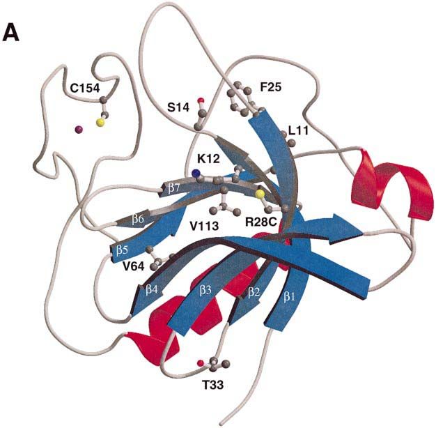
M.Hyvönen and M.Saraste

Table I. Data collection, phasing and refinement statistics

                                                              In-house native               (CH3)3PbAc           BW7B at EMBL-Hamburg c/o DESY

Resolution                                                     2.6                           2.6                  1.6
Wavelength (Å)                                                 1.514                         1.514                0.882
Completeness (highest shell) (%)                              99.8 (98.2)                   97.2 (95.8)          99.9 (99.5)
Rmerge (high shell)a                                           0.060 (0.120)                 0.061 (0.149)        0.054 (0.236)
No. of unique reflections                                     9817                          9319                 42 840
Phasing (2.6 Å)b isomorphous/anomalous                                                       2.77/2.85
Figure of merit                                                                              0.602
No. of sites                                                                                 1

Refinement statistics (XPLOR )
  Resolution (Å)                                          40.5–2.74             2.74–2.17         2.17–1.98    1.98–1.72      1.72–1.60       40.5–1.60
  R-factor (%)                                            18.6                 24.1              26.1         29.8           31.5             23.1
  Rfree(%)                                                25.8                 29.9              27.6         29.5           33.1             28.0
Geometrical statistics (WHAT CHECK)
R.m.s. deviation from ideal bond lenghts (Å)                  0.11
R.m.s. deviation from ideal bond angles (°)                   1.989
aR
  merge  ⫽ Σ|Ii–⬍I⬎|/ΣIi , where Ii is the intensity of an individual reflection and ⬍I⬎ is the mean intensity of the reflection. The value in parenthesis
is for the highest resolution shell.
bPhasing power is the ratio between the root mean square of the heavy atom scattering amplitude and lack of closure error.

Fig. 2. The structure of the Btk PH domain and Btk motif. The figure shows a ribbon representation of the R28C mutant of the human Btk PH
domain and Btk motif. The β-strands and α-helixes are numbered β1–β7 and α1–α2. The zinc ion in the Btk motif is shown in purple. This figure
and Figures 5A and B, 6A and B were prepared using MOLSCRIPT (Kraulis, 1991) and rendered in Raster3d (Bacon and Anderson, 1988; Merrit
and Murphy, 1994).

not be modelled properly. Also, the loop between β-strands                          itself and is held together by a zinc ion. This zinc is
1 and 2 has weak electron density and was partly modelled                           coordinated with a distorted tetrahedral geometry to H143
as polyalanine. There will naturally be differences in our                          and C154, C155 and C165 (Figure 3), which are fully
structure around the mutated residue R28C compared with                             conserved in Btk motifs (Vihinen et al., 1994).
the wild-type protein, but these are not thought to be                                 Database searches using the program DALI (Holm and
significant to our analysis of the structure.                                       Sander, 1993) have revealed no structural homology for
   The Btk motif has a globular core which packs against                            the Btk motif in the Protein Data Bank (PDB). The
β-strands 5–7 of the PH domain. F146 in the Btk motif                               diacylglycerol/phorbol ester-binding domain (PDB entry
is inserted into a hydrophobic pocket formed by residues                            1PTR), LIM repeats (1IMR) and GATA-1 zinc finger
V67, P101, Y112 and F114 (labelled in Figure 4). The                                (1PTR) have a similar zinc coordination with three
core of the Btk motif is connected to the C-terminus of                             cysteines and histidine, and a similar tight turn (formed
the PH domain with a 7 or 8 residue linker. The structure                           by C154 and C155 in the Btk motif) at the zinc-binding
could be described as a long loop which folds back on                               site, but otherwise the structures seem to be unrelated.

3398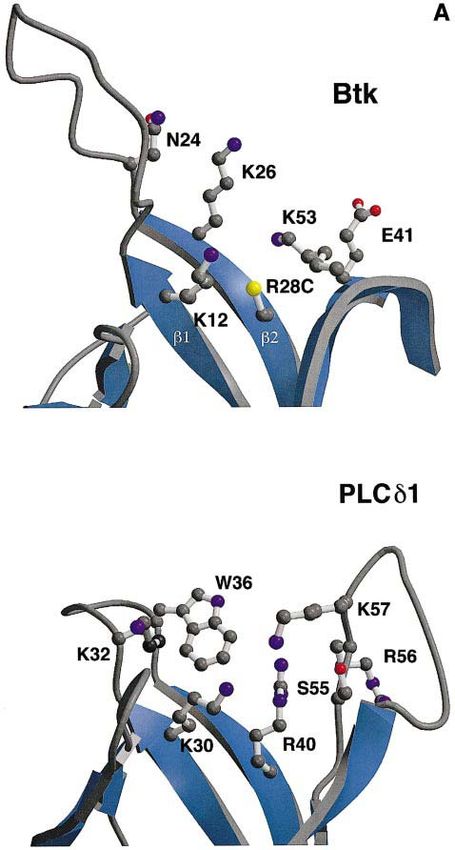
PH domain of Bruton’s tyrosine kinase
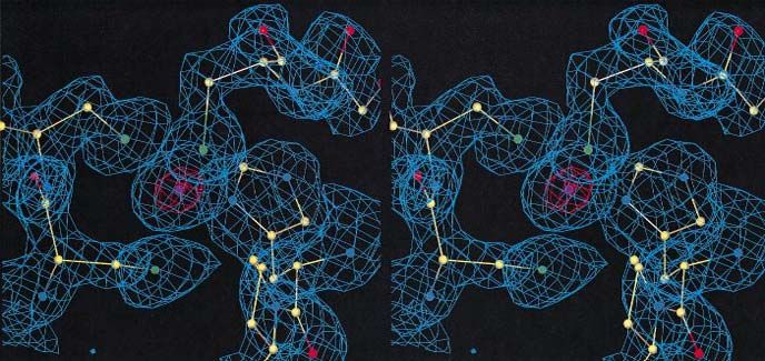
Fig. 3. A stereo view of the electron density in the zinc-binding site of the Btk motif. The 2Fo–Fc omit electron density map around the zinc ion in
the Btk motif is shown. Prior to the map calculation, the zinc ion and residues inside a 5 Å sphere around the zinc ion were removed, and the model
was subjected to a conventional positional refinement in program X-PLOR. A 2Fo–Fc electron density map was calculated with the resulting model
and contoured at 1σ (cyan) and 10σ (orange). The final protein model is shown as a ball-and-stick model and coloured according to the atom type
(C yellow, N blue, O red, S green, Zn purple). The residues shown in the figure are C154 and C155 on the left side of the zinc, C165 on the top and
H143 on the right side of the ion. Bonding distances between the zinc and its ligands are 2.24–2.36 Å for zinc–sulfur and 2.05–2.06 Å for
zinc–nitrogen bonds.
Inositol phosphate-binding site and a rather large conformational change would be required
The three-dimensional structures of Ins(1,4,5)P3 com- to bring these residues close enough to interact with
plexes have been solved for the β-spectrin and PLCδ1 PH a ligand.
domains. These two structures show variant binding sites E41 of the Btk PH domain is close to the predicted
around loop 1–2. Ins(1,4,5)P3 binds to the β-spectrin PH binding site in loop 3–4. In a random mutagenesis study,
domain between loops 1–2 and 5–6 (Hyvönen et al., Li et al. (1995) recently found a constitutively active form
1995), whereas the PLCδ1 PH domain binds the ligand of Btk which is caused by a substitution of this residue
between loops 1–2 and 3–4 (Ferguson et al., 1995). by lysine (E41K). This mutant can transform NIH3T3
Previous sequence- and structure-based alignments and cells and shows enhanced tyrosine phosphorylation and
three-dimensional models of the Btk PH domain made membrane localization as compared with the wild-type
prior to the knowledge of the experimental structure have protein. These results suggest that the E41K mutant has
an error which will position the residues involved in increased affinity for a membrane-bound (lipid) ligand,
the binding of inositol phosphates in the first β-strand supporting our assignment of the binding site.
incorrectly for Btk (Musacchio et al., 1993; Ferguson All PH domain structures show a clear polarization of
et al., 1995; Vihinen et al., 1995). Also, an insertion of a charges, and the binding sites for Ins(1,4,5)P3 are located
gap in the beginning of the second β-strand has led to in the positive ends of the domains. In the Btk PH domain,
incorrect interpretations (Ferguson et al., 1995). the residues within the proposed Ins(1,3,4,5)P4-binding
The revised alignment is shown in Figure 4, in which site are similarly in the most positively charged area of
the residues involved in ligand binding are highlighted. the domain (Figure 5B).
This comparison reveals that the Btk PH domain contains
key residues for both inositol-binding sites. On one side XLA mutations
of loop 1–2, residues R13, F25 and K77 correspond to The database of XLA-causing mutations in Btk, the
the Ins(1,4,5)P3-binding site in the β-spectrin domain, and BTKbase, contains nine point mutations within the PH
on the other side of the loop, residues K12, N24, R28 and domain and one in the Btk motif (Vihinen et al., 1996).
K53 are equivalent to those binding Ins(1,4,5)P3 in the We have constructed and expressed five of them: F25S,
PLCδ1 domain (Figure 5A). R28H, T33P, V64F and V113D. Mutations L11P, K12R,
Although many of the residues that bind Ins(1,4,5)P3 S14F and R28P in the PH domain and C155G in the Btk
in the β-spectrin PH domain are conserved in Btk, it is motif have been reported while our work was in progress.
unlikely that the latter would bind a ligand in this region. Structural analysis of the mutations (T33P, V64F and
R13 of the Btk domain is hydrogen-bonded to the carbonyl V113D) which affect folding and/or stability of the domain
group of W147 in the Btk motif and is not available for shows the following. T33 is in loop 2–3, and mutation to
ligand binding. This site is also partly covered by the Btk a rigid proline would disrupt the structure of the loop.
motif and loop 1–2. It is more likely that the binding site V64 is part of the hydrophobic core and is fully conserved
of Ins(1,3,4,5)P4 in the Btk domain is similar to the site in all proteins containing both the PH domain and the Btk
in the PLCδ1 domain. Many of the ligand-binding residues motif. Introduction of a phenylalanine in this position
are present in Btk, and most of the XLA mutations point cannot be accommodated. V113 is also a conserved, buried
to this site. In the complex of the PLCδ1 PH domain and hydrophobic residue, and its substitution by an aspartate
Ins(1,4,5)P3, additional contacts come from residues 54– will introduce an unfavourable polar residue into the core
57 in loop 3–4. In Btk, this loop has positive residues of the domain.
(R46, R48, R49) but it points away from the binding site, All other XLA-causing mutations in the Btk PH domain
3399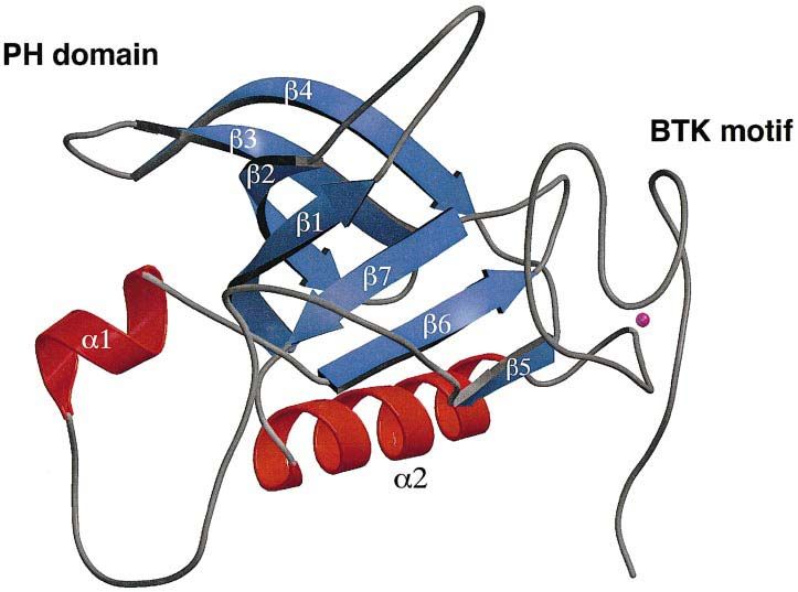
M.Hyvönen and M.Saraste

Fig. 4. Structural alignment of PH domains. Structural alignment of PH domains from Btk, PLCδ1 (Ferguson et al., 1995) (PDB entry 1MAI),
β-spectrin (Hyvönen et al., 1995) (1BTN), dynamin (Ferguson et al., 1994) (1DYN) and pleckstrin (Yoon et al., 1994) (1PLS, the first of the 10
NMR structures in the PDB entry was used for the superpositioning). Superpositioning and the alignment were generated with the MODELLER
program (Sali and Blundell, 1993). Secondary structure elements of the Btk PH domain are shown below the alignment, with blue arrows for
β-sheets and red cylinders for α-helices, and numbered as in Figure 2. Residues which are assigned to the Ins(1,3,4,5)P4-binding site of Btk are
coloured green. Ins(1,4,5)P3-binding residues are coloured blue in β-spectrin and red in the PLCδ1 domain. Residues showing chemical shifts upon
binding to Ins(1,4,5)P3 in pleckstrin and dynamin PH domains are coloured yellow. The boxed areas of the alignment correspond to the well-defined
and superimposable parts of the PH domains. The most conserved hydrophobic residue in each of these blocks appears on a grey background. The
XLA-causing point mutations are marked on top of the Btk PH domain sequence, and the activating mutation E41K is shown in italics. A hash-mark
shows the residues forming the hydrophobic pocket for F146 of the Btk motif. The numbering corresponds to that of the PDB entries and is used in
the text.

are located around loop 1–2 in the area where the predicted                 the following residue K12 by positioning it unfavourably
binding site for inositol phosphates resides (Figure 6A                     for interaction with an inositol phosphate ligand.
and B). K12 of Btk and K30 of PLCδ1 are in equivalent                          In accordance with our interpretations, the mutations
positions, and point to the inositol-binding site of PLCδ1.                 F25S, R28C and R28H have been shown recently to
In a Japanese XLA patient, this residue is mutated to                       reduce the affinity for Ins(1,3,4,5)P4 compared with the
arginine (Hashimoto et al., 1996). In the PLCδ1 PH                          wild-type protein in vitro (Fukuda et al., 1996). In the
domain, K30 binds to both 4- and 5-phosphates of                            same study, the mutants T33P, V64F and V113D showed
Ins(1,4,5)P3, and a guanido group of arginine could not                     no binding to the ligand, as could be expected if the
be accommodated in this position. Similar mutation of a                     protein cannot acquire its native fold.
conserved, phosphate-binding lysine to arginine in the                         The single known XLA mutation in the Btk motif is
ATP-binding sites of protein kinases are known to inactiv-                  C155G. This cysteine is one of the conserved zinc ligands,
ate these enzymes (Hanks et al., 1988).                                     and its substitution will disrupt the metal centre and
   S14 points in the same direction as R12, and the S14F                    prevent folding of the motif.
mutation would sterically block the predicted binding site.
Another mutated residue of the Btk domain is F25 in the
                                                                            Discussion
beginning of the second β-strand. It is equivalent to W23
in the β-spectrin PH domain, one of the Ins(1,4,5)P3-                       All mutations in the PH domain of Btk cause severe forms
binding residues (Hyvönen et al., 1995). F25S mutation                     of XLA and are likely to abolish completely the binding
would disrupt this binding site if Btk bound an inositol                    of a natural ligand. The structure of the Btk PH domain
phosphate in the same site as β-spectrin. The other                         has revealed that, in most cases, the mutations in the PH
possible consequence of this mutation, which we favour,                     domain are located around a putative inositol phosphate-
is destabilization of loop 1–2 and an indirect effect on the                binding site. Previous attempts to explain these mutations
other putative binding site. This is supported by the                       by sequence analysis and modelling have been inaccurate,
reduced solubility of this mutant during expression and                     mainly because of a misalignment of the first β-strand.
purification.                                                                  All functional mutations in the Btk PH domain cluster
   R28 is substituted with a histidine or a proline in human                onto one end of the domain. Is this the only functionally
XLA and with a cysteine in mouse xid. It corresponds to                     important area on the domain? Apart from inositol phos-
R40 in the PLCδ1 domain, a residue in contact with the                      phates, only a few other interaction partners for PH
5-phosphate of Ins(1,4,5)P3. All of these mutations (R28H,                  domains have been suggested, and the data supporting
R28P and R28C) remove a positive charge, and would                          these findings are often rather weak. The βγ-subunits of
abolish binding of the ligand.                                              trimeric G-proteins are believed to interact with some PH
   L11P mutation could affect the folding of the domain,                    domains (Touhara et al., 1994). There is evidence that
although we have not tested its expression in E.coli. This                  this is also the case for the Btk PH domain (Tsukada
residue is at the end of the first β-strand where a proline                 et al., 1994). In the β-adrenergic receptor kinase (βARK),
might be structurally tolerated. It could, however, affect                  the binding site for the βγ-subunits has been mapped to

3400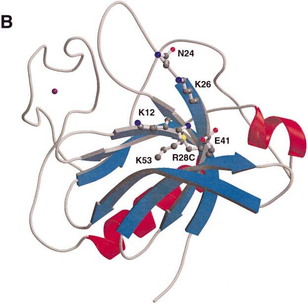
PH domain of Bruton’s tyrosine kinase

                                                              Fig. 5. Bindings site for inositol phosphates in PLCδ1 and Btk PH
                                                              domains. (A) Predicted binding site and the residues thought to be
                                                              involved in Ins(1,3,4,5)P4 binding in the Btk PH domain are shown in
                                                              the upper panel. The corresponding area of the PLCδ1 PH domain
                                                              with residues in direct contact with Ins(1,4,5)P4 is shown in the lower
                                                              panel. The ball-and-stick models of the side chains are coloured
                                                              according to atom types: carbons grey, nitrogens blue, oxygens red and
                                                              sulfurs yellow. The view of the domains is along the C-terminal α-
                                                              helix, which is not visible in the figure. (B) Electrostatic polarization
                                                              of Btk PH domain is shown using a surface representation of the Btk
                                                              PH domain in the same orientation as in Figure 6A and B. The surface
                                                              is coloured according to charge, with positive charge in blue and
                                                              negative in red. The position of residues thought be involved in
                                                              Ins(1,3,4,5)P4 binding are marked on the surface. The figure was
                                                              prepared using GRASP (Nicholls et al., 1991).

the very C-terminus of the PH domain, and residues            development of B cells as it has been recently described
beyond the domain are needed for this interaction. In Btk,    that a PKCβ knock-out mouse has a similar phenotype to
the binding site has not been mapped precisely. The site-     the xid mouse (Leitges et al., 1996). Activation of Btk in
directed mutagenesis and deletions of the possible binding    this mouse is, however, not affected and PKCβ is likely
site in Btk were done without structural considerations,      to act downstream of Btk.
and most of these mutations would certainly affect folding       The binding site for inositol phosphates is predicted to
and stability of the domain (Tsukada et al., 1994). Co-       be similar to that in PLCδ1, but the ligand for Btk does
expression of several different βγ-subunit combinations       not seem to be Ins(1,4,5)P3, but rather Ins(1,3,4,5)P4
with Btk has been shown to activate the kinase in a           (Fukuda et al., 1996; Salim et al., 1996). The same ligand
PH domain-dependent manner, but evidence for a direct         has also been described for Ras-GAP proteins which
interaction is missing (Langhans Rajasekaran et al.,          contain PH domains, namely human IP4BP and murine
1995).                                                        GAP1m (Cullen et al., 1995; Fukuda and Mikoshiba,
   Yao and co-workers (1994) have shown that the Btk          1996). In the case of GAP1m, ligand binding requires
PH domain interacts with various forms of protein kinase      additional elements outside the PH domain. Both of these
C (PKC). The PH domain was found to be phosphorylated         proteins also contain a Btk motif.
on serine residue(s) and interacted with PKCs both in vitro      Most of the binding studies between PH domains and
and in vivo. This interaction was not very strong, but it     inositol phosphates have been conducted with soluble
may nevertheless point to a possible role for PKCs in         head groups of inositol phospholipids, but more careful
regulating Btk. PKC-dependent serine phosphorylation of       studies using phospholipid vesicles are needed to deter-
Btk was shown to down-regulate its kinase activity. PKCβ      mine the role of other lipids in membrane interaction of
seems to be involved in the same pathway as Btk in            PH domains. A 3-fold increase in membrane affinity has

                                                                                                                                3401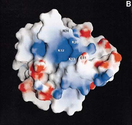
M.Hyvönen and M.Saraste

Fig. 6. Mutations and the predicted inositol-binding site in the Btk PH domain. (A) A ribbon representation of the Btk PH domain and Btk motif
showing residues mutated in XLA patients as ball-and-stick models. Colouring is as in Figures 2 and 5A. (B) Same view of the molecule as in (A),
showing the residues thought to form the Ins(1,3,4,5)P4-binding site as ball-and-stick models.

been demonstrated for PLCδ1 using vesicles containing                       phospholipids in the membrane is thought to affect the
both PtdIns(4,5)P2 and phosphatidylserine (Rebecchi et al.,                 membrane association of other PH domains (Hyvönen
1992). A similar co-operative role for other negative                       et al., 1995).

3402
PH domain of Bruton’s tyrosine kinase

Materials and methods                                                             As high-resolution data became available, original SIRAS phases were
                                                                               extended to 1.6 Å with DM using NCS averaging, histogram matching
Cloning, expression and purification of the domains                             and solvent flattening. Electron density maps calculated with these
A DNA fragment encoding amino acids 1–170 of human Btk (Swiss-                 experimental phases confirmed the correctness of the model and revealed
Prot: Q06187) was amplified by PCR using human Btk cDNA as                     the positions of several previously missing residues.
template (the cDNA was kindly provided by Dr C.I.E.Smith) and cloned
into E.coli expression vector pBAT4 (Peränen et al., 1996). Mutants of        Refinement and structure analysis
the Btk PH domain were created by PCR-directed site-specific muta-             Refinement was carried out using the programs X-PLOR (Brünger,
genesis using the wild-type expression construct as a template. All            1988), TNT (Tronrud et al., 1987) and ARP (Lamzin and Wilson, 1993).
expression constructs were verified by dideoxy sequencing.                     Five percent of the data (2164 reflections in the high-resolution dataset)
   Expression of proteins was carried out in the E.coli strain BL21(DE3)       was set aside for the calculation of the free R-factor (Rfree), which was
carrying plasmid pUBS520 (Brinkmann et al., 1989). In order to obtain          monitored during the refinement. The model was fixed between the
soluble protein, the expression was carried out at 15°C for 20 h after         refinement cycles on a graphics terminal using the program O. In the
induction. The cells were collected by centrifugation and resuspended          beginning of the refinement, the NCS constraints were applied, but were
in 50 mM sodium phosphate buffer pH 7.2. They were lysed by passing            released gradually upon inclusion of high-resolution data. The structure
them twice through a French Press, and the lysate was centrifuged for          was analysed using PROCHECK (Laskowski et al., 1993) and WHAT
30 min at 100 000 g to separate soluble and insoluble fractions. The           CHECK of the WHAT IF program (Vriend, 1990).
solubility of the mutant proteins was analysed by SDS–PAGE (Laemmli,              In the final map, two loops of the PH domain have very weak electron
1970). Soluble wild-type and mutant proteins were purified using               density. The loops between β-strands 1 and 2 are modelled partly as
three chromatographic steps. Cation-exchange chromatography on an              polyalanine, as is loop 5–6 in the second molecule within the asymmetric
S-Sepharose FF column (all columns are from Pharmacia, Sweden) was             unit. In the first molecule, residues 80–88 of this loop are left out
run with the lysis buffer, and protein was eluted with a linear salt           completely. Both of these loops point away from the core of the domain
gradient at 250–300 mM NaCl. Gel filtration in a Superdex 75 16/60             and do not seem to have contacts with it. Several different refinement
preparation grade column was run with 20 mM EPPS buffer (Sigma,                protocols were tried, but none of them was able to improve the quality
USA), 100 mM NaCl, 2 mM dithiothreitol (DTT), pH 8.0. Protein from             of the electron density for these two loops.
gel filtration was diluted 3-fold, and loaded onto a MonoS HR 10/10               The coordinates will be deposited to the Brookhaven Protein Data
cation-exchange column equilibrated with 20 mM EPPS, 2 mM DTT,                 Bank. Before that, the coordinates will be available from the authors by
pH 8.0. Protein eluted at ~250 mM NaCl. Purified protein was concen-           e-mail.
trated to 6–8 mg/ml in 10 mM Tris, pH 8.0, 100 mM NaCl, 2 mM DTT,
and stored in small aliquots at –80°C. The molecular weights of purified
proteins were verified by mass spectroscopy, which showed that the             Acknowledgements
N-terminal methionine was cleaved off. The protein used in all studies
thus corresponds to residues 2–170 of human Btk.                               We wish to thank people at the EMBL-Hamburg Outstation c/o DESY,
   Metal analysis was done by atomic absorption spectroscopy at the            in particular Victor Lamzin, for helping with the data collection, Kristina
University of Saarland, (Saarbrücken, Germany) in the laboratory of Dr        Djinovic-Carugo and Matthias Wilmanns for their generous advice
Michael Zeppezauer. Protein concentration was determined spectrophoto-         during structure determination, Jaime Pascual and Kristina Djinovic-
metrically in 6 M guanididinium–HCl using a calculated molar absorption        Carugo for critically reading the manuscript, Liisa Holm for helping
coefficient of 23 200/M (Gill and von Hippel, 1989).                           with DALI searches, Toby Gibson for discussions, Edvard Smith for
                                                                               providing the cDNA of human Btk, and Michael Zeppezauer and his
Crystallization                                                                colleagues for performing the metal analysis.
Crystallization trials were conducted at room temperature in hanging
drops. Diffraction quality crystals were obtained only with the mutant
R28C with 32.5% (w/v) PEG 3350 (Sigma, USA), 100 mM Tris pH 8.5,               References
200 mM MgCl2, 500 mM NaCl as the well solution. Plate-like crystals
appeared after 24–48 h and were stable for several weeks.                      Bacon,D.J. and Anderson,W.F. (1988) A fast algorithm for rendering
                                                                                 space-filling molecule pictures. J. Mol. Graphics, 6, 219–220.
Data collection and heavy metal derivative search                              Bolen,J.B. (1995) Protein tyrosine kinases in the initiation of antigen
All datasets were collected at 110 K using a cryo-stream cooler from             receptor signaling. Curr. Opin. Immunol., 7, 306–311.
Oxford Cryosystems. In-house data collection was done using a MAR              Brinkmann,U., Mattes,R.E. and Buckel,P. (1989) High-level expression
Research imaging plate detector and a Siemens GX21 generator running             of recombinant genes in Escherichia coli is dependent on the
at 40 kV and 70 mA. Synchrotron data was collected at the EMBL                   availability of the dnaY gene product. Gene, 85, 109–114.
Hamburg Outstation c/o DESY on the BW7B beamline using a MAR                   Brünger,A.T. (1988) X-PLOR Version 3.1 A System for X-ray
Research imaging plate. Data were collected in two passes to obtain              Crystallography and NMR. Yale University Press, New Haven and
reliable low resolution data also. All datasets were processed with              London.
DENZO, and scaled and merged in SCALEPACK (Z.Otwinowski and                    Cullen,P.J., Hsuan,J.J., Truong,O., Letcher,A.J., Jackson,T.R., Dawson,
W.Minor, in preparation).                                                        A.P. and Irvine,R.F. (1995) Identification of a specific Ins(1,3,4,5)P4-
   During the search for the heavy metal derivatives, it became evident          binding protein as a member of the GAP1 family. Nature, 376,
that crystals were often non-isomorphous. An the end, only one heavy             527–530.
metal derivative, trimethyl lead acetate [(CH3)3PbAc], was found, but          de Weers,M., Brouns,G.S., Hinshelwood,S., Kinnon,C., Schuurman,R.K.,
the non-isomorphous nature of the crystals lowered the quality of the            Hendriks,R.W. and Borst,J. (1994) B-cell antigen receptor stimulation
data. To overcome this problem, both native and derivative data sets             activates the human Bruton’s tyrosine kinase, which is deficient in
were collected from the same single crystal. After collecting a high quality     X-linked agammaglobulinemia. J. Biol. Chem., 269, 23857–23860.
native dataset, the crystal was thawed, soaked in 1 mM (CH3)3PbAc in           Divecha,N. and Irvine,R.F. (1995) Phospholipid signaling. Cell, 80,
the mother liquor for 24 h, and a derivative data set was then collected         269–278.
under cryo conditions. The resulting datasets were isomorphous and the         Ferguson,K.M., Lemmon,M.A., Schlessinger,J. and Sigler,P. (1994)
derivative proved to have very good phasing power (Table I).                     Crystal structure at 2.2 Å resolution of the pleckstrin homology
                                                                                 domain from human dynamin. Cell, 79, 199–209.
Phasing and solvent flattening                                                  Ferguson,K.M., Lemmon,M.A., Schlessinger,J. and Sigler,P.B. (1995)
Phasing was done using the PHASES package (Furey and Swaminathan,                Structure of the high affinity complex of inositol trisphosphate with
1996). The position of the heavy metal was determined by Patterson               a phospholipase C pleckstrin homology domain. Cell, 83, 1037–1046.
methods. Phasing and phase refinement was done using both isomorphous          Fukuda,M. and Mikoshiba,K. (1996) Structure–function relationships of
and anomalous differences. Resulting SIRAS phases were already good              the mouse Gap1m. Determination of the inositol 1,3,4,5-
enough to allow partial model building and determination of the non-             tetrakisphosphate-binding domain. J. Biol. Chem., 271, 18838–18842.
crystallographic symmetry (NCS). These phases were then used in the            Fukuda,M., Kojima,T., Kabayama,H. and Mikoshiba,K. (1996) Mutation
program DM of the CCP4 package for solvent flattening, histogram                 of the pleckstrin homology domain of Bruton’s tyrosine kinase in
matching and two-fold NCS averaging. Over 80% of the molecule was                immunodeficiency impaired inositol 1,3,4,5-tetrakisphosphate binding
built into the resulting electron density maps.                                  capacity. J. Biol. Chem., 271, 30303–30306.

                                                                                                                                                  3403
M.Hyvönen and M.Saraste Furey,W. and Swaminathan,S. (1996) PHASES-95: a program package Salim,K. et al. (1996) Distinct specificity in the recognition of for the processing and analysis of diffraction data from phosphoinositides by the pleckstrin homology domains of dynamin macromolecules. Methods Enzymol., in press. and Bruton’s tyrosine kinase. EMBO J., 15, 6241–6250. Gibson,T.J., Hyvönen,M., Musacchio,A., Saraste,M. and Birney,E. (1994) Sato,S. et al. (1994) IL-5 receptor-mediated tyrosine phosphorylation of PH domain: the first anniversary. Trends Biochem. Sci., 19, 349–353. SH2/SH3-containing proteins and activation of Bruton’s tyrosine and Gill,S.C. and von Hippel,P.H. (1989) Calculation of protein extinction Janus 2 kinases. J. Exp. Med., 180, 2101–2111. coefficients from amino acid sequence data. Anal. Biochem., 182, Smith,C.I., Islam,K.B., Vorechovsky,I., Olerup,O., Wallin,E., Rabbani,H., 319–326. Baskin,B. and Hammarstrom,L. (1994) X-linked agammaglobulinemia Hanks,S.K., Quinn,A.M. and Hunter,T. (1988) The protein kinase family: and other immunoglobulin deficiencies. Immunol. Rev., 138, 159–183. conserved features and deduced phylogeny of the catalytic domains. Thomas,J.D., Sideras,P., Smith,C.I., Vorechovsky,I., Chapman,V. and Science, 241, 41–52. Paul,W.E. (1993) Colocalization of X-linked agammaglobulinemia Harlan,J.E., Hajduk,P.J., Yoon,H.S. and Fesik,S.W. (1994) Pleckstrin and X-linked immunodeficiency genes. Science, 261, 355–358. homology domains bind to phosphatidylinositol-4,5-bisphosphate. Touhara,K., Inglese,J., Pitcher,J.A., Shaw,G. and Lefkowitz,R.J. (1994) Nature, 371, 168–170. Binding of G protein βγ-subunits to pleckstrin homology domains. J. Hashimoto,S. et al. (1996) Identification of Bruton’s tyrosine kinase Biol. Chem., 269, 10217–10220. (Btk) gene mutations and characterization of the derived proteins in Tronrud,D.E., Ten Eyck,L.F. and Matthews,B.W. (1987) An efficient 35 X-linked agammaglobulinemia families: a nationwide study of Btk general-purpose least-square refinement program for macromolecular deficiency in Japan. Blood, 88, 561–573. structures. Acta Crystallogr., A43, 489–501. Holm,L. and Sander,C. (1993) Protein structure comparison by alignment Tsukada,S. et al. (1993) Deficient expression of a B cell cytoplasmic of distance matrices. J. Mol. Biol., 233, 123–138. tyrosine kinase in human X-linked agammaglobulinemia. Cell, 72, Hyvönen,M., Macias,M.J., Nilges,M., Oschkinat,H., Saraste,M. and 279–290. Wilmanns,M. (1995) Structure of the binding site for inositol Tsukada,S., Simon,M.I., Witte,O. and Katz,A. (1994) Binding of βγ phosphates in a PH domain. EMBO J., 14, 4676–4785. subunits of trimeric G proteins to the PH domain of Bruton tyrosine Kraulis,P. (1991) MOLSCRIPT: a program to produce both detailed and kinase. Proc. Natl Acad. Sci. USA, 91, 11256–11260. schematic plots of proteins. J. Appl. Crystallogr., 24, 946–950. Vetrie,D. et al. (1993) The gene involved in X-linked agammaglobulin- Laemmli,U.K. (1970) Cleavage of structural proteins during the assembly emia is a member of the src family of protein-tyrosine kinases. Nature, of the head of bacteriophage T4. Nature, 227, 680–685. 361, 226–233. Lamzin,V. and Wilson,K. (1993) Automated refinement of protein Vihinen,M., Nilsson,L. and Smith,C.I. (1994) Tec homology (TH) models. Acta Crystallogr., D49, 129–147. adjacent to the PH domain. FEBS Lett., 350, 263–265. Langhans Rajasekaran,S.A., Wan,Y. and Huang,X.Y. (1995) Activation Vihinen,M., Zvelebil,M.J., Zhu,Q., Brooimans,R.A., Ochs,H.D., of Tsk and Btk tyrosine kinases by G protein βγ subunits. Proc. Natl Zegers,B.J., Nilsson,L., Waterfield,M.D. and Smith,C.I. (1995) Acad. Sci. USA, 92, 8601–8605. Structural basis for pleckstrin homology domain mutations in X-linked Laskowski,R.A., MacArthur,M.W., Hutchinson,E.G. and Thornton,J.M. agammaglobulinemia. Biochemistry, 34, 1475–1481. (1993) PROCHECK: a program to check the stereochemical quality Vihinen,M., Iwata,T., Kinnon,C., Kwan,S.P., Ochs,H.D., Vorechovsky,I. of protein structures. J. Appl. Crystallogr., 26, 283–291. and Smith,C.I. (1996) BTKbase, mutation database for X-linked Leitges,M., Schmedt,C., Guinamard,R., Davoust,J., Schaal,S., Stabel,S. agammaglobulinemia (XLA). Nucleic Acids Res., 24, 160–165. and Tarakhovsky,A. (1996) Immunodeficiency in protein kinase Cβ- Vriend,G. (1990) WHAT IF—a molecular modelling and drug design deficient mice. Science, 273, 788–791. program. J. Mol. Graphics, 8, 52–56. Lemmon,M.A., Ferguson,K.M., O’Brien,R., Sigler,P.B. and Yao,L., Kawakami,Y. and Kawakami,T. (1994) The pleckstrin homology Schlessinger,J. (1995) Specific and high-affinity binding of inositol domain of Bruton tyrosine kinase interacts with protein kinase C. phosphates to an isolated pleckstrin homology domain. Proc. Natl Proc. Natl Acad. Sci. USA, 91, 9175–9179. Acad. Sci. USA, 92, 10472–10476. Yoon,H.S., Hajduk,P.J., Petros,A.M., Olejniczak,E.T., Meadows,R.P. and Li,T., Tsukada,S., Satterthwaite,A., Havlik,M.H., Park,H., Takatsu,K. Fesik,S.W. (1994) Solution structure of a pleckstrin-homology domain. and Witte,O.N. (1995) Activation of Bruton’s tyrosine kinase (BTK) Nature, 369, 672–675. by a point mutation in its pleckstrin homology (PH) domain. Immunity, 2, 451–460. Received on January 21, 1997; revised on February 25, 1997 Merrit,E.A. and Murphy,M.E.P. (1994) Raster3D version 2.0—a program for photorealistic molecular graphics. Acta Crystallogr., D50, 869–873. Musacchio,A., Gibson,T., Rice,P., Thompson,J. and Saraste,M. (1993) The PH domain: a common piece in the structural patchwork of signalling proteins. Trends Biochem. Sci., 18, 343–348. Nicholls,A., Sharp,K.A. and Honig,B. (1991) Protein folding and association: insights from the interfacial and thermodynamic properties of hydrocarbons. Proteins: Struct. Funct. Genet., 11, 281–296. Park,H., Wahl,M.I., Afar,D.E., Turck,C.W., Rawlings,D.J., Tam,C., Scharenberg,A.M., Kinet,J.P. and Witte,O.N. (1996) Regulation of Btk function by a major autophosphorylation site within the SH3 domain. Immunity, 4, 515–525. Paterson,H.F., Savopoulos,J.W., Perisic,O., Cheung,R., Ellis,M.V., Williams,R.L. and Katan,M. (1995) Phospholipase Cδ1 requires a pleckstrin homology domain for interaction with the plasma membrane. Biochem. J., 312, 661–666. Peränen,J., Rikkonen,M., Hyvönen,M. and Kääriäinen,L. (1996) T7 vectors with modified T7lac promoter for expression of proteins in Escherichia coli. Anal. Biochem., 236, 371–373. Rawlings,D.J. et al. (1993) Mutation of unique region of Bruton’s tyrosine kinase in immunodeficient XID mice. Science, 261, 358–361. Rawlings,D.J., Scharenberg,A.M., Park,H., Wahl,M.I., Lin,S., Kato,R.M., Fluckiger,A.C., Witte,O.N. and Kinet,J.P. (1996) Activation of BTK by a phosphorylation mechanism initiated by SRC family kinases. Science, 271, 822–825. Rebecchi,M., Peterson,A. and McLaughlin,S. (1992) Phosphoinositide- specific phospholipase C-δ1 binds with high affinity to phospholipid vesicles containing phosphatidylinositol 4,5-bisphosphate. Biochemistry, 31, 12742–12747. Sali,A. and Blundell,T. (1993) Comparative protein modelling by satisfaction of spatial restraints. J. Mol. Biol., 234, 779–815. 3404
You can also read


