LUNG HEALTH The International Workshop on - Rising Stars Abstracts A story lasting since 2016
←
→
Page content transcription
If your browser does not render page correctly, please read the page content below
The International Workshop on
        LUNG HEALTH

     Rising Stars Abstracts
   A story lasting since 2016

              1
Circulating biomarkers in asthmatic pregnancy

Andras Bikov1; Aniko Bohacs1; Noemi Eszes1; Janos Rigo2; Gyorgy Losonczy1; Ildiko
Horvath1,3; Lilla Tamasi1
1
 Semmelweis University, Department of Pulmonology, Budapest, Hungary; 2Semmelweis
University, First Department of Obstetrics and Gynecology, Budapest, Hungary; 3National
Koranyi Istitute for TB and Pulmonology, Budapest, Hungary

Background/Aims: Asthma is one of the most common disorders which may complicate
pregnancy. Because of the complex immunological interrelation between the mother and
foetus, asthma during gestation represents a distinct inflammatory phenotype. However,
its pathophysiology is not fully known. Elevated levels of vasculogenic vascular endothelial
growth factor (VEGF), anti-apoptotic survivin and elements of complements system were
previously reported in induced sputum samples in non-pregnant asthmatics. We aimed to
analyse these markers in systemic samples in order to have a better picture on asthma
during gestation.

Methods: 31 asthmatic pregnant (AP), 28 healthy pregnant (HP), 29 asthmatic non-
pregnant (ANP) and 29 healthy non-pregnant (HNP) women were enrolled. In addition to
lung function and exhaled nitric oxide measurements, venous blood was collected in EDTA
tubes for VEGF, survivin, complement 5a (C5a) and complement H factor (CHF) analyses
which were performed with commercially available ELISA kits.

Results: Plasma VEGF and survivin levels were significantly lower, while CHF levels were
elevated in pregnancy (p0.05). In contrast, circulating C5a levels were higher in AP compared to all other groups
with a significant relationship with FEV1 (r = -0.44, p = 0.04).

Conclusions: Pregnancy itself alters the levels of circulating vasculogenic, anti-apoptotic
and complement markers; but these are only markedly influenced by asthma. These results
suggest that asthma-related inflammatory processes are mainly localised in the airways. To
understand the background of asthmatic pregnancy further studies with exhaled breath
measurements are required, as other, potentially harmful techniques (i.e. BAL or induced
sputum) cannot be performed during gestation.

The study was supported by Hungarian Scientific Fund (OTKA 68808) and Hungarian
Respiratory Society

                                              2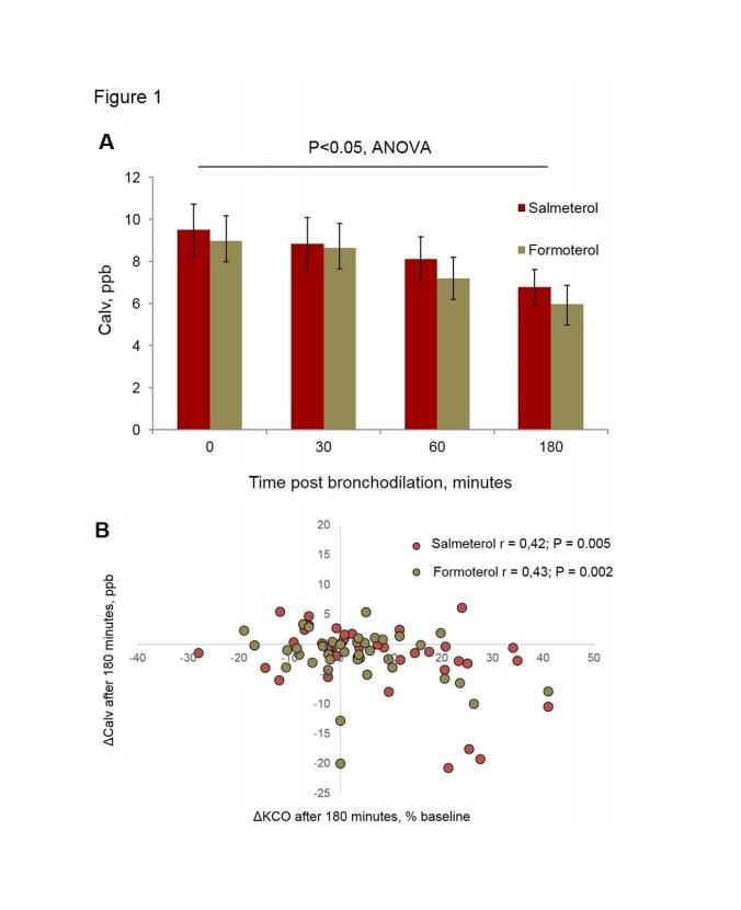
SERUM CYTOKINES PATTERNS IN LUNG TRANSPLANT RECIPIENTS

Letizia Corinna Morlacchi1
1
 U.O. Broncopneumologia, Dipartimento di fisiopatologia e dei trapianti, IRCCS Fondazione
Ca’ Granda Ospedale Maggiore Policlinico di Milano, Università degli Studi di Milano.,
Milan, Italy

BACKGROUND & AIMS

End stage COPD is currently the first indication for lung transplantation worldwide (ISHLT
Registry, 2015); however, little is known about chronic rejection, which is the the leading
cause of long-term mortality, accounting for approximately 40% of deaths.

The               aims             of             this             study            were:
- to evaluate cytokine profiles in serum of lung transplant (LTx) recipients
- to evaluate the relationship between cytokine levels and the development of chronic lung
allograft dysfunction (CLAD).

METHODS

A prospective, observational study was performed on consecutive patients who underwent
LTx from May 2010 to June 2012. Systemic inflammatory response was evaluated detecting
cytokine levels on samples of blood using a high sensitivity immunoassay (RandoxTM Kit).
Two groups of patients were identified: those who developed CLAD and those who did not.
Comparisons between groups were performed using Anova test and Wilcoxon-Mann-
Whitney test; Spearman's rank correlation coefficient was also calculated.

RESULTS

24 patients were considered. General characteristics of the population are shown in table 1:
no significant difference was found among the two groups. Some cytokines showed a linear
increase on serum over time, whilst IFNgamma remained stable (Figure 1). IL1 was
significantly higher in the group of patients who developed CLAD (p=0,004) (Figure 2).

CONCLUSIONS

Our study showed a temporal trend for several cytokines, suggesting a progressive
rearrangement of inflammatory response and intracellular communication after LTx.
Conversely, IFNgamma did not show any significant change over time: this may be due to
immunosuppressive therapy with tacrolimus, which inhibits IL2 transcription and T-cells

                                              3
proliferation and thus expression of IFN; therefore, this cytokine might play a role as a
marker of effective immunosuppression. Finally, circulating IL1 concentrations may also
help in predicting the risk of CLAD: IL1 is a crucial cytokine in innate immunity response and
its production can be elicited by external stimuli, like viral or bacterial infection and GERD,
which are well-known risk factors for CLAD.
4Risk factors for anaphylaxis and the differences in B3GAT1 and ITGB1 gene expression in
patients with mastocytosis.
Aleksandra Górska1; Marek Niedoszytko1; Agnieszka Maciejewska2; Marcin Skrzypski3;
Marta Chełmińska1; Magda Lange4; Ryszard Pawłowski2; Marek Słomiński5; Ewa Jassem1
1
Allergology and Pneumonology Dept, Medical University of Gdansk, Gdansk, Poland;
2
Forensic Medice Dept, medical University of Gdansk, Gdansk, Poland; 3Oncology Dept,
Medical University of Gdansk, Gdansk, Poland; 4Dermatology, Wenerology and Allergology
Dept, Medical University of Gdansk, Gdansk, Poland; 5Pneumonology Dept, Medical
University of Gdansk, Gdansk, Poland
Mastocytosis is an uncommon group of disorders classified as an myeloproliferative
neoplasm, characterized by an abnormal proliferation and accumulation of atypical mast
cells in different organs.
Aims 1. to find differences in gene expression in peripheral blood cells of patients with
mastocytosis, compared to healthy controls; 2. to analyze any differences in gene
expression in peripheral blood cells between patients with IVA compared to patients
without anaphylaxis in history; 3. to analyze the prevalence of the anaphylactic reactions
and to identify causative and risk factors of anaphylaxis in mastocytosis patients depending
on the variant of the disease.
Methods: Study group included 152 adult patients with mastocytosis; 57 patients were
qualified to assess gene expression analysis using RT-PCR; 19 healthy persons in control
group.
Results: Significant differences in gene expression were found for B3GAT1 (p=0,006) and
ITGB1 (p=0,02) in mastocytosis patients compared to controls. Furthermore significant
differences were found in gene expression for B3GAT1 (p=0,003) and ITGB1 (p=0.02) in
patients with IVA compared to patients without anaphylaxis. The prevalence of
anaphylactic reactions was 50%. Such reactions were more prevalent in patients with
indolent systemic mastocytosis than in cutaneous. The most frequent triggers of
anaphylaxis were food (29%), insect stings (22%) and drugs (15%). Serum tryptase levels
were higher in patients with anaphylaxis (p=0.029), also in physical factors related
syndromes (p=0.002). The frequency of those symptoms was reported in 112 (74%)
patients and was higher in systemic mastocytosis patients compared with cutaneous
mastocytosis (p=0.026).
Conclusions: We found significant differences: 1. in gene expression in peripheral blood
cells of patients with mastocytosis compared to controls, 2. in patients with a history of IVA
compared to those without anaphylaxis. We reported significant incidence of MCAS in
mastocytosis patients with anaphylaxis in history. For anaphylaxis resulting from physical
factors, sBT was a significant predictor. Gene expression assessment may be useful in
clinical practice to predict the presence of mastocytosis and the risk of anaphylaxis
5Fluticasone propionate alters the lower respiratory tract microbiota and impairs lung
anti-viral and anti-bacterial host-defence
Aran Singanayagam1; Glanville Nicholas1; Cox Michael1; James Phillip1; Moffatt Miriam1;
Cookson William1; Bartlett Nathan1; Johnston Sebastian1
1
Imperial College, London, United Kingdom
Background: Inhaled corticosteroids are frequently used to treat airways diseases but have
limited effect on exacerbation frequency and are associated with increased pneumonia risk.
The aim of this study was to evaluate effects of fluticasone propionate(FP) on airway anti-
viral and anti-bacterial host-defence.
Methods: C57BL/6 mice were treated with intranasal FP or vehicle control. 16S rRNA
sequencing was used to evaluate lung tissue microbiota. Using mouse models of
rhinovirus(RV)-1B and S.pneumoniae D39 challenge, effects of FP administration on
immune responses and pathogen control were evaluated.
Results: Mice treated with FP had increased lung bacterial loads versus vehicle controls at 8
hours post administration(p=0.015). 16S sequencing showed a significant increase in
bacterial diversity and Stenotrophomonas genera at 8 hours post-administration in mice
treated with FP (fig 1).
In mice challenged with S.pneumoniae, high dose FP(1mg/kg) suppressed a range of anti-
bacterial responses and increased bacterial loads by quantitative culture in lung tissue (8
hours post-infection;pThe role of bronchodilation on airway mechanical stress, lung hyperinflation and NO
production in stable COPD
Dejan Radovanovic1; Marco Contoli2; Susanna Mascetti2; Matteo Pecchiari3; Alberto Papi2;
Stefano Centanni4; Pierachille Santus4
1
School of Respiratory Diseases, University of Milan, Milano, Italy; 2Research Centre on
Asthma and COPD, Section of Internal and Cardio-Respiratory Medicine, University of
Ferrara, Ferrara, Italy; 3Department of Physiopathology and Transplantation, University of
Milan, Milano, Italy; 4Department of Health Sciences, University of Milan, Milano, Italy
Background
Patients with severe chronic obstructive pulmonary disease (COPD) experience cyclic
opening and closure of the airways even at tidal volume (Pecchiari M et al. Respir Physiol
Neurobiol 2016). The effect of this mechanical stress on lung inflammation and its
reversibility are still poorly understood. The aim of our study is to investigate the acute
effect of long acting beta-agonists (LABAs) on lung inflammation and its relationship with
lung function in patients with moderate to very severe COPD.
Methods
This was a phase IV, multicenter, randomized, interventional, double blind, crossover study.
Stable patients with COPD and a forced expiratory volume in 1 second (FEV1)8
9
Using Optical Coherence Tomography to evaluate airway dynamics in vivo
Margit Szabari1,4; Vanessa J Kelly1; Matthew Applegate1; David C Adams1,4; Lida P Hariri3,4;
Chunmin Chee1,4; Khay Tan1,4; Robert S Harris1; Tilo Winkler2; Melissa J Suter1,4
1
 Department of Medicine, Pulmonary and Critical Care Unit, Massachusetts General
Hospital and Harvard Medical School, Boston, USA; 2Department of Anesthesia, Critical Care
and Pain Medicine, Massachusetts General Hospital and Harvard Medical School, Boston,
USA; 3Department of Pathology, Massachusetts General Hospital and Harvard Medical
School, Boston, USA; 4Wellman Center for Photomedicine, Massachusetts General Hospital
and Harvard Medical School, Boston, USA

Background/Aims

Airway hyperresponsiveness is a hallmark feature of asthma. To better understand this
condition, it is essential to visualize airway behavior in vivo. Optical coherence tomography
(OCT) is a high-resolution imaging modality that can be used to provide real-time
visualization of airway dynamics in vivo. Our aim was to use OCT to investigate the
structure and function of airways in the healthy and constricted lung in both dependent
and non-dependent airway regions during mechanical ventilation.

Methods

N=3 sheep were anesthetized and mechanically ventilated. A total of 6 dependent and 6
non-dependent airways were imaged at baseline and following Methacholine
administration (Mch: 1 mg/hr). OCT imaging was used to acquire cross-sectional images
during Regular Tidal Breathing (RTB) (tidal volume (TV): 10 ml/kg) and in a response to one
minute of Double TV ventilation (DTV). The airway-areas during the breathing cycle and
pre- and post-DTV maneuvers were calculated from the OCT images using semi-automated
image processing algorithms.

Results

Dependent airway-area increased more than non-dependent airway-area during RTB in the
healthy airways (p
Supported by the Center for Improving Medicine with Innovation & Technology.
Presenting author has nothing to disclose.
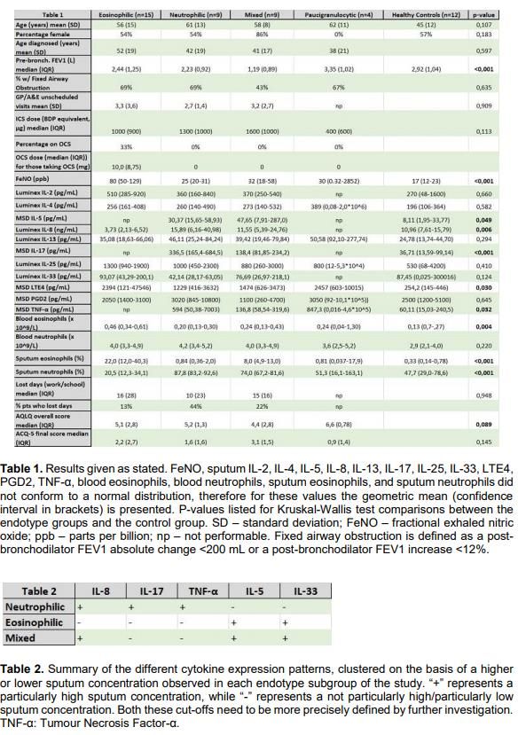
11Assessing inflammatory patterns in asthma endotypes:
new diagnostic and therapeutic perspectives
Matteo Bradicich1,2; Ian Pavord2; Gareth Hynes2; Rahul Shrimanker2
1
Respiratory Pathophysiology and Rehabilitation Department, Cisanello University Hospital,
University of Pisa, Pisa, Italy; 2Respiratory Medicine Unit, Nuffield Department of Clinical
Medicine, NDM Research Building, University of Oxford, Oxford, United Kingdom
Background
Bronchial asthma is a chronic airway disease affecting more than three hundred million
patients worldwide. Given the high prevalence of this disease and the social and economic
burden resulting from under- or mistreatment, the optimal management of this condition
represents therefore a key goal. In order to investigate the pathophysiological
heterogeneity of this disease, a rather new trend in the Literature suggests categorising
different asthmatic patient subpopulations on the basis of their specific molecular patterns,
which are supposed to represent the key pathological determinant leading to one particular
group of symptoms and signs – i.e. a phenotype – rather than another one. Therefore,
asthma endotypes are the key for understanding and treating asthmatic patients with a
precision medicine approach.
Overview
A clinical study led by Oxford (United Kingdom) and Pisa (Italy) Universities quantifies the
differences in the expression levels of multiple sputum inflammatory molecules between
different subpopulations of asthmatic patients, previously labelled on the basis of their
sputum differential cell count as affected by eosinophilic, neutrophilic, mixed, or
paucigranulocytic asthma.
Methods
37 asthmatic patients and 12 healthy controls were recruited. Assessment of symptom
burden and medication usage as well as laboratory measures including blood and sputum
cell count and cytokine levels in sputum supernatant – measured using MSD® and Luminex®
– was performed. The asthmatic patients were subdivided in four subgroups (eosinophilic,
neutrophilic, mixed, and paucigranulocytic) on the basis of their sputum differential cell
count. A comparison of the sputum concentration of the inflammatory molecules taken
into consideration (IL-2, IL-4, IL-5, IL-8, IL-13, IL-17, IL-25, IL-33, PGD2, LTE4, TNF-α) between
the aforementioned subgroups was subsequently carried out.
12Results
The study shows that sputum IL-8, IL-17 and TNF-α are leading molecules in the
neutrophilic asthma endotype, whilst sputum IL-5 and IL-33 underlie eosinophilic asthma.
The mixed endotype is defined by high levels of sputum IL-5, IL-8 and IL-33. There are no
significant results regarding paucigranulocytic asthma.
Conclusions
These pathophysiologic differences might be used in a compact, multi-cytokine assessment
test that defines univocally the specific patient’s inflammatory pattern from a single sample
of induced sputum. Such results shed light on the multifaceted inflammatory environment
in bronchial asthma and might promote further research in order to define new targeted
therapy strategies for those patients with difficult-to-treat asthma.
1314
Circulating MDSC modulate IPF progression by orchestrating immunosuppressive
                             and pro-fibrotic networks

Isis E. Fernandez1; Flavia Greiffo1; Marion Frankenberger1; Jurgen Behr2,3; Alistair Forrest4;
Oliver Eickelberg1,5
1
 Comprehensive Pneumology Center, Helmholtz Zentrum Munchen; Member of the German
Center for Lung Research, Munich, Germany; 2Asklepios Fachkliniken Munchen-Gauting,
Munich, Germany; 3Comprehensive Pneumology Center, Medizinische Klinik und Poliklinik V,
Klinikum der Ludwig-Maximilians- Universität, Munich, Germany; 4Harry Perkins Institute of
Medical Research, QEII Medical Centre and Centre for Medical Research, the University of
Western Australia, Perth, Australia; 5Division of Respiratory Sciences and Critical Care
Medicine, Department of Medicine, University of Colorado, Denver, USA

Rationale:

Idiopathic pulmonary fibrosis (IPF) is a fibroproliferative lung disease with irreversible loss
of lung function. Myeloid-derived suppressor cells (MDSC) are pathologically activated
immature myeloid cells, which suppress immune responses in cancer, autoimmunity, and
other inflammatory conditions. Recent literature supports that aberrant immune responses
contribute to IPF pathogenesis. We reported, for the first time, that MDSC are increased in
numbers, functionally active, and reflect disease status in IPF, in cross-sectional and
longitudinal analysis serving as potent biomarker for IPF progression. Monocytic MDSC are
the predominant subtype in IPF, and yet, differences between mature monocytes and
monocytic MDSC, and their interaction in IPF have not been explored. Here we hypothesize
that MDSC creates an immunosuppressive and pro-fibrotic environment in IPF,
perpetuating disease.

Methods and results:

We included 170 patients, including patients with IPF (n=69), non-IPF ILD (n=56), COPD
(n=23), and healthy controls (n=22). We detected increased circulating MDSC in IPF
compared to controls (30.99±15.61 vs 18.96±8.17%, p=
respectively). In total, we identified and quantified more than 7000 proteins. Principal
component analysis unequivocally discriminated both cell types, showing that proteome
differences between them are larger than the biological variations between the donors.
Comparing the sets of proteins identified in the two cell types we found 502 MDSC enriched
and 1224 monocyte enriched proteins (2 to >30 log10-transformed LFQ intensity ratios).
Next, we examined the potential for these two cell types to communicate with each other,
by identification of the receptors and ligands expressed by each, and considering known
receptor-ligand interactions, compiled from published datasets. In the combined dataset
200 ligands and 153 receptors were detected. From the cell-to-cell communication analysis
we identified both autocrine signaling edges from monocyte to monocyte (339), MDSC to
MDSC (290), and paracrine signaling edges from monocyte to MDSC (311) and MDSC to
monocyte (316). Specific ligands predicted to signal from monocyte to MDSC included:
ANXA1, CCL18, CXCL2, HSP90AA1, ICAM1, TGFB2, amongst others. While ligands from
MDSC to monocyte included: COL1A1, FN1, HLA-C, HSPG2, MMP1, S100A8-9, TGFB1,
amongst others. Finally, FACS staining confirmed the surface expression of the cognate
expressed receptors in both populations.
Conclusions:
In summary, this study explores for the first time the MDSC proteome in fibrosis. We
detected an increase in MDSC in peripheral blood from IPF patients. We further detected a
correlation between MDSC and FoxP3+ T cells, and a decrease in the transcript levels of
CD28, ICOS, ITK, and LCK in PBMC of IPF patients, suggesting that elevated MDSC might
cause a blunted immune response. MDSC inversely correlate with lung function, as such
MDSC may serve as potent biomarker for IPF progression. Using network analysis, our
proteome data shows an autocrine and paracrine signals from and between monocytes and
MDSC. MDSC signals include strong pro-fibrotic molecules, supporting a pro-fibrotic
modulation. Furthermore, confirmation by flow cytometry of exclusively expressed surface
receptors, might lead to identification of novel proteins useful for therapeutic targeting of
MDSC and monocytes in IPF.
16Differences in Tobacco smoke associated and Biomass fuel associated Chronic Obstructive
Pulmonary disease (COPD) – A unique disease of the Indian Subcontinent
Arjun Khanna1
1
Pulmonologist & Intensivist, Delhi, India
Differences in Tobacco smoke associated and Biomass fuel associated Chronic Obstructive
Pulmonary disease (COPD) – A unique disease of the Indian Subcontinent
Objective
The objective of this study was to differentiate between the various epidemiological,
clinical , radiological, laboratory and prognostic characteristics of Tobacco smoke (TS)
associated and Bio mass fuel(BMF) associated COPD. BMF associated COPD is a major cause
of morbidity and mortality in the developing world. A large population especially in rural
India still uses coal and indigenous gas stoves which lead to a large amount of air pollution.
Current western literature does not distinguish between the characteristics of TS and BMF
associated COPD.
Method
80 patients with COPD, defined using the GOLD spirometry criteria, were studied. 40
patients had significant exposure to smoked tobacco. 40 patients were never smokers, but
had been exposed to BMF for at least 5-10 years, mostly for domestic cooking exposure.
Both the groups were compared for various parameters.
Results
92.5%(37/40) in the smoking group were males. 85%(34/40) in the BMF group were
females. The average age of the TS group at the time of diagnosis was 58 years, in the BMF
group the average age of diagnosis was 69 years. All females from the BMF group were
uneducated and 88.2(30/34) were brought to medical attention for the first time with an
acute exacerbation. In the TS group the average FEV1 value(%age predicted ) was 52%. In
the BMF group the average FEV1 was better at 64% predicted.65%(26/40) patients were on
inhaled therapy for their disease as compared to 30%(12/40) in the BMF were on any kind
of inhaled therapy. 97.5(39/40) in the TS group knew that Tobacco smoking could lead to
COPD. In the BMF group only 17.5%(7/40) knew that BMF exposure could lead to
respiratory disease. 72.5(29/40) in the TS group had associated Cardiovascular and
metabolic disorders, this was 22.5(9/40) in the BMF group. The patients in both the groups
were equally dyspnic at presentation, with an average MMRC of 3, depicting that most
patients with COPD present to health care facilities late in our country. Both the groups had
an average exacerbation of 2 / year , over the 2 years of study period, However, the TS
group had a higher number of ICU admissions and worse exacerbations ( 10 in TS group vs 3
in BMF group).On HRCT Thorax BMF COPD had lesser percentage emphysema (low
attenuation areas). The BMF group had a higher prevalence of associated Bronchiectasis on
CT scans(53% vs 18%). Post discharge the TS group had higher adherence to the prescribed
drugs and hospital follow up as compared to the BMF group ( 70% vs 45%).
Conclusions
BMF associated COPD is a unique group of COPD patients encountered in rural India.
Majority of these patients are females, who are exposed to large a corpus of inhaled smoke
via indigenous stoves and wood fire used for cooking. The health seeking behaviour of this
group is worse than the classical TS COPD, as indicated by late age of first presentation ,first
17presentation in exacerbation and poor post discharge follow up. Despite having better lung
functions and less cardiovascular morbidities most of these patients are equally dyspnic and
exacerbate at the same rates as their smoker counterparts. This special subgroup of COPD
patients needs to be identified and treated early, for more favourable outcomes.
18The IL-17s/IL-17Rs axis in airway defense and immunopathology during chronic
respiratory disease associated to Pseudomonas aeruginosa infections.
Giulia Rizzo1,2; Alessandro Migliara2,3; Barbara Sipione1,2; Fabio Saliu1; Alessandra Bragonzi1;
Angelo Lombardo2,3; Cristina Cigana1; Nicola Lorè1,2
1
Division of Immunology, Transplantation and Infectious Diseases, IRCCS San Raffaele
Scientific Institute, Milan, Italy; 2Vita-Salute San Raffaele University, Milan, Italy; 3San
Raffaele Telethon Institute for Gene Therapy (SR-Tiget), IRCCS San Raffaele Scientific
Institute, Milan, Italy
The pathophysiological mechanisms driving exaggerated inflammation and tissue damage
associated to un-resolved airway infections during chronic lung diseases remain to be
elucidated. Recent reports prompt the hypothesis that IL-17 immunity, through IL-17RA
mediates host defense and immunopathology during chronic lung disease associated to
persistent infections, such as P. aeruginosa. Thus, we are dissecting the pleiotropic
activities of IL-17RA by the interaction with other IL-17 receptors (IL-17RC, IL-17RB, IL-17RE)
during long term chronic infection by P. aeruginosa in murine models and human
respiratory samples.
When C57Bl/6 mice were challenged with P. aeruginosa embedded in agar beads, we found
that IL-17 cytokine family (IL-17A, IL-17F, IL-17E, IL-17B or IL-17C) increased during the early
(2 days) and late (28 days) phases of chronic respiratory infection. We are evaluating the
dynamic expression of IL-17 receptors by flow cytometry in C57BL/6 mice chronically
infected by P. aeruginosa; preliminary data suggest that IL-17 receptors are differently
expressed among stromal and immune cells in the lung. To directly address the
contribution of each IL-17 receptor during chronic respiratory infections, three new
knockout (KO) murine models for IL-17Rs were generated by CRISPR/Cas9 indel-mediated
gene KO for each IL-17 receptors (IL-17RC, IL-17RB, IL-17RE). To date, functional validation
of the three IL-17 receptors KO murine lines both in vitro and in vivo is in progress.
In respiratory samples from Cystic Fibrosis patients, we are evaluating the levels IL-17
cytokine family associated to both early and advanced stages of P. aeruginosa infection,
strengthening the importance of IL-17 cytokine family mediated response in the overall
progression of chronic airways disease.
In conclusion, targeting selectively IL-17s/IL-17Rs axis may provide a novel potential host-
based intervention to limit immunopathology during chronic respiratory illnesses, mediated
by opportunistic infections.
Supported by “ Fondazione Cariplo”.
19Antonia Saktiawati1; Tjip S Werf2 1GadjahMada University, Yogyakarta, Indonesia; 2University Medical Center Groningen, Groningen, Netherlands Background/Aims One third of TB suspects has difficulty to collect an adequate quality sputum sample. Therefore, a non-sputum based test would be a tremendous asset. Due to infections, the host metabolism changes and produces distinct volatile organic compounds (VOCs). In addition, Mycobacterium tuberculosis (MTB) also produces several VOCs. These VOCs can be detected from the breath. Breath tests have several advantages, which are non-invasive, potentially point-of-care, easy-to-perform, fast, and convenient. A gas chromatography combined with mass spectrometry (GC/MS) was used to analyse specific VOCs in breath specimens, but it requires complex equipment, operation skills, and a well-conditioned environment; moreover, different studies report different VOCs. The electronic-nose has an array of sensors that identifies a pattern of VOCs without considering the specific composition of VOCs. We investigated the potency of a hand-held electronic-nose to diagnose pulmonary tuberculosis (PTB) among those suspected of PTB. We also measured the time needed to generate the results of breath test. To our knowledge, this is the first study testing the electronic-nose to diagnose PTB among patients with suspected TB. Methods We recruited patients with suspected PTB and healthy controls in Yogyakarta, Indonesia. The participants breathed through an electronic nose for 5 minutes. Patients with suspected PTB were classified into active PTB, probably active PTB, probably no PTB, and no PTB based on sputum-smear-microscopy, culture, chest- radiography, and follow-up for 1.5-2.5 years. After building a breath model based on active PTB, no PTB, and healthy controls (Calibration phase), we validated the model in all patients with suspected PTB (Validation phase). We evaluated several variables that may associate with the breath prints, which were age, sex, body mass index, co- morbidities, smoking status, use of antibiotics, consumption of alcohol, flu symptoms, stress, food and drink intake. In each variable, one stratum’s Receiver Operating Characteristic (ROC)-curve indicating sensitivity and specificity of the breath test was compared with another stratum’s ROC-curve. An association between the variable and sensitivity - specificity of the breath test was shown by differences between Area-under-the-Curve between strata (p
analysis was performed using STATA (version 15 SE; Stata Corporation, College
Station, TX, USA).

Results

We enrolled 400 participants; 73 were excluded due to extra-pulmonary TB,
incomplete data, previous TB, and cancer. Calibration phase involved 182 subjects,
and Validation phase involved 287 subjects. Sensitivity was 85% (95%CI: 75-92%) and
specificity was 55% (95%CI: 44-65%) in calibration phase. In validation phase,
sensitivity was 78% (95%CI: 70-85%), and specificity was 42% (95%CI: 34-50%). The
test was significantly less sensitive and specific for men than for women. The analysis
time with electronic-nose took approximately two weeks as the data needed to be
sent to the device producer.

Conclusions

Among patients with suspected TB, the electronic-nose showed modest sensitivity
and low specificity. To improve the sensitivity, a larger calibration group needs to be
involved. To give real-time measurements, the pattern recognition technique
algorithm should be fully trained. With its portable form, the electronic-nose could
be used for TB screening in remote rural areas.

Conflict of Interest:

A.S. and T.S W report grant from the e-Nose company (support to conduct a study
with the equipment of this company).

                                            21
NEBULISED AMPHOTERICIN B VERSUS ORAL ITRACONAZOLE IN PULMONARY
ASPERGILLOMA : A PARALLEL GROUP RANDOMIZED CONTROLLED TRIAL
Animesh Ray1; Naveet Wig1; Surabhi Vyas1; Manish Soneja1
1AIIMS, New Delhi, New Delhi, India
Background: Pulmonary aspergilloma is a common problem in tuberculosis-
endemic countries where the fungus colonizes the post-tubercular cavities resulting
in myriad symptoms. While the definitive therapy is considered to be surgical,
treatment with oral anti-fungals is often used as a temporizing measure as access to
surgery may be delayed due to long waiting period, scarcity of trained personnel or
inoperability of some patients due to poor lung function. In this trial, the
effectiveness of daily oral itraconazole for 6 months was compared with nebulised
amphotericin B given for a week. Here we present the interim analysis at 1 month
of therapy.
Methodology
Hypothesis: Nebulised amphotericin B given for treatment of pulmonary
aspergilloma , as assessed by clinical resolution and radiological parameters , is non-
inferior to daily oral itraconazole given for 6 months
Objectives:
1) To compare the effectiveness of nebulised amphotericin B given for 7 days
versus oral itraconazole given for 6 months , in reducing symptoms and size of the
fungal ball at 1 and 6 months of therapy.
2) To compare the side-effects observed in patients receiving nebulised
amphotericin B and oral itraconazole
Study design and duration: It was a parallel group randomized controlled trial (RCT)
with a non-inferiority design conducted over 2 years. It was registered in Clinical
Trial Registry India (reference number CTRI2018/11/016266)
Sample size:
The calculated sample size was 26, i.e 13 in each arm
Inclusion criteria:
22• Patients having pulmonary aspergilloma on CT thorax with symptoms
attributed to the fungal ball.
• Age more than 18 years
• Patients not having concomitant respiratory infections like tuberculosis
Exclusion criteria
• Patients having known hypersensitivity to azoles or amphotericin B.
• Patients not giving consent for receiving nebulised therapy or prolonged oral
therapy.
Results
The two groups were age and sex matched and the mean age was 43±12
years with 92 % being males. The most common symptoms
were hemoptysis(96%) , cough (85%) and weight loss (12%). The mean size of the
fungal ball was 1.5 ± .4 cm in the largest axis with 90% located in the upper lobe.
Fungal culture of brochoalveolar lavage was positive most commonly
for Aspergillus fumigatus(19%). At 1 month , there was no difference in clinical
resolution and radiological parameters between the itraconazole and amphotericin
B arm. However, the amphotericin B treatment was more cost effective as it’s cost
was 6 times less than the itraconazole arm. There was no significant difference in
the incidence of side-effects in the two arms. The most common side effect in the
itraconazole group was gastric intolerance while it was cough in the amphotericin B
group.
Conclusion
In patients with pulmonary aspergilloma , nebulised amphotericin B given is non-
inferior to oral itraconazole in terms of clinical resolution and radiological
parameters. Both the therapies were well tolerated but amphotericin B was more
cost-effective.
23You can also read