Larval hemolymph feeding and hemolymph taps in the ant Proceratium itoi (Hymenoptera: Formicidae) - Biotaxa
←
→
Page content transcription
If your browser does not render page correctly, please read the page content below
Myrmecological News
                                                                                                         ISSN 1997-3500
                                                                                                   myrmecologicalnews.org

                  Myrmecol. News 29: 21-34               doi: 10.25849/myrmecol.news_029:021                  6 February 2019
                  Original Article

Larval hemolymph feeding and hemolymph taps in the ant Proceratium itoi
(Hymenoptera: Formicidae)
Keiichi Masuko

         Abstract

         Queens of the ant species Proceratium itoi (Forel, 1918) ordinarily obtain their nutrition through a phenomenon
         known as larval hemolymph feeding (LHF), whereby they feed on the hemolymph of final (4th-) instar larvae of their own
         offspring. Each larva has four pairs of specialized structures on the dorsal integument on the 2nd through 5th abdominal
         segments. Each structure consists of a small cuticular area with a shallowly cracked surface, resembling the surface of
         a corncob. Because of these cracks, the queen can break open the cuticular surface by mandibular pinching and intake
         the hemolymph from the resulting openings. This structure can be considered the P. itoi larval hemolymph tap and is
         the second confirmed case of such an organ in ants (the first was in Leptanilla). The queen in a developed colony de-
         pends exclusively on larval hemolymph for her nutrition, but in an incipient colony, with a small population, the queen
         suppresses LHF and feeds only on prey. Meanwhile, the workers feed on prey and seldom perform LHF except when the
         colony is starving. The wounds on the larval integument close shortly after LHF by hemolymph coagulation, and the
         larvae are repeatedly subjected to LHF, exhibiting multiple scars characteristic of LHF on their dorsa. Similar scars are
         also found on the dorsa of many prepupae, indicating that they can survive LHF and mature.

         Key words: Ant, Proceratium itoi, queen, larva, hemolymph feeding, hemolymph tap, coagulation.

         Received 17 October 2018; revision received 7 December 2018; accepted 12 December 2018
         Subject Editor: Evan Economo

         Keiichi Masuko, Biological Laboratory, Senshu University, 2-1-1 Higashimita, Tama-ku, Kawasaki,
         Kanagawa Prefecture 214-8580, Japan. E-mail: kmasuko@isc.senshu-u.ac.jp

Introduction
Larval hemolymph feeding (LHF), or simply hemolymph                involves a specialized hemolymph-discharging organ on
feeding, is the term coined to denote the phenomenon in            the larval body surface. Histological analysis in a previ-
which adult ants, especially queens, feed regularly on the         ous study revealed that this organ is a duct-like structure
hemolymph of their own larval progeny (Masuko 1986).               connecting the outer surface and the internal body cavity
Only older larvae in the final instar are affected and,            (hemocoel), and the author called the organ a “larval
despite the apparent harm resulting from LHF, many of              hemolymph tap” (Masuko 1989). This type of specialized
them mature and successfully eclose as adults since most           LHF has only been observed in the ant genus Leptanilla. In
prepupae have the scars of LHF on their dorsal surface             contrast, the first type has been observed in several genera,
(Masuko 1986). This phenomenon may be thought of ei-               mostly belonging to the poneroid subfamily Amblyoponi-
ther as nondestructive cannibalism by adults on larvae or          nae (sensu Borowiec & al. 2017), namely, Stigmatomma
a form of nutrient transfer from larvae to adults in a social      (= Amblyopone) (see Masuko 1986), Myopopone (see
insect colony. Two forms of hemolymph feeding can be               Ito 2010), Mystrium (see Wheeler & Wheeler 1988),
distinguished: a crude, perhaps ancestral form and a more          Onychomyrmex (F. Ito, unpubl.), Prionopelta (see Ito &
derived / specialized form. In the crude form, the queen           Billen 1998), and Adetomyrma (see Saux & al. 2004).
uses the mandibles to wound or puncture the integument             This puncturing type of hemolymph feeding has also
of the dorsum of a larva and imbibes the hemolymph                 been documented in the formicoid subfamilies (sensu
leaking from the resulting openings. However, the injuries         Borowiec & al. 2017); in Calyptomyrmex in the subfamily
close rapidly, likely due to hemolymph coagulation, and            Myrmicinae (see Ito 2001) and Gnamptogenys in the sub-
the same larvae can be repeatedly employed for feeding;            family Ectatomminae (see Ito & Gobin 2008), workers and
consequently, the larval dorsum has dark scars character-          queens, respectively, were observed to perform this behav-
istic in appearance and location. The other form of LHF            ior. Finally, hemolymph feeding has been described briefly

© 2019 The Author(s).                                                                      Open access, licensed under CC BY 4.0
                                                                                       https://creativecommons.org/licenses/by/4.0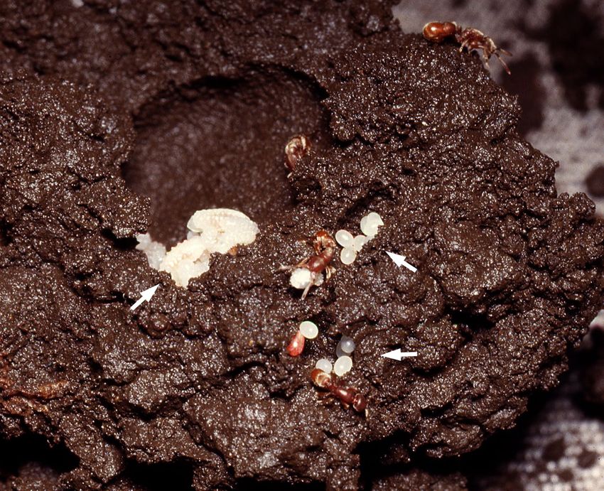
Fig. 1: Spheroidal chambers of a Proceratium itoi nest excavated
in the soil. Nests of this species consist of multiple subterranean
chambers like these, where the ant brood (left arrow) and prey
eggs (right arrows) are placed separately.

only for three Japanese species in the poneroid subfamily
Proceratiinae, that is, Proceratium itoi (Forel, 1918), P.
japonicum Santschi, 1937, and P. watasei (Wheeler,
1906) (Masuko 1986), but no further details were reported
in the study. Therefore, the aims of the present study were
to detail hemolymph feeding in Proceratium ants, using P.               a
itoi. We found that, although workers feed on prey, queens
are dependent exclusively on hemolymph feeding for their
nutrition once a colony has developed, and that each larva
has four pairs of hemolymph taps on the dorsal integument
of the abdominal segments. These specialized structures
are initially intact (closed), but are broken open by the
queen’s mandibular gnawing. This is the second instance
of hemolymph taps confirmed in ants; however, the tap
structures are completely different between Leptanilla
and Proceratium. In this study, we characterized LHF in
P. itoi, and report on the larval stage subjected to LHF,
behavior of the queen, and morphological structure of the               b
hemolymph taps.
                                                                      Fig. 2: Scars on the larval integument of Proceratium itoi. (a)
Material and methods
                                                                      Dorsal view of a fourth-instar larva (prepupa). A pair of scars
Ants: Proceratium itoi is a reddish-brown ant; the work-              are present in each posterior region of the 2nd to 5th abdominal
ers are approximately 3 mm and the queen 4 mm long                    segments (marked as II - V). Scale bar 0.5 mm. (b) Whole-mount
(Onoyama & Ogata 1989, Japanese Ant Database Group                    preparation of a fourth-instar larva. The left hemolymph tap
2003). In this study, colonies of P. itoi were collected              is broken open on the 4th abdominal segment. Stained with
from an evergreen broad-leaved forest in Cape Manazuru                acid fuchsin.
(35.144° N, 139.154° E), Kanagawa Prefecture, central
Japan. The ants were brought to the laboratory, counted,
and then cultured or preserved either in 80% ethanol                  the genus Proceratium are known as specialized predators
or Kahle’s solution (Barbosa 1974), depending on the                  of arthropod eggs (Brown 1957, 1958), and the field data
intended purpose.                                                     from Manazuru forest revealed that P. itoi predates on
    This species is subterranean and the nest is constructed          eggs of Chilopoda, Hemiptera, and Opiliones (K. Masuko,
directly in the soil, mostly at depths of 10 - 15 cm (Fig. 1).        unpubl.). This predation and other aspects of the ecology
The organization of most colonies at Manazuru is mon-                 of this ant will be published elsewhere.
ogynous, but approximately 10% of the collected colonies                  Ant rearing: Three monogynous colonies collected
contained multiple queens (K. Masuko, unpubl.). Ants of               from Manazuru were cultured in the laboratory to inves-

22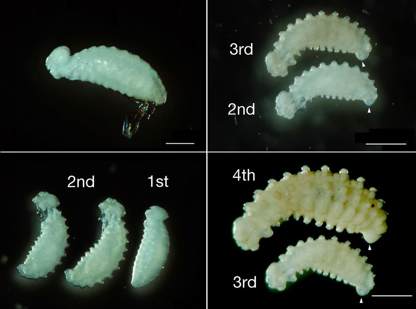
Tab. 1: Percentage of time spent by Proceratium itoi queens in each behavior. a Percentage of time in which prey was present in
the brood chamber compared with the total sample time. b Composition of the colonies when the behavior of the queens was
observed in the laboratory.

                                                     A                      B                     C                  D
 Queen                                                   81-68                  84-36                  14-5                14-5
 Colony stage                                Developed              Developed             Incipient           Developed
 Behavior
  Larval hemolymph feeding                                  2.6                    3.2                  0.0                 4.8
  Feeding on prey                                           0.0                    0.0                  3.6                 0.0
  Resting                                                  53.3                   44.5                 58.6                51.8
  Self-grooming                                             4.4                    7.8                  3.8                 9.6
  Being groomed                                             1.0                    2.0                  0.0                 1.0
  Others                                                   38.7                   42.5                 34.0                32.8
  Total                                                  100.0                  100.0                 100.0               100.0
 Sample time (h)                                           18.4                   15.2                 15.0                15.0
 Prey availability (%)a
                                                         100.0                  100.0                 100.0               100.0
 Colony compositionb
  Number of queens                                             1                     1                    1                   1
  Number of workers                                            35                   31                    3                  31
  Number of larvae                                         c. 40                  c. 50               11-12               c. 100

tigate the behavior of the queens. Two colonies (nos. 81-68         workers, 20 eggs, and 165 larvae; the other (no. 84-114)
and 84-36) were collected in 1981 and 1984, respectively,           was collected on 26 July 1984 and contained one queen,
and queen behavior was analyzed in 1984. An additional              199 workers, 47 eggs, 290 larvae, 255 prepupae, and 12
colony (no. 14-5) was collected in 2014 and studied in 2015.        pupae (11 gynes and one worker). All these specimens
The ants were housed in styrene observation nests (105 ×            were preserved in Kahle’s solution (later transferred to
113 × 20 mm or 65 × 100 × 22 mm, depending on the size              80% ethanol) immediately after being counted in the
of the housed population); the bottom of each nest was              laboratory. From each colony, 60 - 70 fourth-instar larvae
covered with plaster of Paris mixed with activated carbon           (not including prepupae) were examined for head width
powder. Brood chambers were excavated in the center of              at 160× and the width of the 3rd abdominal segment at
the plaster floor, and the tops of the terraria and brood           63× magnification, using a stereomicroscope (Olympus
chambers were covered with clear glass. The ants were               Model X-II) equipped with an ocular micrometer with a
easily reared on eggs of the spiders Pardosa astrigera L.           precision of 12.8 μm and 32.3 μm, respectively (based on
Koch, 1878 and Parasteatoda tepidariorum (L. Koch,                  calibration). As most of these larvae had multiple scars
1841). When the spider eggs were unavailable, the hatch-            characteristic of LHF on their dorsa, and the scars varied
lings (the first-instar larvae) of the mealworm Tenebrio            in size or area (Fig. 2a), it was necessary to quantitate the
molitor Linnaeus, 1758 were used as prey. When such                 total extent of scarring for each larva. For this, degrees
small soft-bodied mealworms were supplied alive in the              of scarring were classified into three arbitrary classes:
foraging arena, they were immediately retrieved into the            light, medium, and heavy, scored as + 1, + 2, and + 3,
brood chamber by workers and consumed by the larvae.                respectively. Thus, each larva had a total score according
Prepupae and pupae of other ant species like Myrmecina              to the number of scars and degree of scarring. In addition
nipponica Wheeler, 1906 were also used as prey, al-                 to the scars included in these three classes, faint scratches
though infrequently. The colonies were kept at 21 - 26 °C           could be observed on the larval body surface; these were
in the laboratory.                                                  minute, but seemingly caused by biting of the queen (see
    Measurement of head and body width of                           below). As they appeared to be superficial injuries that
scarred larvae and quantification of scars: As the                  did not puncture the integument, they were not included
larger, fourth-larval instar individuals are invariably the         in the scoring.
ones subjected to LHF (see below), the body size and the                Queen behavior: From a previous study on Stig-
degree of LHF-induced scarring, which reflect the age and           matomma (= Amblyopone) silvestrii Wheeler, 1928
frequency of LHF, were examined in the fourth-instar lar-           (Masuko 1986), it was suggested that, as in the case of
vae present in the two colonies when they were collected            S. silvestrii, the Proceratium itoi queen of a developed
in the field. Both colonies were mature. One (no. 84-72)            colony may also depend exclusively on hemolymph feeding
was collected on 7 July 1984 and contained one queen, 34            for her nutrition; in contrast, the queen of a founding or

                                                                                                                              23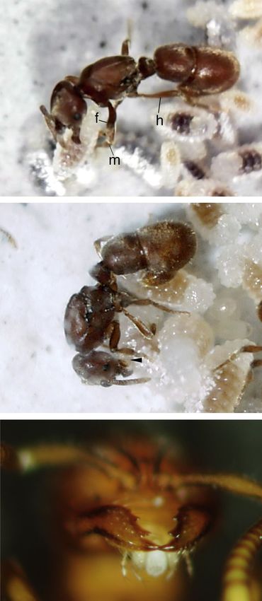
a                                                                   c

  b                                                                   d

Fig. 3: Larval instars of Proceratium itoi. The larval stage of P. itoi consists of four instars. Between-instar differences in the
morphology of the 10 th abdominal segment (arrowheads) and the dorsal and ventral protuberances are apparent in side-by-side
comparisons of the entire lateral view. (a) First larval instar. The egg membrane is attached to the posterior end of the first instar.
(b) First and second larval instars. (c) Second and third larval instars. (d) Third and fourth larval instars. Scale bars: 0.25 mm
in (a), and 0.5 mm in (c) and (d). No scale in (b).

incipient colony would feed on prey and suppress LHF to              microcomputer. In this procedure, behavior was recorded
enhance the development of the larvae. To test this, a be-           on a worksheet at intervals of 30 s during each 1 h sampling
havioral time budget was performed for each of the three             session, making a total of 120 data points per h. These
P. itoi queens that occupied colonies at different stages of         sessions were repeated 15 times from 18 March to 4 April
development. The census of these colonies at the time of             2015; therefore, the behavioral time budget of the queen
the behavioral studies is given in Table 1. Observations             was based on 15 h of observation. This queen was collected
were made under a stereomicroscope attached to a swing               on 11 August 2014 with a colony at a relatively early stage
arm (10 - 40× magnification). The behavioral repertoire              of development containing the queen, one adult worker,
of P. itoi queens had been established by recording notes            seven eggs, eight larvae, one prepupa, and four worker
on audio tapes before starting the quantitative analysis.            pupae. A subsequent shortage of arthropod eggs to feed the
Thereafter, the observed behaviors were logged directly              colony retarded colony development in the laboratory and,
onto a portable microcomputer (Epson HC-20). For the                 when the behavioral study was started (18 March 2015),
queens of developed colonies (nos. 81-68 and 84-36),                 the colony contained only the queen, three adult workers,
behavior was assayed by the “focal animal sampling”                  two eggs, 12 larvae, and one prepupa. Consequently, the
method (Altmann 1974) over a total of more than 15                   colony was still in the incipient stage.
h, consisting of 57 - 110-min episodes (repeated during                  After the initial behavioral analysis, colony 14-5 de-
11 - 22 September 1984, for queen 81-68, and 16 - 30 June            veloped successfully, containing more than 30 workers by
1984 for queen 84-36).                                               August 2017. To confirm the change in the feeding charac-
     When the queen of an incipient colony (no. 14-5) was            teristics of the queen, her behavior was again observed for
studied, a different method, “point sampling” (Dunbar                15 h with the same procedure used for the incipient stage.
1976), was used due to technical reasons related to the              During the study period (14 - 22 August 2017), the colony

24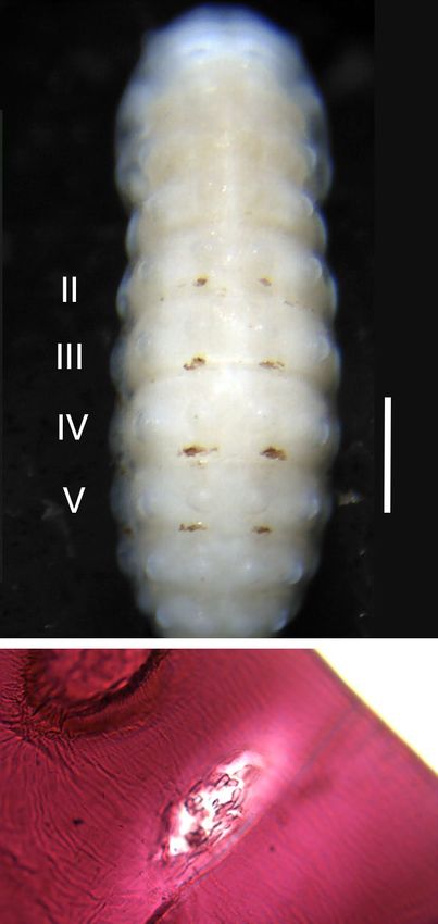
contained the queen, 31 adult workers, and approximately                              20        Colony 84-72
15 eggs and 100 larvae; no pupae or prepupae were ob-                                           N=71
served. Data for all these behavioral studies were collected                                    R 0.5536
between 09:00 and 23:00 hours.                                                        15

                                                               Score of scar extent
    SEM, histology, and whole-mount prepara-
tions: For morphological and anatomical studies, the lar-
                                                                                      10
val specimens were obtained from colonies collected from
1981 to 1987; they were preserved first in Kahle’s solution,
then transferred to 80% ethanol. Some specimens were                                  5
examined with a scanning electron microscope (SEM)
(Keyence VE-8800). Before the SEM examination, the
specimens were further dehydrated with a series of ethanol                            0
                                                                                           20                  25                  30         35
dilutions and treated with hexamethyldisilazane (Nation
1983, Masuko 1990). After air-drying, the specimens              a                                          Width of 3rd-abdominal segment

were mounted on stubs and coated with gold-palladium.
A total of 52 larvae were observed (eight first-instar, nine                                     Colony 84-114
second-instar, 14 third-instar, and 21 fourth-instar).                                20         N=61
    For histological examination of the hemolymph taps                                           R 0.5898
on the larval body, the preserved larvae were further de-
                                                                                      15
hydrated in a series of ethanol dilutions with propylene
oxide, before embedding in Agar low viscosity resin (Agar      Score of scar extent
Scientific). Sections 0.5 - 0.8 μm thick were obtained using                          10
a diamond knife, then stained in a 1% methylene blue / 1%
borax solution. Whole-mount preparations were also made
to examine closely the wounds in the cuticular integument                              5
(the method in Masuko 2017). Sections and whole-mounts
were examined using compound microscopes (Olympus
                                                                                       0
Vanox and BH2) and morphological details were imaged                                       20                  25                  30         35
with a digital camera (Shimadzu Moticam-2500).                   b                                           Width of 3rd-abdominal segment

Results
                                                                 Fig. 4: Quantified extent of scarring and widths of the 3rd ab-
LHF-subjected larvae and scar quantification: The                dominal segment in fourth-instar larvae. Preserved specimens
larval stage of Proceratium itoi consists of four instars        from two colonies (a, 84-72; b, 84-114) were studied. The x-axis
(Fig. 3). In some species of the genus Proceratium, the lar-     is represented by divisions of the ocular micrometer used (1
vae have numerous protuberances or bosses on the dorsal,         division = 32.3 μm). The larvae present scars characteristic of
lateral, and ventral body surfaces (Wheeler & Wheeler            LHF when they reach 0.78 mm (24 divisions) measured as the
1952). The four instars of P. itoi are easily distinguished      width of the 3rd abdominal segment.
by a combination of differences in the morphology of these
protuberances and the 10th abdominal segment, and total
body length and size of head capsule (Fig. 3). Figure 2a             Figure 4 shows the quantification of the extent of
depicts a mature fourth-instar larva (prepupa) with nu-          scarring on the dorsal abdominal segments of the larval
merous pigmented scars on its dorsum, and all the scars          specimens collected from the two field colonies. In both
are assumed to have been made by hemolymph feeding.              colonies, larvae in the early fourth instar stage, that is,
Cuticular melanization is known to occur at wound sites of       smaller in size, were not subjected to LHF; instead, once
insect integuments (Nation 2016). In this larva, the dark        they had reached a certain size, that is, 0.78 mm, meas-
scars are present on the dorsum of the 2nd to 5th abdomi-        ured as the width of the 3rd abdominal segment (Fig. 4a,
nal segments that present an invariable pattern in P. itoi       b), they were repeatedly utilized in LHF, and the extent
larvae. This scar distribution is more extensive than in         of scarring increased. Most or all the scars are likely to
Stigmatomma silvestrii, where the scars are distributed          have been made by the queen as hemolymph feeding in a
in the two intersegmental grooves between the 1st and 3rd        developed colony is almost exclusively performed by the
abdominal segments (Masuko 1986) (but see Discussion             queen (see below).
for the previous misidentification of the larval body seg-           Description of LHF in Proceratium: When the
ments for this species). However, the scars are typically        behavior of the queen was observed in the laboratory, each
present in, or close to, the intersegmental grooves in both      bout of hemolymph feeding was always initiated with a
species. The protuberances on the P. itoi larvae, especially     restless walk, with active antennation, over an aggregation
the dorsal ones, are currently not thought to be related to      of larvae. In many instances, the queen did not proceed to
hemolymph feeding (see below), and the function of these         hemolymph feeding but started self-grooming near the lar-
structures remains unknown.                                      val pile, followed by immobility (rest). In other instances,

                                                                                                                                               25
sum of the larval body, this initial handling was performed
haphazardly; more specifically, when the larva was lying
on its back or side, the queen initially licked and gnawed
the ventral or lateral side of the larva. On this occasion,
the queen did not even distinguish between the anterior
and posterior ends of the larval body. In response to such
tactile stimuli, it was likely that the larvae discharged a
small amount of fluid from the mouth and proctodeum as,
even though the droplets were hardly visible, the queen
actively licked the vicinities of the mouthparts and proc-
todeum of the larva for a short time.
When the queen handled a larva, she did not only use
the forelegs, but also extended the middle legs forward
a along with the forelegs and applied all of them to the lower
or lateral sides of the larva (Fig. 5a); when not handling a
larva, the middle legs, along with the hind legs and gastral
end, were placed on the substratum to maintain posture,
and the queen ceaselessly moved round the larva while
licking and gnawing its surface (Fig. 5a). The mandibular
gnawing was directed not only at the larval dorsum but
also the ventral side; and when the larva was grasped from
the side, one of the mandibles of the queen was applied
to the larval dorsum and the other to the venter (Fig. 5a).
Although the distal ends of the mandibles are pointed
(Fig. 5c), most of the gnawing movements slid unsuccess-
fully over the larval surface. During this repetitive man-
dibular biting, the mandibular tips finally stuck and were
marginally inserted into the surface of the larval dorsum.
The queen immediately pinched tightly several times, and
b the tap was likely broken open at this time because the
queen instantly stopped the mandibular movement and
attached the lower mouthparts to the site (Fig. 5b). The
hemolymph taps could not be observed directly with the
binocular magnification used when observing this behav-
ior, but the site where the queen attached the mouthparts
corresponded exactly to the position of the hemolymph
taps. Observed from the side, the lapping movement of
the lower mouthparts was evident and it was likely that
the queen ingested hemolymph oozing from the puncture.
Each bout of hemolymph ingestion lasted for more than
5 min (mean ± SD = 464 ± 109 s, n = 7, range 305 - 610;
observations on queen 14-5). After hemolymph ingestion,
the queen moved a short distance from the larva and ei-
c ther started grooming herself or was groomed by a nearby
worker.
Fig. 5: A Proceratium itoi queen performing hemolymph feed- Under well-fed laboratory conditions, most scarred
ing. (a) Queen biting the lateral part of a larva while holding it larvae were observed to continue prey feeding and suc-
with the forelegs (f) and middle legs (m); h, hind leg. (b) Queen cessfully pupated, in agreement with the observation that
licking the dorsal part of a larva with her lower mouthparts many prepupae collected from field colonies exhibited
(arrowhead). (c) Frontal view of the mandibles of a founding scarring characteristic of LHF.
queen of P. itoi collected from Manazuru. LHF by queens of developed colonies: A quan-
titative behavioral study of the Proceratium itoi queens
(81-68 and 84-36) in the laboratory revealed that nutrition
however, immediately after antennating a larva, the queen of the queens in developed colonies is exclusively depend-
put the fore tarsi on the larva and tried to seize or gnaw the ent on larval hemolymph (Tab. 1A, B). The colonies of both
larval body with her mandibles, or licked its body surface. queens contained approximately 30 workers and 40 - 50
These movements were obvious signs for the start of LHF. larvae when their behavior was studied. The total obser-
Despite the hemolymph taps being present only on the dor- vation time of more than 15 h for each queen consisted of
26a d
b e
c f
Fig. 6: Dorsal protuberances and hemolymph taps on a fourth-instar larva. Hemolymph taps are located between the dorsal
protuberances and the intersegmental grooves with the following somite. No hemolymph taps exist on the 6th and 7th abdominal
segments. (a) Dorsal view of a complete larva. The 2nd to 5th abdominal segments are marked as II - V. (b) Second abdominal seg-
ment. (c) Third abdominal segment. (d) Fifth abdominal segment. (e) Sixth abdominal segment. (f) Seventh abdominal segment.
60 - 110-min episodes, repeated within two weeks. During eggs or trophallaxis with workers, was observed during the
each observation episode, spider eggs were always present study period (both the queen and workers of P. itoi have
in the brood chamber, giving the queens the opportunity to three ovarioles per ovary, K. Masuko, unpubl.).
feed freely on the prey. However, both queens continuously Another queen, 14-5, also showed total dependence on
ignored the spider eggs and performed only LHF. No other larval hemolymph for nutrition in a colony with approx-
food exchange activity, for example, feeding on worker-laid imately 30 workers (Tab. 1D). During this observation,
27a c
b d
Fig. 7: Damaged hemolymph taps of a fourth-instar larva. The gaps between the cobble-like structures are filled with an unknown,
uniform substance (asterisks). Arrows point to the anterior. (a) Left hemolymph tap on the 2nd abdominal segment. (b) Right
hemolymph tap on the 2nd abdominal segment. (c) Right hemolymph tap on the 3rd abdominal segment. (d) Right hemolymph
tap on the 5th abdominal segment.
prey (first-instar mealworms) was also always accessible were examined occasionally under a stereomicroscope
to the queen. Larvae in the vicinity fed on the prey that after the behavioral study on the incipient colony. No LHF
were initially bitten and injured by the workers and then scarring was found on the dorsa of the three prepupae
placed on the head of the larvae. present in the colony on 2 June 2015, when the colony
Observation of these three queens indicates that contained eight workers. Similarly, no scars were observed
hemolymph feeding is the exclusive mode of feeding for on another two prepupae on 15 June 2015, when the col-
the queens of developed Proceratium itoi colonies. During ony again contained only eight workers; however, on 8
the observations, workers always fed on prey, and were August 2015, when the colony contained 19 workers and
never observed to perform hemolymph feeding. one prepupa, the prepupa presented LHF scars on its
LHF by the queen of an incipient colony: In dorsum.
contrast, queen 14-5 from the incipient colony that con- Structure of the hemolymph taps: The presence
tained only three workers and approximately 10 larvae of a pre-formed tap structure on the Proceratium itoi
did not perform LHF, only prey feeding (Tab. 1C). At the larval body has been overlooked, even though it has long
start of the observation, the brood consisted of a prepupa, been confirmed that P. itoi queens perform LHF (Masuko
two eggs, and 12 fourth-instar larvae (the number was 1986). Instead, Proceratium queens were thought to im-
reduced to 11 during the study period because one larva bibe larval hemolymph from new punctures made in
pupated). Thus, the feeding characteristics of the queens the larval dorsum by the queen herself, as observed for
differ greatly between early-stage colonies with small pop- Stigmatomma silvestrii (Masuko 1986). The possibility
ulations and developed colonies with larger populations. that hemolymph taps may be present in Proceratium
To know when, and at what colony size, hemolymph larvae was indicated when whole-mount preparations
feeding by the queen starts, the body surfaces of prepupae were analyzed.
28a d
b e
c f
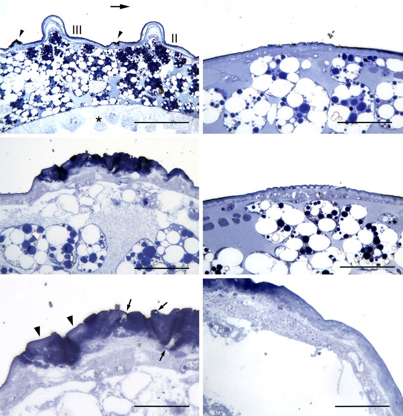
Fig. 8: Semithin sections of hemolymph taps on fourth-instar larvae. (a) Longitudinal section of the left dorsum of the 2nd and
3rd abdominal segments (marked as II and III). Arrow points to the anterior. The left hemolymph taps of the two segments are
indicated by arrowheads. Asterisk marks the midgut. (b) Cross-section of the left hemolymph tap on the 3rd abdominal segment.
A damaged hemolymph tap from a strongly scarred larval specimen. (c) A damaged integument. Enlargement of (b). Arrowheads
indicate intact regions of the integument; strongly stained substances fill the spaces between them. Arrows point to crevices in
the cuticular layer. (d, e) Cross-sections of right hemolymph taps on the 2nd and 3rd abdominal segments. Intact hemolymph taps
from an unscarred larval specimen. (f) Cross-section of the periphery of the damaged integument (upper left). Same series as in
(b). Small dark particles are distributed in both the cuticular layers and epidermis. Scale bars: 200 μm in (a), 50 μm in (b), 25
μm in (c), 33 μm in (d) and (e), and 20 μm in (f).
Whole-mount preparations: When whole-mount immersion in KOH solution, numerous granule-like struc-
preparations were made with Stigmatomma silvestrii lar- tures remained inside the openings in the whole-mounts
vae, vacant openings appeared in the cuticular integument of Proceratium itoi larvae; the structures were connected
at the locations containing LHF scars (K. Masuko, un- to the inner rim of the openings (Fig. 2b), and evidently
publ.). Despite anticipating this, and even after prolonged formed part of the cuticular integument. Consequently,
29a d
b e
c f
Fig. 9: Dorsal surface of the abdominal segments of a fourth-instar larva. All micrographs are from the same individual. Arrows
point to the anterior. (a) First and second abdominal segments (marked as I and II). No noticeable structures exist in the posterior
region of the dorsal protuberances on the 1st abdominal segment. The body surface of the larva is not fully extended soon after
ecdysis. (b) Second and third abdominal segments (marked as II and III). A pair of hemolymph taps (arrowheads) are present near
the intersegmental groove. (c) Surface of the left hemolymph tap on the 2nd abdominal segment. (d) Left hemolymph tap of the 4th
abdominal segment. (e) Left hemolymph tap of the 5th abdominal segment. (f) Dorsal protuberances of the 6th abdominal segment. No
hemolymph taps like those on the preceding somites are observed, except for faint modifications of the body surface (arrowheads).
the surface structure and internal anatomy of LHF scars SEM and histology: A dorsolateral view of a com-
were investigated closely with a SEM and with histological plete fourth-instar larva is shown in Figure 6a. The entire
sections, respectively. body surface is covered with circular rounded protuber-
30a b
c d
e f
Fig. 10: First- to third-instar larvae of Proceratium itoi. Arrows point to the anterior. (a) Laterodorsal view of a first-instar larva.
(b) Left hemolymph tap (arrowhead) on the 3rd abdominal segment of a first-instar larva. Same specimen as in (a). (c) Dorsal view
of a second-instar larva. Boxed is the location of the right hemolymph tap on the 2nd abdominal segment. (d) Right hemolymph tap
on the 2nd abdominal segment. Same specimen as in (c). (e) Laterodorsal view of a third-instar larva. Boxed is the location of the
left hemolymph tap on the 3rd abdominal segment. (f) Left hemolymph tap on the 3rd abdominal segment. Same specimen as in (e).
ances or bosses. Conspicuous scars are located on the dor- and regularly situated behind a pair of the most dorsal
sal surface from the 2nd through 5th abdominal segments bosses, and slightly anterior to the intersegmental groove.
(Figs. 2a, 6a - d). They are not present on the three thoracic Each scar extends laterally and is raised above the body
segments or on the 1st, 6th, and subsequent abdominal seg- surface like a pomegranate (Fig. 6b - d). Enlarged scars are
ments (Fig. 6a, e, f). The scars on each segment are paired shown in Figure 7. The surface structure has an irregular
31tessellated or cobblestone appearance; the “cobbles” vary Examination of semithin sections revealed that numer-
in shape and size, and are identical to the granule-like ous small dark particles were present both in the epidermis
structures observed in the whole-mount preparations and in the cuticular layers (Fig. 8f), and they appeared to
(Fig. 2b). Notably, the gaps between the cobbles are filled be more concentrated in the damaged regions or periphery
with an unknown, uniform substance (Fig. 7b, c, asterisks). of the integument; in contrast, few or none were observed
To further characterize the scars, histological sections in non-scarred regions.
were prepared and analyzed (Fig. 8).
Discussion
Despite the regular spatial relationships between the
dorsal protuberances and the scars (Fig. 8a, arrowheads), Larval hemolymph feeding was first reported for Stigmat-
examination of the histological sections did not reveal omma silvestrii under the taxonomic name Amblyopone
any specific structural or functional connection between silvestrii (see Masuko 1986). In a recent phylogenetic
them. In cross-sections, the entire surface of a heavy scar analysis based on morphology (Yoshimura & Fisher
was raised and lumpy, and the integument was strongly 2012), approximately 70 species (including S. silvestrii)
deformed and thickened (Fig. 8b). Gaps or cracks could be were transferred from Amblyopone to Stigmatomma as
seen in this thickened cuticular layer (Fig. 8c, arrows), and new or revived combinations, while the other species re-
dark-stained materials filled the gaps between the original mained in Amblyopone. Additionally, a recent molecular
cobble-like structures (Fig. 8c, arrowheads). phylogenetic analysis (Ward & Fisher 2016) showed that
To identify structures existing before being damaged Amblyopone and Stigmatomma belong to two distinct
(no special structures were predicted to be present on the clades. LHF is likely to be also present in true Amblyo-
body surface), fourth-instar larvae were observed with pone as the larvae of A. australis Erichson, 1842 have
a SEM shortly after ecdysis from the previous instar, LHF-characteristic scars on their dorsal integument (K.
thus presenting no dorsal scarring (Fig. 9). Unexpectedly, Masuko, unpubl.). To date, LHF has been observed in
some surface structures, somewhat like the surface of a twelve ant genera from five subfamilies: Adetomyrma, Am-
corncob, were apparent and were located exactly where blyopone, Calyptomyrmex, Gnamptogenys, Leptanilla,
scars typically appear on each of the 2nd to 5th abdominal Myopopone, Mystrium, Onychomyrmex, Prionopelta,
segments (Fig. 9b - e); no such structure was apparent on Proceratium, Stigmatomma, and Typhlomyrmex (see
the three thoracic segments or the 1st abdominal segment below). Among them, hemolymph taps were found only
(Fig. 9a). Incomplete or vestigial structures could be ob- in Leptanilla and Proceratium. However, closer morpho-
served on the 6th abdominal segment of some individual logical and anatomical observations, as in the present
larvae (Fig. 9f, arrowheads). Histological observation of study, may lead to hemolymph taps or other LHF-related
the structures (Fig. 8d, e) indicated that the surface of the organs or structures being discovered in other genera. For
integument neither rose nor thickened like the damaged instance, LHF using specialized organs was reported for
ones; instead, the cuticular layer was regularly uneven, Typhlomyrmex, another genus of Ectatomminae, although
suggesting that the regularly interspersed crevices or the structural details remain unknown (Lacau & al. 2007).
depressions could be broken open by the mandibular In Leptanilla japonica Baroni Urbani, 1977, only the
gnawing of adult ants. Notably, the epidermal layer in that 4th abdominal segment has a hemolymph tap on each side
region is initially thicker than in the surrounding regions of its posterior region (Masuko 1989). In Proceratium itoi,
(Fig. 8d, e). The results of the behavioral observations, four pairs of hemolymph taps are present in the posterior
as well as the SEM and histological studies, indicate that region of the dorsal integument from the 2nd through 5th
these four pairs of structures on the dorsa of the 2nd to 5th abdominal segments. In Stigmatomma silvestrii, LHF-
abdominal segments are likely to be the organs through related scarring was originally reported as being present in
which adult ants imbibe hemolymph from the larval hemo- the two intersegmental grooves between the 2nd and 4th ab-
coel. Consequently, these organs can be described as larval dominal segments (Masuko 1986). However, a later study
hemolymph taps like those previously described for Lep- on the larval morphology of this species (Masuko 1990)
tanilla (see Masuko 1989). Nevertheless, the structures showed that the two most anterior body regions, which
are somewhat different between these two genera (see had been erroneously considered as the 1st and 2nd thoracic
below). segments, were in fact the anterior and posterior regions
The behavioral observations and examination of the of the 1st thoracic segment. From this, it follows that the
scars on the body surface of all larval instars confirmed true 1st abdominal segment was previously mistaken for
that only the fourth instars are subjected to LHF. To de- the 2nd abdominal segment and, therefore, LHF-related
termine whether hemolymph taps pre-exist in younger scars in S. silvestrii are now correctly considered to be
instars, the dorsal body surfaces of first, second, and third located at the two intersegmental grooves between the 1st
instars were analyzed with a SEM (Fig. 10). Intriguingly, and 3rd abdominal segments. Consequently, the locations
all the younger instars possessed hemolymph taps on each of hemolymph taps and the regions subjected to LHF are
of the 2nd to 5th abdominal segments. Although the taps in all limited to the anterior half of the larval abdomen (the
the first instar appeared imperfect or vestigial (Fig. 10b), abdomen of ant larvae consists of ten somites, Wheeler
those on the third instar appeared to be almost as complete & Wheeler 1976). However, the reason for this bias in
as those of the fourth instar (Fig. 10f). spatial distribution is unclear.
32The structure of hemolymph taps in Proceratium Indeed, several forms of nutrient exchange have
itoi clarified in the present study differs greatly from evolved among members of the same colony, especially in
Leptanilla. In the latter, a pair of naked bilateral circular ants of the formicoid subfamilies. One form is trophallaxis,
areas is present on the 4th abdominal segment. In the which denotes the transfer of nutrients, mostly in liquid
center of this area, there is a slit-like opening that extends form, through mouth-to-mouth or anus-to-mouth feeding;
dorsoventrally and is attached internally to a short duct the former is referred to as stomodeal trophallaxis, and the
that is sharply bent and opens internally directly into the latter as proctodeal trophallaxis (Hölldobler & Wilson
hemocoel. Hemolymph flow in the duct may initially be 1990). Stomodeal and proctodeal trophallaxis in ants have
interrupted somewhere between the internal body cavity been best studied in Myrmicinae species. Ingestion of lar-
and the surface opening, but the queen’s biting would val saliva (most likely from labial glands) by the queen was
initiate hemolymph flow in the first instance of LHF. In frequently observed in a myrmicine ant, Temnothorax (=
contrast, the larval hemolymph tap of P. itoi appears to Leptothorax) curvispinosus (Mayr, 1866) (Wilson 1974).
consist only of a cuticular modification on the dorsal body Similarly, workers of Myrmica rubra (= laevinodis) (Lin-
surface. The most recent molecular phylogenetic analyses naeus, 1758) and Monomorium pharaonis (Linnaeus,
(Ward & Fisher 2016, Borowiec & al. 2017) have placed 1758) stimulate the larvae to discharge saliva and rectal
the Leptanillinae or the Leptanillinae plus Martialinae as fluid (Ohly-Wüst 1977); biochemical analyses of these
a sister or basal group to all other ant lineages (including fluids revealed that larval saliva contains amino acids,
Proceratium). Therefore, the hemolymph taps in Lepta- proteases, and carbohydrases, and the rectal fluid contains
nilla and Proceratium likely evolved independently. amino acids and proteins in addition to uric acids (lipases
Our behavioral study using the three Proceratium and a small amount of uric acids are also present in Myr-
itoi queens in the developed colonies revealed that their mica larval saliva). Moreover, the volume of these fluids
nutrition was almost exclusively dependent on the larval in a single discharge is not negligible (Ohly-Wüst 1977).
hemolymph. However, observation of queen behavior also Finally, in ants like Stigmatomma and Proceratium
revealed that in each bout of LHF, the queen obtained some that present LHF, a breach of the larval integument is
substance from the larvae prior to feeding on hemolymph. not accidental but occurs regularly and repeatedly in the
From the behavior of the queen, it is likely that a small same larva. Histological analysis revealed numerous dark-
quantity of fluid was discharged from the mouth and stained particles near the cuticular region damaged by
proctodeum of the larvae in response to the queen’s biting. LHF. They are likely proteins that, through melanization
The amount of fluid transferred might be negligible as the and polymerization, may seal the injuries in combination
fluid present between those larval body regions and the with localized coagulation of hemolymph (Nation 2016).
queen’s mouthparts cannot be seen, and the queen only However, the physiology and biochemistry of LHF remains
licked the larvae briefly. Nevertheless, the presence of unexplored.
such behavior may be important when considering the
Acknowledgments
origin of LHF. Similarly, Stigmatomma silvestrii larvae
were observed to discharge a transparent droplet from the I would like to thank Kazuhiro Katoh (the Open University
mouth or proctodeum under a strong contact stimulus, or of Japan) for permission to conduct SEM examinations in
while being pinched during LHF, especially shortly after his laboratory, and Kazuki Tsuji (Ryukyu University) for
feeding (Masuko 1986). In another Proceratium species, collection of a Proceratium itoi colony. I also thank Su-
that is, P. croceum (Roger, 1860), workers were observed sumu Izumi (Kanagawa University) for helpful discussions
to lick the larval mouth and 1st thoracic segment regularly, on the insect integument, and Margarete Ohly-Wüst for
supposedly to obtain a minute quantity of saliva (Haskins allowing me to read her dissertation.
1930). Also in this species, the larvae were observed to be This work was partly supported by KAKENHI Grant
“eagerly licked for exudates” and even pinched “to hasten Number JP15K07805 from the Japan Society for the Pro-
the flow” (Haskins 1930). Even though there was no men- motion of Science.
tion of which members of the colony licked and pinched
References
the larvae, and “the exudates” were not identified, this
behavior observed in P. croceum is very similar to that ex- Altmann, J. 1974: Observational study of behavior: sampling
hibited by P. itoi queens prior to, or during, LHF. All these methods. – Behaviour 49: 227-267.
observations imply that nondestructive ways of obtaining Barbosa, P. 1974: Manual of basic techniques in insect histol-
larval hemolymph, such as LHF performed by extant ogy. – Autumn Publishers, Amherst, MA, 245 pp.
ants, may have had behavioral origins; it is unlikely that a Borowiec, M.L., Rabeling, C., Brady, S.G., Fisher, B.L.,
behavior resulting in a harmful breach of the integument Schultz, T.R. & Ward, P.S. 2017: Compositional heter-
ogeneity and outgroup choice influence the internal phy-
evolved immediately when the need arose for the queen to logeny of the ants. – , retrieved on 5 September 2018.
Instead, a starving queen may have stimulated the larvae Brown, W.L. Jr. 1957: Predation of arthropod eggs by the ant
using the mandibles (pinching) or the antennae (palpat- genera Proceratium and Discothyrea. – Psyche 64: 115.
ing) to obtain fluids from the larval mouth and anus, as ob- Brown, W.L. Jr. 1958: Contributions toward a reclassification
served in extant species of Proceratium and Stigmatomma. of the Formicidae. II. Tribe Ectatommini (Hymenoptera).
33– Bulletin of the Musem of Comparative Zoology, Harvard Masuko, K. 2017: Larval instars of the myrmicine ant, Manica
118: 173-362. yessensis Azuma (Hymenoptera: Formicidae). – ARI (Journal
Dunbar, R. 1976: Some aspects of research design and their of the Myrmecological Society of Japan) 38: 1-11.
implications for the observational study of behaviour. – Be- Nation, J.L. 1983: A new method using hexamethyldisilazane
haviour 58: 78-98. for preparation of soft tissues for scanning electron micro
Haskins, C.P. 1930: Preliminary notes on certain phases of scopy. – Stain Technology 58: 347-351.
the behavior and habits of Proceratium croceum Roger. – Nation, J.L. 2016: Insect physiology and biochemistry. 3rd
Journal of the New York Entomological Society 38: 121-126. edn. – CRC Press, Boca Raton, FL, 644 pp.
Hölldobler, B. & Wilson, E.O. 1990: The ants. – Belknap Ohly-Wüst, M. 1977: Soziale Wechselbeziehungen zwischen
Press of Harvard University Press, Cambridge, MA, 732 pp. Larven und Arbeiterinnen im Ameisenstaat, mit besonderer
Beachtung der Trophallaxis. – Doctoral dissertation, Jo-
Ito, F. 2001: Specialized predation on arthropod eggs in a myr-
hann-Wolfgang-Goethe-Universität zu Frankfurt am Main,
micine ant, Calyptomyrmex sp. (Hymenoptera: Formicidae),
Frankfurt, Germany, 138 pp.
in the Oriental tropics. – Tropics 10: 405-407.
Onoyama, K. & Ogata, K. 1989: The genus Proceratium. In:
Ito, F. 2010: Notes on the biology of the Oriental amblyoponine The Myrmecological Society of Japan (Ed.): A guide for the
ant Myopopone castanea: queen-worker dimorphism, worker identification of Japanese ants (I): Ponerinae, Cerapachyinae,
polymorphism and larval hemolymph feeding by workers Pseudomyrmecinae, Dorylinae and Leptanillinae (Hymeno
(Hymenoptera: Formicidae). – Entomological Science 13: ptera: Formicidae). – The Myrmecological Society of Japan,
199-204. Tokyo, Japan, pp. 14-15. (in Japanese)
Ito, F. & Billen, J. 1998: Larval hemolymph feeding and Saux, C., Fisher, B.L. & Spicer, G.S. 2004: Dracula ant phy-
oophagy: behavior of queen and workers in the primitive logeny as inferred by nuclear 28S rDNA sequences and
ponerine ant Prionopelta kraepelini (Hymenoptera: Formi- implications for ant systematics (Hymenoptera: Formicidae:
cidae). – Belgian Journal of Zoology 128: 201-209. Amblyoponinae). – Molecular Phylogenetics and Evolution
Ito, F. & Gobin, B. 2008: Colony composition and behaviour 33: 457-468.
of a queen and workers in the Oriental ectatommine ant Ward, P.S. & Fisher, B.L. 2016: Tales of dracula ants: the
Gnamptogenys cribrata (Emery) 1900 in West Jawa, Indo- evolutionary history of the ant subfamily Amblyoponinae
nesia. – Asian Myrmecology 2: 103-107. (Hymenoptera: Formicidae). – Systematic Entomology 41:
Japanese Ant Database Group. 2003: Ants of Japan. – Gakken, 683-693.
Tokyo, Japan, 224 pp. Wheeler, G.C. & Wheeler, J. 1952: The ant larvae of the
Lacau, S., Villemant, C., Jahyny, B., Ramos-Lacau, L.S., subfamily Ponerinae, Part I. – The American Midland Nat-
Delabie, J.H.C. & Bueno, O.C. 2007: O “larval hemolymph uralist 48: 111-144.
feeding”: um comportamento alimentar original das formi- Wheeler, G.C. & Wheeler, J. 1976: Ant larvae: review and
gas (Hymenoptera: Formicidae). – Biológico, São Paulo 69, synthesis. – Memoirs of the Entomological Society of Wash-
Brazil, Supplement 2: 121-128. ington 7: 1-108.
Masuko, K. 1986: Larval hemolymph feeding: a nondestructive Wheeler, G.C. & Wheeler, J. 1988: An additional use for ant
parental cannibalism in the primitive ant Amblyopone sil- larvae (Hymenoptera: Formicidae). – Entomological News
vestrii Wheeler (Hymenoptera: Formicidae). – Behavioral 99: 23-24.
Ecology and Sociobiology 19: 249-255. Wilson, E.O. 1974: Aversive behavior and competition within
Masuko, K. 1989: Larval hemolymph feeding in the ant Lepta- colonies of the ant Leptothorax curvispinosus. – Annals of
nilla japonica by use of a specialized duct organ, the “larval the Entomological Society of America 67: 777-780.
hemolymph tap” (Hymenoptera: Formicidae). – Behavioral Yoshimura, M. & Fisher, B.L. 2012: A revision of male ants
Ecology and Sociobiology 24: 127-132. of the Malagasy Amblyoponinae (Hymenoptera: Formicidae)
Masuko, K. 1990: The instars of the ant Amblyopone silvestrii. with resurrections of the genera Stigmatomma and Xymmer.
– Sociobiology 17: 221-244. – Public Library of Science One 7: art. e33325.
34You can also read