Abdominal Trauma WWW.RN.ORG
←
→
Page content transcription
If your browser does not render page correctly, please read the page content below
Abdominal Trauma
                        WWW.RN.ORG®
   Reviewed December, 2020, Expires December, 2022
Provider Information and Specifics available on our Website
            Unauthorized Distribution Prohibited
       ©2020 RN.ORG®, S.A., RN.ORG®, LLC
                By Wanda Lockwood, RN, BA, MA

 Purpose        The purpose of this course is to outline the different
                types of abdominal injuries that may result from blunt
or penetrating trauma, including diagnosis, symptoms, associated
injuries, and treatment approaches.

Goals Upon completion of this course, the healthcare provider should
be able to:
   • Describe 3 types of blunt injuries.
   • Discuss penetrating injuries.
   • Explain the 3 sections of the abdominal cavity and the organs in
      each.
   • Discuss primary and secondary surveys.
   • Describe physical signs of bleeding, including Cullen’s sign and
      Grey Turner’s sign.
   • Describe the 4 classes of hemorrhagic shock.
   • Explain the APVU and AMPLE methods of neurological
      assessment.
      Describe key information needed during history for motor vehicle
      accidents and penetrating trauma.
   • Explain FAST and DPL.
   • Discuss hepatic trauma, including grading.
   • Discuss splenic trauma, including grading.
   • Discuss gastric trauma, including grading.
   • Discuss intestinal trauma, including at least 6 signs of peritonitis.
   • Discuss pancreatic trauma, including grading.
   • Discuss bladder trauma and at least 4 indications of bladder
      rupture.
   • Describe the difference between intraperitoneal bladder rupture
      and extraperitoneal, including implications.
   • Discuss renal trauma, including grading.
   • Discuss adrenal trauma.
   • Discuss aortic trauma.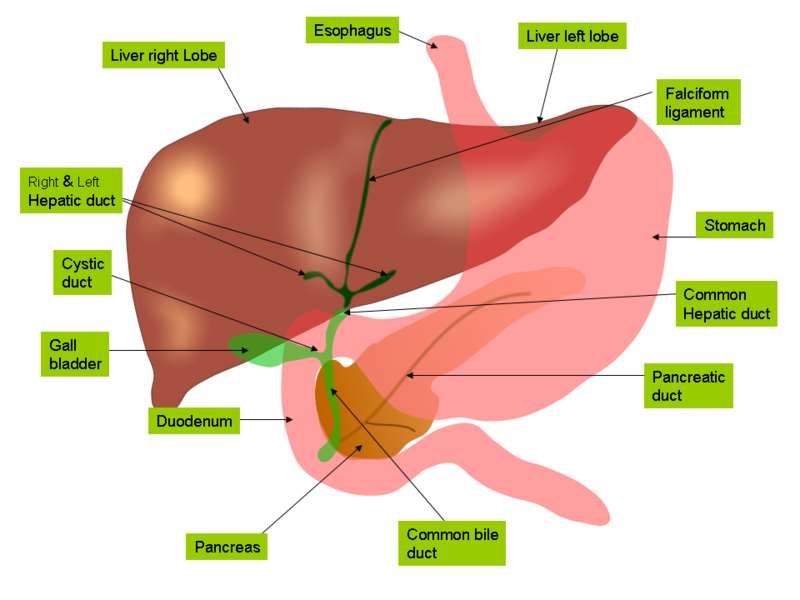
Introduction

Wikimedia Commons

Abdominal trauma is responsible for about 10% of all deaths related to
trama. Abdominal trauma may involve penetrating or blunt injuries.
Penetrating injuries include gunshot and shrapnel injuries,
impalements, and knifings.

   •   Penetrating injuries often result in injury to hollow organs, such
       as the intestines. The liver is the most commonly injured solid
       organ. Gunshot wounds are classified as high energy and may
       result in extensive damage, especially if the bullet ricochets off
       of bone. Patients are at increased risk for both hemorrhage and
       peritonitis, especially with intestinal injury.

       The most common injuries from gunshot wounds include:
         o Small intestines: 50%.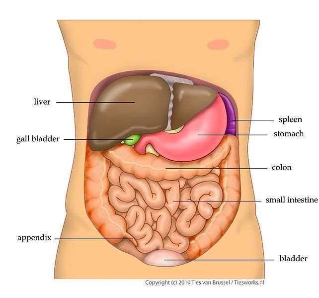
o Colon: 40%.
         o Liver: 30%.
         o Vascular structures: 25%.

      The most common injuries from stab wounds include:
        o Liver: 40%.
        o Small bowel: 30%.
        o Diaphragm: 20%.
        o Colon: 15%.

  •   Blunt trauma is more common than penetrating and may result
      from motor vehicle accidents, sports accidents, blows, falls, or
      explosions. Blunt trauma is more difficult to assess because
      injuries are less obvious, so massive blood loss may occur before
      injuries are detected.

      There are 3 types of blunt injuries: 1) crush, which results from
      compression, 2) shear, which involves tearing, and 3) burst,
      which relates to sudden increased pressure (such as from an
      explosion). Motor vehicle accidents, the most common cause of
      blunt abdominal injuries, often result in hepatic injury to the
      passenger if impact is on the passenger’s side and splenic injury
      to the driver if impact is on the driver’s side.

A tremendous force is needed to fracture a pelvis, so any time a
trauma patient presents with pelvic trauma, abdominal trauma should
be suspected.

Abdominal cavity
Thoracic         Liver, spleen,        The ribs provide some
abdomen          diaphragm,            protection although the organs
                 stomach               are vulnerable to both blunt and
                                       penetrating trauma.
Retroperitoneal    Kidneys, ureters,   Organs are generally better
abdomen            pancreas,           protected than organs in the
                   duodenum            thoracic abdomen as they are
                                       behind other organs.
True abdomen       Small intestines,   The large and small intestines
                   colon, bladder      are vulnerable to rupture with
                                       blunt trauma. The bladder is
                                       somewhat protected behind the
                                       symphysis pubis, but a full
                                       bladder is especially vulnerable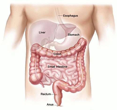
to rupture.

Common abdominal injuries include:
  • Hepatic lacerations.
  • Splenic rupture.
  • Bladder rupture.
  • Mesenteric artery tears.
  • Great vessel tears.
  • Diaphragmatic rupture.
  • Gastric rupture.
  • Renal injury.
  • Pancreatic injury.
  • Retroperitoneal hematoma.

Multiple injuries are common, so any type of abdominal injury should
raise suspicions of associated injuries.

Initial assessment

 Primary survey: ABCs          As with all trauma patients, assessment
                               should begin with the primary survey,
during which the patient is at least partially disrobed for examination,
placed on monitors (BP, cardiac, pulse oximeter) as indicated, and two
large-bore intravenous lines placed. It IV access cannot be achieved,
then a large-bore central line may be placed in the femoral vein,
subclavian, or internal jugular vein. In some cases, venous cutdown
may be indicated. An interosseous line may also be placed, especially
in children, if other access is not possible.

Airway, breathing, and circulation (ABCs) are checked immediately,
often while resuscitation efforts are occurring. The patient must be
assessed for blood loss. With blunt trauma, severe bleeding may
occur rapidly, so the patient must be observed for signs of bleeding
and blood loss estimated.

Any impaled items, such as a knife stuck into the abdomen, should be
stabilized with bulky dressings until scans are completed and/or the
patient is taken for surgical removal. Protruding organs or
eviscerations should be covered with sterile saline dressings.

Oxygen is usually administered with a non-rebreather mask, and NG
tube inserted (if there is no facial trauma), and blood samples (type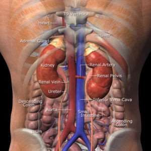
and crossmatch and CBC) and urine specimen obtained per indwelling
catheter.
Physical signs of internal bleeding include:
• Abdominal pain.
• Guarding, rigidity.
• Bruising, crepitus, swelling (especially across chest and pelvis
from seat belt and or shoulder harness).
• Abdominal distention, deformity.
• Tachycardia, hypotension.
• Pallor.
• Evisceration.
• Cullen’s sign: Bruising about the umbilicus (may indicate
hemoperitoneum or retroperitoneal bleeding but may take 12
hours to develop).
• Grey Turner’s sign: Bruising over flank (may indicate
retroperitoneal bleeding but may take 12 hours to develop).
• Hematuria.
• Blood or semen at urethral meatus (from injury to prostate).
• Inability to urinate.
Hemorrhagic shock classification
Class Blood loss Signs and symptoms
I 15% Mild tachycardia (90-100 bpm), localized
swelling, and frank bleeding.
II 15 to 25% Tachycardia, prolonged capillary refill and
increased diastolic BP.
III 25 to 50% Above signs (any) as well as hypotension,
confusion, decreased urinary output, and
acidosis.
IV >50% Hypotension and acidosis unresponsive to
resuscitation.
Geriatric patients must be observed carefully as they may have less
obvious signs of shock for a variety of reasons. Cardiac response to
hypovolemia is often lessened because of myocardial pathology or
medications, such as -blockers and calcium channel blockers.
Metabolic acidosis (decreased serum bicarbonate and increased base
deficit (> -6) or increased serum lactate) may result from hemorrhage
and hypovolemia.
Secondary survey: DE,
ABPU, and AMPLE The patient is examined more thoroughly to
determine which diagnostic tests are indicated (often after initial
standard x-rays of later cervical spine, supine chest, and pelvis).

D: Neurological status (D for disability) is assessed. A quick
assessment may be done using the AVPU method:
   • A: Is the patient alert?
   • V: Is the patient verbal?
   • P: Is the patient responding only to verbal stimuli?
   • U: Is the patient completely unresponsive.

If the patient is alert (A) and verbal (V), then the AMPLE method may
be used to ask a series of basic informational questions:
    • A: Do you have any allergies?
    • M: Are you taking any medications?
    • P: What is your past medical history and (if applicable) are you
       pregnant?
    • L: When did you last eat?
    • E. What events led to the trauma, and when did you have your
       last tetanus vaccination?

E (for exposure): The patient is completely disrobed, if possible. If
there is any danger of cervical trauma or the patient cannot be moved,
the clothing should be cut off. If forensics are involved (as in
shootings and knifings), care must be used to avoid cutting through or
damaging areas of clothing that may provide evidence, such as where
a bullet has entered clothing. Protocols for collection of evidence
should be carefully followed.

NOTE: In reality, the Primary and secondary surveys may be done
concurrently or overlap, depending on the patient’s condition.

Key information needed during history
Motor vehicle Crash mechanism (what happened).
accidents      Use of seatbelt and chest restraints.
               Amount of external damage to the car.
               Airbag deployment.
               Integrity of windshield.
               Deformity of steering wheel
               Loss of consciousness.
               Ambulatory status.
               Other passenger injuries or fatalities.
               Ejection from vehicle.
Penetrating    Type of weapon involved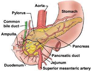
trauma             Number of injuries (gunshots, stabbings).
                   Victim’s distance from weapon.
                   Patient’s position at the time of injury.

 Diagnostic procedures:       If time is not a critical factor, the CT
                              scan is the best tool for assessing all
types of abdominal injuries, including bleeding. CT is usually done with
contrast. Fast-scanning and image-reconstructing helical CT scanners
have reduced the turnaround time for CTs to as little as 10 minutes,
but older scanners may require more time.

X-rays, while often done, provide little useful information as they may
not identify free air and fluid collections must be large (>800 mL) to
be detected by standard x-ray. Angiograms are indicated if injury to
vessels is suspected in order to identify the site of bleeding.

However, if the patient is unstable or severe bleeding is suspected,
then focused abdominal sonography in trauma (FAST)
examination should be done.

                               FAST is able to identify intra-abdominal
                               fluid in about 98% of cases. With
                               multiple trauma patients, FAST may also
                               be used if the CT scan is in use and wait
                               time is extended. However, it’s
                               important to remember that while FAST
                               will identify intra-abdominal fluid or
                               bleeding, it is not sufficiently sensitive to
show disruption of an internal organ.

Additionally, a single negative finding by FAST does not preclude
bleeding or other injury as it is sensitive to >300 mL of blood. For
example, a bowel perforation may result in limited intraabdominal fluid
in the initial period. Additionally, intracapsular bleeding or delayed
organ rupture may not be captured at the time of FAST but may be
identified on later examination or CT scan. Additionally, a positive
FAST finding does not necessarily indicate a need for surgical
intervention.

Diagnostic peritoneal lavage (DPL) may also be used, but FAST is
preferred and has generally replaced DPL, as it is relatively fast and
less invasive. DPL is especially insensitive to colonic wounds, which
require early diagnosis and treatment.

                                     DPL is done by inserting an
                                     abdominal catheter under local
                                     anesthetic. Aspiration is done to
                                     determine if free blood is present.
                                     If aspirate contains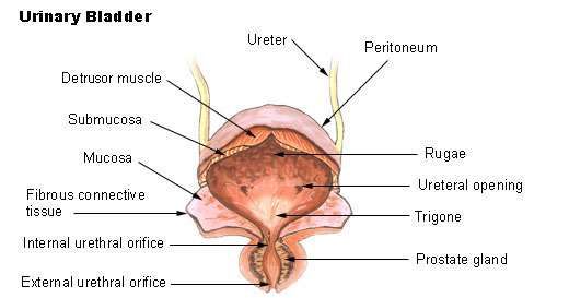
Stab wounds that are superficial may require extending the wound
opening and exploring to determine the extent of injury. Laparoscopy
may be used to determine if the peritoneum has been violated.

Hepatic trauma

The liver is especially vulnerable to trauma because of its anterior
position in the abdomen. It may be lacerated or avulsed by either
blunt or penetrating injuries. Liver injury should be suspected if a
patient has rib fractures on the right side or has abdominal pain,
especially in the right upper quadrant.

After the spleen, hepatic injury is the second most common abdominal
injury, with injury to the posterior segment of the right lobe occurring
most frequently. Because of this, bleeding may occur into the
retroperitoneal area rather than the peritoneal. Injuries that are
commonly associated with hepatic trauma include right lung contusion,
right pneumothorax, right-sided rib fractures, right kidney and/or
adrenal gland injuries. About 45% of those with hepatic injuries also
have injury to the spleen.
Injury of the left lobe is less common and most often associated with
direct blow to the epigastric area and is associated with injury to the
duodenum, pancreas, and transverse colon.
Mortality rates with hepatic injury are high (8% to 25%), and it is the
most common cause of death related to abdominal trauma. With
severe injuries, death can occur within minutes. Hepatic injuries can
include lacerations, contusions, subcapsular hematoma, and
intrahepatic hematoma.
Hepatic injury grading scale
Grade I Laceration(s) < 1cm deep.
Subcapsular hematoma 10 cm deep.
Subcapsular or central hematoma >10 cm diameter.
Lobar maceration or devascularization.
Grade V Bilobar tissue maceration or devascularization.
While in the past, surgical management of liver injury was common, it
was found that those undergoing surgery tended to have more
complications and required more transfusions than those treated more
conservatively, so presently only about 20% are treated surgically. In
about 70% of cases, bleeding stops spontaneously. Even grade IV
injuries may be treated non-surgically if there is no bleeding into the
peritoneal or retroperitoneal cavities.
However, patients must be carefully monitored as delayed
complications, including hemorrhage, abscess, and biloma
(encapsulated collection of bile in the peritoneal cavity), may occur.
Additionally, patients may require replacement fluids and blood
products, such as plasma and platelets for coagulopathy.
Hepatic contusions usually heal within a week, but subcapsular
hematomas may enlarge initially before slowly clearing. Lacerations
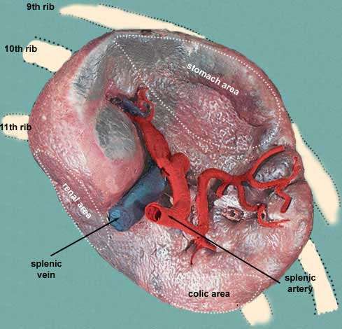
may require weeks to heal, and small residual bilomas are common.Splenic trauma
The spleen lies posteriorly in the left upper quadrant, behind the 9th,
10th, and 11th ribs with the convex surface under the left
hemidiaphragm. The tail of the pancreas contacts the spleen in about
30% of people and is within 1 cm of the spleen in about 70%. The
splenic artery provides the major blood supply to the spleen. The
spleen filters approximately 10 to 15% of the total blood volume every
minute and usually contains 40 to 50 mL of blood, but the spleen can
expand to hold much more blood.
Splenic injuries are
the most common
solid organ injuries,
accounting for about
a fourth of total
injuries. Splenic
injuries may occur
along with injuries to
other abdominal
organs as well.
Penetrating injuries
are not common, but
can occur, especially
with gunshot
wounds.
Blunt trauma may
occur from
compression or deceleration injuries, including motor vehicle
accidents, blows to the abdomen (kick injuries), and falls.
The most common injury associated with splenic injury is fracture of
the left lower rib as this indicates sufficient force to the LUQ to cause
organ damage. The triad of symptoms associated with splenic rupture
includes:
• Elevation of the left hemidiaphragm.
• Left lower lobe atelectasis.
• Pleural effusion.
Patients may present with various clinical symptoms. Indications of
splenic trauma can include left upper quadrant or left flank pain, Kehr
sign (referred pain to the left shoulder). Some patients may beessentially asymptomatic. Indications of shock may be present,
including tachycardia, tachypnea, anxiety, and hypotension.
If blood begins to pool in the intraperitoneal area, more diffuse
abdominal pain and rebound tenderness may be evident. Hypotension
occuring with splenic injury is cause for concern as it may indicate
rupture of the spleen with profound hemorrhage.
If shock cannot be compensated, immediate surgical exploration is
indicated as the delay needed to confirm hemorrhage by CT scan may
result in the patient bleeding out. Patients with compensated shock
may be treated with angioembolization if this can be done quickly.
Because of the spleen’s importance to the immune system, splenic
retention is the goal for those who are hemodynamically stable. Over
90% of children can be treated nonsurgically, regardless of the grade
of injury and up to 65% of adults.
Patients who are unstable with suspected injury to the spleen and
intra-abdominal hemorrhage may require exploratory laparotomy and
repair or removal of the spleen. Additionally, patients with blunt
trauma and hemodynamic instability that does not respond to
administration of intravenous fluids may be considered to have a life-
threatening splenic injury requiring surgery.
Splenic injury grading scale
Grade I Laceration(s) < 1cm deep.
Subcapsular hematoma 10 cm deep.
Subcapsular or central hematoma >10 cm diameter.
Grade V Splenic tissue maceration or devascularization.
The goal with splenic injury is to conserve the spleen whenever
possible. Splenectomy increases the risk of postsplenectomy sepsis
although it may be necessary with multiple injuries or severe
hemodynamic instability.
Postoperatively, recurrent bleeding may occur with splenorrhaphy,
especially during the first 24 to 48 hours. Complications in the earlypostoperative period for splenectomy or splenorrhaphy can include bleeding, gastric distention, gastric necrosis, pancreatitis, and subphrenic abscess. Later complications can include deep vein thrombosis and overwhelming post splenectomy infection (OPSI) (usually at 1 to 6 weeks). OPSI can occur within 2 years of splenectomy, especially in children, so prophylactic antibiotics may be given for 2 years. OPSI occurs in about 3% of splenectomy patients with about 50% developing pneumonia or meningitis. Symptoms are often rapid and about half die within 2 days of onset. Gastric Trauma Because the stomach has 3 muscle layers, blunt trauma perforations are rare although risk increases if a person suffers a severe blunt force trauma with a full stomach. Forceful blunt trauma may result in rupture of the left hemidiaphragm, causing the stomach to herniate into the left hemithorax. The areas most prone to rupture include: • Anterior wall: 40%. • Greater curvature: 23%. • Lesser curvature: 15%. • Posterior wall: 15%. Perforation may result from abdominal impact such as when a pedestrian is struck by a motor vehicle or during a motor vehicle accident in which a person is ejected from the car or the seat belt is improperly applied. Other injuries can include hematomas and contusions, but these injuries are often essentially asymptomatic and may resolve over time although large hematoma of the distal portion of the stomach may narrow the lumen and prevent emptying. Because the stomach has a rich supply of arteries, damage to the arteries may result in life- threatening hemorrhage.
It’s important to note that when blunt trauma is severe enough to
cause perforation of the stomach, 95% of patients also have another
serious injury. The most common injuries associated with gastric
perforation include:
    • Spleen: 27% to 43%.
    • Left chest: 18% to 29%
    • Liver: 18%.
    • Small intestine: 18%.

Penetrating trauma of the stomach, such as from a knife or gunshot
wound, is more common and should be suspected when penetration is
inferior to the nipples or 4th intercostal space anteriorly or inferior to
the tips of the scapulae posteriorly.

Symptoms of perforation include severe abdominal pain, abdominal
rigidity, hematemesis and bloody nasogastric drainage. While the
stomach usually contains few bacteria, with perforation, stomach acids
begin to pour into the peritoneal cavity, resulting in chemical
peritonitis. Most patients present with shock and pain in the abdomen,
but some patients may have no signs of an acute abdomen in the
initial period.

Gastric injury grading scale
I     Intramural hematoma OR                May be observed or drain
      superficial laceration                placed with Lembert,
                                            imbricated suturing.
II
GE junction OR 5 cm laceration pyloroplasty or total
of proximal third of stomach OR gastrectomy (usually for GE
10 cm laceration of distal two- junction injury) as indicated.
thirds of stomach.
IV Perforation or devascularization Subtotal or total gastrectomy
of two-thirds of stomach. reconstruction).
Upright x-rays may show free air in the abdomen, but only about 50%
to 66% develop enough free air in the abdomen to be detected by
upright x-ray. DPL may show food particles or bilious fluids in the
abdomen. However, if the patient is hemodynamically stable, CT with
contrast provides a definitive diagnosis.
Intestinal trauma
Intestinal trauma may occur as the result of blunt or penetrating
trauma, such as from gunshot wounds or knifings. Penetrating trauma
may result in evisceration of the small intestines through the
abdominal wall. Falls from great heights or crush injuries may result
in evisceration through the rectum or perineum. The small intestines
are especially vulnerable to penetrating wounds as they cover theabdominal surface. Motor vehicle accidents also frequently result in
injury to the small intestines.
The location of the duodenum and its attachments make it one of the
most commonly injured sites, especially with steering wheel injuries
that force the duodenum against the spine. Patients often present with
back pain when they have duodenal injuries.
Blunt trauma can result from two different types of forces:
• Compression: Increases intraluminal pressure inside of the bowel
and compresses the fluid-filled bowel against the vertebrae or
other solid structures.
• Deceleration: Stretches and tears the bowel.
Injuries of the small intestine occur about 4 times more frequently
than injuries of the colon. Injury to the colon occurs in 2% to 15% of
those with blunt abdominal injuries, often motor vehicle accidents.
Intestinal trauma may also result from diagnostic or therapeutic
procedures, such as colonoscopy, laparoscopy, and radiotherapy, as
well as from ingestions, such as from swallowing a toothpick.
Because injury to the colon requires considerable force, other
abdominal injuries are frequently present, especially with injury to the
transverse colon. Other injuries include:
• Hepatic: 64%.
• Spleen: 52%.
• Small intestinal mesentery: 48%.
Intestinal injuries results in mortality rates of 10% to 30%, but death
may be caused by other injuries rather than just the intestinal trauma.
Delay in diagnosing and treating intestinal perforation results in
mortality rates of 25% to 35%.
As with other abdominal injuries, the CT is the best diagnostic tool and
is about 97% accurate in providing evidence of bowel injury; however,
CT findings may be compromised if a patient first has DPL, as this
procedure may introduce intraperitoneal fluid and air. CT is not always
effective in identifying the exact location of an injury to the bowel, and
CT in general is less effective in identifying injuries to hollow organs
than to solid organs.
Typically, patients have non-specific symptoms or are asymptomatic
initially but return with a few hours or a couple of days with signs of
peritonitis, including:
• Abdominal distention, rigidity.• Guarding, rebound tenderness.
• Abdominal pain.
• Lack of bowel sounds, paralytic ileus.
• Elevated WBC counts.
• Fever.
• Tachycardia.
• Dyspnea.
• Nausea and vomiting.
If gross blood is found with a rectal exam, this is usually indicative of
severe intestinal injury.
Intestinal perforation may progress to abscess, fistula, and/or sepsis.
The site of perforation is an important risk determinant. Little bacteria
are present in the proximal segments of the small intestines, but the
distal segments contain both aerobic and anaerobic bacteria, which
can rapidly result in severe infection. According to some studies, the
most commonly perforated areas of the small intestine are the
proximal jejunum and the distal ileum.
Treatment of mild intestinal injury may be conservative, but with
perforation, surgical intervention with peritoneal lavage is indicated as
well as antibiotics. In some cases, primary closure is avoided and the
wound left open to heal by secondary intention.
Pancreatic trauma
The pancreas is located in a fairly protected part of the abdomen, high
in the retroperitoneum, so when it is injured as the result of blunt
trauma, then 90% to 95% of the time, there are injuries to other
organs as well. Because of this, injury to the pancreas may be
overlooked. Common associated injuries include:
• Liver: 32% to 42%.
• Spleen: 25% to 40%.
• Stomach: 20% to 40%.
• Major vessel: 25% to 35%.
• Thorax: 22% to 31%.
• Intestines: 10% to 29%.
• Central Nervous System: 25%.
• Duodenum: 18%.
While injury to the pancreas occurs in fewer than 10% of all cases of
blunt abdominal trauma, the pancreas is injured in 20% to 30% of
cases of penetrating trauma. Blunt trauma may result from motor
vehicle or pedestrian accidents. Direct blows to the epigastrium may
crush the pancreas against the spine.Penetrating injuries most often are associated with gunshot wounds or
stabbings to the back, flank, or abdomen.
Because vascular
structures, such as
the aorta and the
superior mesenteric
artery lie close to
the head of the
pancreas, injury to
that area may
include adjacent
vascular injury and
life-threatening
hemorrhage.
Symptoms specific
to pancreatic
trauma are often missing or nonspecific but can include abdominal
pain, flank pain, epigastric pain and nausea and vomiting (bile).
Diagnosis is often delayed because patients are treated for other
injuries and then further testing is done when the patients fail to
improve or continue to deteriorate.
As with other injuries, the CT scan provides the most definitive
diagnosis as the scan may show lacerations or peripancreatic fluids.
Blunt trauma may result in retroperitoneal hematoma and fluid, free
abdominal fluid and pancreatic edema. In the early phases after
injury, CT scans may not detect injury, so ongoing evaluation is
necessary. If the CT scan is inconclusive, then magnetic resonance
cholangiopancreatography may be indicated, as it more clearly outlines
damage to the ducts.
Standard x-rays are not effective for diagnosis of pancreatic injury but
findings of fractures of the lower thoracic or upper lumbar vertebrae
should raise suspicion of pancreatic or duodenal injury. FAST may
show free abdominal fluid but the position of the pancreas and the
overlying of the pancreas by the colonic gas make visualization
difficult. DPL has not been found useful because of the retroperitoneal
location.Serum amylase levels have been used to help diagnose pancreatic
injury, but studies show that levels don’t elevate for about 3 hours, so
serum amylase levels prior to 3 hours are not diagnostic. Additionally,
patients with brain injuries also have elevations in serum amylase, so
if a patient suffers both brain and abdominal trauma, elevation does
not necessarily indicate pancreatic trauma. Pancreatic enzymes
(amylase and lipase) may be abnormal because of shock rather than
direct pancreatic injury, but they may remain normal even with severe
pancreatic injury.
There are 3 different grading scales used to classify pancreatic injury.
Although they are similar, this can lead to some confusion. The
following grading system is that of the Organ Injury Scaling
Committee of the American Association for the Surgery of Trauma.
Pancreatic injury grading scale
I Simple contusion.
II Major contusion or laceration without tissue loss or involvement
of the main pancreatic duct.
III Complete transection of the pancreas or a parenchymal injury
with involvement of the major duct to the left of the superior
mesenteric vein.
IV Ductal transection or a major parenchymal injury to the right of
the superior mesenteric vein.
V Massive disruption of the head of the pancreas.
Most injuries to the pancreas are relatively minor and can be treated
by inserting external drains until healing occurs. Distal pancreatectomy
may be necessary with traum to the body, neck and/or tail with duct
disruption. Trauma to the head of the pancreas is usually treated with
external drainage even in the presence of ductal damage.
Pancreaticoduodenectomy is usually done only when the head of the
pancreas has been severely damaged and devitalized.
Post-operative complications can include pancreatic abscess, fistula,
pseudocysts, and diabetes mellitus. The most common complication is
development of a pancreatic fistula. These usually resolve
spontaneously in 1 to 2 weeks with adequate drainage and nutrition.
Octreotide is sometimes given perioperatively to prevent fistula
development or when a pancreatic fistula occurs. Complications occur
in 20% to 40% of those who are treated surgically for pancreatic
trauma with 30% of deaths related to sepsis and multi-organ failure.
About 10% to 25% develop abscesses. Pseudocysts may developweeks or months after trauma. About 18% of those with surgical repair of pancreatic trauma develop mild transient pancreatitis. Bladder trauma The urinary bladder is a muscular hollow distensible organ that lies at the floor of the pelvis, protected by the symphysis pubis. The bladder is most vulnerable in children because it is suspended above the symphysis pubis. By about age 20, the bladder descends into the pelvis. Older males are also more vulnerable because an enlarged prostate displaces the bladder upward. The most common injury is bladder contusion, an incomplete or partial tear of the bladder mucosa, causing localized injury and hematoma. Contusion usually presents as gross hematuria, but the condition usually resolves after a period of bedrest. Blunt trauma is more likely to result in rupture of the bladder if the bladder is distended, causing it to rise above the symphysis pubis. People who are inebriated at the time of injury in a motor vehicle injury should be carefully examined for possible bladder rupture as excessive drinking often results in a distended bladder. Most blunt trauma results from motor vehicle accidents (85%) with falls (7%) and assaults (6%) causing the rest of the injuries. Fractures of the pelvis (commonly the anterior pubic arch) or symphysis pubis may also result in rupture either from bone fragments or shear injury. Damage to the bladder may also result from orthopedic trauma when orthopedic pins or screws used to stabilize
pelvic fractures perforate the bladder. Up to a quarter of those with pelvic fractures also have urethral trauma. The bladder may also be ruptured through penetrating trauma, such as gunshot wounds (85%) and knifings (15%) usually resulting in multiple organ injuries. While non-traumatic bladder perforation can occur, about 82% of bladder rupture relates to external trauma with 60% to 85% from blunt trauma and 15% to 40% from penetrating. Approximately 50% to 71% of traumatic bladder perforations are extraperitoneal (usually related to pelvic fractures) while 25% to 43% are intraperitoneal (usually related to a direct blow to a distended bladder, such as with a seatbelt injury) and 7% to 14% are combined (often related to gunshot wounds), a much more serious situation with a 60% mortality rate. Intraperitoneal perforation may remain undiagnosed for extended periods because the urine continues to drain into the abdomen. In this case, the patient may be anuric and may develop electrolyte imbalances as urine is reabsorbed. Urinary ascites may occur with diagnosis per paracentesis. Associated injuries are common and include: • Gunshot wounds: 83% have bowel injuries. • Stab wounds: 33% have colon injuries. • Penetrating trauma in general: 82% have vascular injuries. While symptoms of bladder injury may be non-specific, a common triad includes hematuria, suprapubic pain or discomfort, and difficulty urinating or anuria. Gross hematuria occurs in about 90% of those with bladder rupture with 88% having pelvic fractures. Other signs of rupture include abdominal distention, guarding, and rebound tenderness. With intraperitoneal rupture, bowel sounds may be absent. Blood in the urethral meatus may indicate trauma to the urethra, an indication for retrograde urethrography prior to insertion of a Foley catheter because passing a catheter may exacerbate a small tear. If a patient has a posterior urethral injury, a suprapubic catheter should be inserted. Urethral injuries are rare in females but may occur in males because of the longer length and positioning of the urethra. Ureters are rarely injured by blunt trauma but may be damaged by penetrating trauma.
The CT cystogram provides the most definitive diagnosis of bladder
rupture. Once a catheter is in place, dilute water soluble contrast is
instilled followed by an abdominal/pelvic CT. Note that water soluble
contrast is less likely to result in peritonitis if the solution leaks into
the abdomen. In some cases, standard cystography may also be
indicated.

Treatment depends on the type of rupture:

   •   Extraperitoneal ruptures: Foley catheter (SP or urethral) for 7
       to 10 days with a cystogram to determine if laceration has
       healed. Almost all heal within 21 days. If extensive extravasation
       occurs, then surgical exploration may be indicated.
• Intraperitoneal: Most require surgical repair as prolonged
catheterization alone rarely brings about healing. However, some
authorities recommend conservative treatment for small
perforations, but there is no consensus regarding this approach.
Note that gunshot wounds are almost always explored surgically, and
in that case, even extraperitoneal ruptures may be sutured closed.
After surgical exploration, a pelvic drain is usually left in place for 2 to
3 days, and IV antibiotics are administered.
Post-surgical complications can include urinary extravasation (usually
treated by extended catheter drainage), wound dehiscence,
hemorrhage, infection, and impaired bladder function.
Remember that while only 10% of pelvic fracture patients have a
ruptured bladder, 90% of ruptured bladders relate to pelvic fractures,
so bladder rupture should always be suspected with pelvic fractures.
Renal trauma
Generally, the kidneys are paired organs in the retroperitoneal space
on the posterior abdominal wall extending from the 12th thoracic
vertebrae to the 3rd lumbar vertebrae in the adult. The kidneys are
well protected by the rib cage and the muscles of the back and
abdomen; however, the lower portions of the kidneys extend belowthe 12th ribs. Fatty deposits that protect them against jarring surround
the kidneys. An adrenal gland sits on top of each kidney.
The kidneys receive 20% to 25% of the total cardiac output, and all of
the body’s blood circulates through the kidneys about 12 times each
hour, so damage to the kidney’s vasculature may result in
hemorrhage.
About 10% of those with abdominal trauma sustain renal injuries with
blunt trauma injuries about 9 times more frequent than penetrating
trauma injuries. Blunt injuries include renal contusion, renal laceration,
and renal vascular injury. Blunt injuries usually result from motor
vehicle accidents, falls, and pedestrian accidents or sport injury that
result in a direct blow to the flank area.
Symptoms may be very nonspecific but can include abdominal or flank
pain and gross or microscopic blood in the urine. In the renal injury
grading system, grades I and II are classified as minor injuries and
grades III, IV, and V as major. Major renal trauma is usually caused
by penetrating injuries (40%) rather than blunt (15%).
Renal injury grading system
I Contusion: Microscopic or gross hematuria. Urological studies
normal.
Hematoma: Subcapsular, nonexpanding without parenchymal
laceration.
II Hematoma: Nonexpanding perirenal hematoma confined to
renal retroperitoneum.
Laceration: 1 cm depth of rental cortex, without collecting
system rupture or urinary extravasation.
IV Laceration: Parenchymal laceration extending through the
renal cortex, medulla, and collecting system.
Vascular: Main renal artery or vein injury with contained
hemorrhage.
V Laceration: Completely shattered kidney.
Vascular: Avulsion of renal hilum, which devascularizes
kidneys.
Diagnosis is based on a number of factors. Urinalysis is standard, but
some types of renal injury (avulsion, renal artery laceration) may not
result in hematuria, so the absence of blood does not rule out damage
to the kidneys. In the past the intravenous pyelogram (IVP) was usedfor diagnosis, but the CT scan with contrast is now the procedure of choice. While in the past surgical repair was the standard, with low-grade blunt trauma, the kidneys actually usually heal with bedrest and observation. Most injuries to the kidneys (grades I to IV) are managed non-surgically, but patients must be monitored carefully after treatment as late bleeding may occur, and some patients develop hypertension because of kidney damage. With high-grade trauma or penetrating injuries, surgical exploration may be indicated, especially if the patient has other abdominal injuries and is hemodynamically unstable. Only about 9% of renal injuries require surgical intervention and about 11% of these result in nephrectomy, usually because of hemorrhage or severe renovascular injury. Complications can also include perirenal abscess and or urinary leakage. Adrenal trauma Trauma to the adrenal glands is rare (
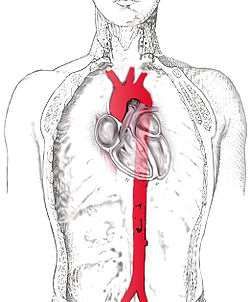
Adrenal trauma is associated with:
   • Hepatic injury.
   • Splenic injury.
   • Renal injury.
   • Rib fractures.
   • Lumbar fractures.
   • Pelvic fractures.
In many cases, patients also have multiple other injuries, such as head
injuries and injuries to the extremities.

Hematoma or laceration of the adrenal gland often is found incidentally
with CT scan to evaluate other abdominal injuries. One study found
that left adrenal hematomas were most commonly associated with left
rib fractures and splenic and left renal injuries while right adrenal
hematomas were associated with right rib fracture and hepatic and
right renal injuries.

One study of pediatric patients with adrenal trauma showed that those
with unilateral injury rarely showed evidence of adrenal insufficiency,
but adrenal insufficiency should be suspected in bilateral injuries.

About 78% of adrenal hematomas occur on the right, possibly because
the area about the gland is more confined because of the mass of the
liver on the right. Additionally, high deceleration pressures may be
transmitted through the inferior vena cava and the short right adrenal
gland. The right adrenal gland lies directly behind the inferior vena
cava.

Diagnosis is by CT but may be impacted by the presence of benign
adenomas, which are common but can be misdiagnosed as
hematomas. FAST may also be used as a screening tool.

Treatment is usually conservative as hematomas usually resolve over
time. In severe injuries, such as with gunshot wounds, adrenalectomy
may be indicated. Hemorrhage may require transfusions. Acute
adrenal insufficiency (usually related to bilateral injury and
hemorrhage) must be quickly diagnosed and treated with
glucocorticoids.

Aortic trauma
The aorta can be injured with both blunt and penetrating trauma.
Tearing may occur from sudden deceleration, as in motor vehicle
accidents, and impact with the steering wheel may result in
compression that ruptures the aorta. About 80% to 90% of great
vessel trauma is fatal with 15% of deaths related to motor vehicle
accidents caused by aortic trauma. Those with small tears or partial
thickness tears may survive.

A free rupture typically present with widened mediastinum,
                                           hemothorax and
                                           hemodynamic instability and
                                           is almost always fatal;
                                           however, a controlled
                                           rupture in which there is no
                                           hemothorax and the patient
                                           remains hemodynamically
                                           stable has about a 90%
                                           survival rate with prompt
                                           surgical intervention.

                                          The most common site of
                                          aortic trauma is distal to the
left subclavian at the level of the ligamentum arteriosum as this is the point of maximum stress and tearing from deceleration forces. Therefore, the aorta is at risk in frontal and side impacts as well as falls from heights. If aortic injury is not treated promptly the patient may develop hypoxia and hypovolemia with anoxic encephalopathy and ischemic damage to other viscera. Diagnosis of aortic injury is through CT scan. If the scan is not definitive, than an aortogram may be done to identify small tears. In some cases intravascular ultrasound (IVUS) or transesophageal echocardiography (TEE) may be done instead of aortogram, as these procedures are less invasive. CT scan may be preceded by chest x-ray (with head elevated) and insertion of an NG tube and/or chest tube. The CT may show a contained rupture, which is referred to as a traumatic pseudoaneurysm. Chest x-ray is not diagnostic but may show an abnormal mediastinum, distortion of the aorta, and depressed left main stem bronchus as well as deviation of a nasogastric tube. These finding are suggestive of aortic injury. Bleeding may result from tears of mediastinal veins, but this is an indirect indication of possible aortic injury. If patients with suspected aortic injury are hemodynamically unstable, priority is given to controlling hemorrhage while avoiding over- resuscitation. FAST of DPL may be utilized to identify concealed areas of hemorrhage. Patients with aortic tear and impending rupture may develop a cyclical pattern of responding to fluid resuscitation and then
exhibiting hypotension. A cycle of repeated fluid resuscitations can lead to rupture, especially if other signs, such as widened mediastinum and left-sided hemothorax, are present. Aortic injury is associated with diaphragmatic rupture, so any patient with aortic injury should be assessed for injury to the diaphragm and vice versa. Aortic injuries are almost always repaired surgically. In some cases thoracic endovascular aortic repair (TEVAR) with insertion of stents may be used. Recent studies indicate that delaying surgery for up to 4 days in non-acute cases, allowing other injuries to be treated and the patient stabilized, has resulted in a lower overall mortality rate than rushing patients into surgery. If the patient must undergo a craniotomy or exploratory laparotomy because of other injuries and the aortic injury is not acute, -blockers may be administered to reduce heart rate and force of contractions. Nitroprusside may also be used to control blood pressure and prevent rupture. Conclusion Time is a critical factor with abdominal trauma. Patients must be assessed and evaluated immediately, and a finding of pelvic fracture or trauma to one abdominal organ should always raise suspicion of associated injuries. The diaphragm is the broad muscle that separates the thoracic cavity from the abdominal cavity. The diaphragm is rarely (
•   Abdominal trauma: Role of CT. (n.d.). The Radiology Assistant.
    Retrieved November 9, 2011, from
    http://www.radiologyassistant.nl/en/466181ff61073
•   American Academy of Orthopaedic Surgeons. (2009). Advanced
    Assessment and Treatment of Trauma. Sudbury, MA: AAOS.
•   Bjerke, HS. (2009, August 6). Splenic rupture. Medscape
    Reference. Retrieved November 9, 2011, from
    http://emedicine.medscape.com/article/432823-overview
•   Bjerke, HS. (2010, January 2). Pancreatic Trauma. Medscape
    Reference. Retrieved November 9, 2011, from
    http://emedicine.medscape.com/article/433177-overview
•   Chest trauma: Traumatic aortic injury. (2004, April).
    Trauma.org. Retrieved November 9, 2011, from
    http://www.trauma.org/archive/thoracic/CHESTaorta.html
•   Degiannis, E, Glapa, M, Loukogeorgakis, SP, & Smith, MD.
    (2008). Management of pancreatic trauma. International Journal
    of the care of the Injured, 39: 21-29. Retrieved November 9,
    2011, from
    http://www.smmemx.org/Documentos/Trauma/PancreaticTraum
    a.pdf
•   Fotheringham, T. (2008). Pancreatic injuries. Nordic Trauma
    Society. Retrieved November 9, 2011, from
    http://www.nordictraumarad.com/Syllabus08/pancreas.pdf
•   Havens, J, & Lieberman, G. (2002, November 17). Blunt thoracic
    aortic trauma. Harvard Medical School. Retrieved November 9,
    2011, from
    http://eradiology.bidmc.harvard.edu/LearningLab/cardio/Havens
    .pdf
•   Hepatic trauma: Liver laceration. (2004).
    LearningRadiology.com. Retrieved November 9, 2011, from
    http://www.learningradiology.com/notes/ginotes/livertraumapag
    e.htm
•   Kawashima, A, et al. (2001, May). Imaging of renal trauma: A
    comprehensive review. RadioGraphics. Retrieved November 9,
    2011, from http://radiographics.rsna.org/content/21/3/557.full
•   Kidney (renal) trauma. (2011, January). AUAFoundation.
    Retrieved November 9, 2011, from
    http://www.urologyhealth.org/urology/index.cfm?article=61
•   Lusaya, DG. (2011, October 20). Renal trauma. Medscape
    Reference. Retrieved November 9, 2011, from
    http://emedicine.medscape.com/article/440811-
    overview#showall
•   McPhee, SM, & Papadakis, MA. (2009). Current Medical
    Diagnosis & Treatment. San Francisco: McGraw Hill Medical.
• Mick, MW, Peter, JR, Egan, D, & Nadel, ES. (2006). Blueprints
Emergency Medicine. 2nd ed. Baltimore, MD: Lippincott Williams
& Wilkins.
• Mitchell, EL, & Medson, R. (2005). Introduction to Emergency
Medicine. Philadelphia: Lippincott Williams & Wilkins.
• Mukhopadhyay, M. (2009). Intestinal injury from blunt
abdominal trauma: A study of 47 cases. OMJ, 24: 256-459.
Retrieved November 9, 2011, from
http://www.omjournal.org/OriginalArticles/FullText/200910/FT_I
ntestinalInjuryfromBlunAbdominalTraumaAStudy.html
• Rackley, R. (2009, August 17). Bladder trauma. Medscape
Reference. Retrieved November 9, 2011, from
http://emedicine.medscape.com/article/441124-overview
• Rana, AI, et al. (2004, March). Adrenal gland hematomas in
trauma patients. Radiology, 230: 660-675. Retrieved November
9, 2011, from http://radiology.rsna.org/content/230/3/669.full
• Seldman, C. (n.d.). Renal trauma. Trauma.org. Retrieved
November 9, 2011, from
http://www.trauma.org/archive/abdo/renal/intro.html
• Smeltzer, SC, Bare, BG, Hinkle, JL, & Cheever, KH. (2008).
Brunner & Suddarth’s Textbook of Medical-Surgical Nursing, 11
ed., Philadelphia: Wolters Kluwer/Lippincott, Williams, & Wilkins.
• Stanton-Maxey, KJ. (2011, August 9). Penetrating abdominal
trauma. Medscape Reference. Retrieved November 9, 2011, from
http://emedicine.medscape.com/article/2036859-
overview#a0104
• Tritos, NA. (2011, July 13). Adrenal hemorrhage. Medscape
Reference. Retrieved November 9, 2011, from
http://emedicine.medscape.com/article/126806-overviewYou can also read