Alteration of spatial patterns at the network-level in facial synkinesis: an independent component and connectome analysis
←
→
Page content transcription
If your browser does not render page correctly, please read the page content below
Original Article
                                                                                                                                     Page 1 of 13

Alteration of spatial patterns at the network-level in facial
synkinesis: an independent component and connectome analysis
Zhen-Zhen Ma1,2#, Ye-Chen Lu1,2#, Jia-Jia Wu2, Si-Si Li1,2, Wei Ding3, Jian-Guang Xu1,2,4
1
School of Rehabilitation Science, Shanghai University of Traditional Chinese Medicine, Shanghai, China; 2Department of Rehabilitation Medicine,
Yueyang Hospital of Integrated Traditional Chinese and Western Medicine, Shanghai University of Traditional Chinese Medicine, Shanghai, China;
3
Department of Plastic and Reconstructive Surgery, Shanghai Ninth People Hospital Affiliated to Shanghai Jiaotong University School of Medicine,
Shanghai, China; 4Department of Hand Surgery, Huashan Hospital, Fudan University, Shanghai, China
Contributions: (I) Conception and design: JG Xu, YC Lu; (II) Administrative support: JG Xu, W Ding; (III) Provision of study materials or patients:
W Ding; (IV) Collection and assembly of data: All authors; (V) Data analysis and interpretation: ZZ Ma, YC Lu, JJ Wu; (VI) Manuscript writing: All
authors; (VII) Final approval of manuscript: All authors.
#
These authors contributed equally to this work.
Correspondence to: Jian-Guang Xu, MD, PhD. School of Rehabilitation Science, Shanghai University of Traditional Chinese Medicine, No.
1200 Cailun Road, Shanghai, China. Email: xjg@shutcm.edu.cn; Wei Ding, MD, PhD. Department of Plastic and Reconstructive Surgery,
Shanghai Ninth People Hospital, Affiliated to Shanghai Jiaotong University School of Medicine, No. 639, Zhizaoju Road, Shanghai, China.
Email: drdingwei@outlook.com.

                 Background: The treatment of post-facial palsy synkinesis (PFPS) remains inadequate. Previous studies
                 have confirmed that brain plasticity is involved in the process of functional restoration. Isolated activation
                 has been well studied, however, the brain works as an integrity of several isolated regions. This study aimed
                 to assess the alteration of the brain network topology with overall and local characteristics of information
                 dissemination. Understanding the neural mechanisms of PFPS could help to improve therapy options and
                 prognosis.
                 Methods: Patients with facial synkinesis and healthy controls (HCs) were estimated using functional
                 magnetic resonance imaging (fMRI) of resting-state. Subsequently, an independent component analysis (ICA)
                 was used to extract four subnets from the whole brain. Then we used the measurements of graph theory and
                 calculated in the whole-brain network and each sub-network.
                 Results: We found no significant difference between the patient group and the HCs on the whole-
                 brain scale. Then we identified four subnetworks from the resting-state data. In the sub-network property
                 analysis, patients’ locally distributed properties in the sensorimotor network (SMN) and ventral default
                 mode network (vDMN) were reduced. It revealed that γ (10,000 permutations, P=0.048) and S (10,000
                 permutations, P=0.022) within the SMN progressively decreased in patients with PFPS. For the analysis of
                 vDMN, significant differences were found in γ (10,000 permutations, P=0.019), Elocal (10,000 permutations,
                 P=0.008), and β (10,000 permutations, P=0.011) between the groups.
                 Conclusions: Our results demonstrated a reduction in local network processing efficiency in patients with
                 PFPS. Therefore, we speculate that decreased characteristics in the intra-vDMN and intra-SMN, rather
                 than the whole-brain network, may serve distinct symptoms such as facial nerve damage or more synkinetic
                 movements. This finding of the alteration of network properties is a small step forward to help uncover the
                 underlying mechanism.

                 Keywords: Post-facial palsy synkinesis (PFPS); independent component analysis (ICA); default mode network
                 (DMN); sensorimotor network (SMN); graph theory analysis

                 Submitted Jun 21, 2020. Accepted for publication Oct 18, 2020.
                 doi: 10.21037/atm-20-4865
                 View this article at: http://dx.doi.org/10.21037/atm-20-4865

© Annals of Translational Medicine. All rights reserved.                   Ann Transl Med 2021;9(3):240 | http://dx.doi.org/10.21037/atm-20-4865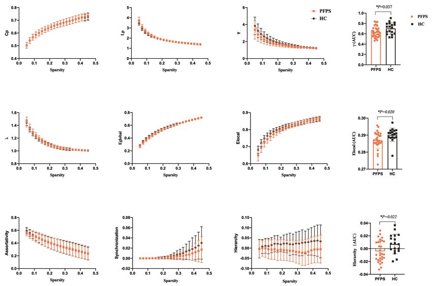
Page 2 of 13                                                                        Ma et al. Spatial patterns alteration in facial synkinesis

Introduction                                                              rather than the whole-brain network, can better explain the
                                                                          abnormal movement, collectively inducing neural plasticity
Post-facial palsy synkinesis (PFPS) is a sequela of facial
                                                                          of pathologic involuntary activity. This study used a high-
nerve injury. It manifests as involuntary abnormal facial
                                                                          accuracy network analysis that was able to detect mild
movements accompanied by volitional movement in
                                                                          changes to a limited extent from the perspective of the
another area of the face, such as eye closure, triggered by
                                                                          connectome; this is useful for increasing our understanding
the volitional movement of the mouth (1,2). However, the
                                                                          of PFPS.
common therapeutic modalities for synkinesis, including
                                                                             We present the following article in accordance with
facial neuromuscular retraining with physical therapy,
                                                                          the MDAR reporting checklist (available at http://dx.doi.
targeted chemodenervation, and surgical procedures, are
                                                                          org/10.21037/atm-20-4865).
not sufficiently efficacious. Therefore, synkinesis remains a
significant clinical challenge owing to its unclear causative
mechanisms (3-5). In particular, it is unclear how abnormal               Methods
information exchanges occur within the architecture of
                                                                          Participants
the sub-networks. This hinders our understanding of
the full spectrum of pathophysiologic interactions in this                Thirty-one patients with PFPS (20 females; age range: 12–
debilitating movement disorder.                                           49 years; mean age: 33.61 years) and 19 HCs (17 females;
   Previous neuroimaging studies on movement disorders                    age range: 23–42 years; mean age: 33.21 years) participated
h av e d e m o n s t r a t ed b ra i n reg i o ns res p o ns i ble for    in this cross-sectional study (Table 1). Patients with Bell’s
sensorimotor control, aberrant excitation, inhibition of the              palsy were included, who were suffered from unilateral
basal ganglia circuitry, and abnormal integration of the basal            facial synkinesis, as well as participants that were recruited
ganglia, thalamus, insula, and cerebellum, with the primary               via two experienced plastic surgeons from the Department
sensorimotor engaged in disorder-specific alterations                     of Plastic Surgery. Inclusion criteria were medical history
(6-9). Also, many studies of task-dependent on facial                     of 9 months or over and signed informed consent for
synkinesis lead us to conclude that there is one particular               the study. Participants with one or more of the following
module that focuses on the projection area in the brain                   characteristics were excluded: Hunt syndrome or other
map, which specifically corresponds to ocular-oral or oral-               causes (e.g., trauma, iatrogenic, or parotid tumors); central
ocular synkinesis (10). This isolated activation has been well            nervous system pathology; recurrent BP or concurrent
studied. However, the brain works as an integrity of several              peripheral neuropathy; a history of nerve transposition,
regions but not several isolated modules (7,11). The brain                and contraindications to an investigation by MRI. All fMRI
network recruits neurons to complete various functional                   measurements and clinical assessments were performed on
activities, such as sensorimotor, speech, and cognition, in an            the same day. The whole procedure lasts about 75 minutes.
integrated circuit (12). Instead of exploring large-scale and             Besides, a neurological examination (normal vision and
distributed properties for information processing, research               the capability to follow instructions) and an interview
on facial synkinesis research has started to focus on regional            (the Sunny-Brook facial grading system for patients) were
alterations patterns or deficits (13).                                    conducted. The HCs had to be free of facial palsy or other
   To examine the brain plasticity changes associated                     neurological impairments. All participants signed informed
with facial paralysis, we used an independent component                   consent forms before proceeding to the trial according to
analysis (ICA) to compare patients with PFPS and healthy                  the Helsinki declaration (as revised in 2013). The study was
controls (HCs) in resting-state functional magnetic                       approved by the local ethics committee (approval number
resonance imaging (fMRI). We built a graph theory                         2017-365-T267), and the study protocol was registered in
analysis based on the results of the ICA that identified the              the Chinese Clinical Trial Registry (ChiCTR1800014630).
topological changes in patients with PFPS compared to
HCs on the whole-brain network scale and functional sub-
                                                                          Data acquisition and preprocessing
network range, to expand our understanding of global and
focal alterations. PFPS is a sequela of facial palsy, which               All subjects went through a whole-brain scan of resting-
generally does not cause a strong reaction in the whole                   state fMRI (rs-fMRI). fMRI data were taken on a 3 T MRI
brain. However, most researchers still prefer to target an                scanner (GE Signa VH/I) using a 32-channel phased-array
area (14). We hypothesize that functional sub-networks,                   head coil. The rs-fMRI in whole-brain used a gradient

© Annals of Translational Medicine. All rights reserved.                 Ann Transl Med 2021;9(3):240 | http://dx.doi.org/10.21037/atm-20-4865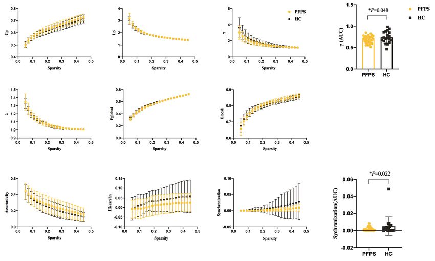
Annals of Translational Medicine, Vol 9, No 3 February 2021                                                                        Page 3 of 13

 Table 1 Demographic material of facial synkinesis patients and healthy controls
 Items                                           PPFS (n=31)                         HC (n=19)                           P value

 Age                                              33.61±8.55                        33.21±6.75                            0.863a

 Sex (male/female)                                   11/20                             2/17                               0.105b

 Duration                                       30.82±9.79 m                             –                                  –

 Affected side                                 13 (right)/18 (left)                      –                                  –

 Sunny-brook scores                              41.45±14.07                             –                                  –
 a                                   b
  , two independent sample t-test; , Chi-square test. PFPS, post-facial palsy synkinesis; HC, healthy controls.

echo-planar imaging sequence (interleaved scanning order,                   dimensionally reduced using principal component analysis
slice number =43, TR =2,000 ms, matrix size =64×64, FOV                     (PCA), then temporally concatenated and reduced to extract
=220×220 mm, voxel size 3.4×3.4×3.2 mm 3, number of                         40 spatial components using the expectation-maximization
acquisitions =240). The preprocessing and analysis of fMRI                  algorithm at a group level. In addition, we performed the
data was performed using SPM12 (http://www.fil.ion.ucl.                     infomax ICA algorithm in ICASSO for 100 repetitions to
ac.uk/spm) and the graph-theoretical network analysis was                   validate its robustness. After the aggregated spatial maps
developed on the GRaph thEoreTical Network Analysis                         were estimated, subject-specific spatial patterns and time
(GRETNA) (http://www.nitrc.org/projects/gretna/)                            courses were extracted through the back-reconstruction
toolbox (15). Briefly, after discarding the first 10 volumes of             approach. A time course for all subjects as well as spatial
each fMRI run, slice timing was performed to correct the                    weight maps were produced, which revealed the possibility
inconsistency of temporal collection. Then the data were                    of one voxel belonging to one particular component.
motion-corrected, normalized to stereotactic Montreal                       Then we thresholded these maps at group level after a
Neurological Institute (MNI) space via a standard EPI                       Z-transformation of the spatial weight map.
template, and spatially smoothed (6×6×6 mm3 Gaussian                           We selected meaningful ICs through spatial sorting
kernel). Point-to-point head motion and mean head motion                    an d visual in spection . Four ICs- of- in ter e s t w ere
was estimated for subjects to control the motion-induced                    identified in this study: the sensorimotor network (SMN)
artifacts. Therefore, one patient with excessive head motion                (Figure 1A), frontoparietal network (FPN) (Figure 1B),
(cumulative translation or rotation >3 mm or 3°) was                        dorsal default mode network (dDMN) (Figure 1C), and
excluded (16).                                                              ventral default mode network (vDMN) (Figure 1D). For
                                                                            these sub-networks, we specified each voxel as the node
                                                                            and functional connectivity of the voxel–voxel as the edge.
Node and edge definition
                                                                            Besides, we resampled the data and adjusted the voxel size
Topological analyses in whole-brain network and functional                  to 6×6×6 mm3 to decrease the large scale of the connectivity
sub-network were conducted in this study. We used an                        matrix, which reduced the computational work significantly.
AAL90 structure-labeled atlas for parcellation in the whole-                   After the functional connectivity matrices were obtained,
brain network analysis. Thus, nodes were defined as the                     we calculated the network metrics of the whole-brain and
parcellated 90 brain regions. Subsequently, Pearson’s                       the sub-networks separately using a pre-selected sparsity
correlations of the meantime series between all pairs of the                value (the ratio of the number of actual edges divided by
90 nodes were characterized as the edges.                                   the maximum possible number of edges in a network)
   For the sub-network analysis, preprocessed rs-fMRI                       to ensure the relative network organization. Specifically,
data were decomposed into independent components                            the topological organization of whole-brain networks
(ICs) using GIFT software (Group ICA of fMRI Toolbox,                       was analyzed over a wide range of network sparsity
version 4.0, http://icatb.sourceforge.net) to construct                     (0.05–0.45) (19), where the small-world metrics were
the sub-networks. Generally, the group ICA for this                         analyzed (20). Network density was applied to each adjacent
multi-subject analysis utilized a concatenation approach                    matrix with an increment of 0.01 for the whole-brain
plus back-reconstruction (17). The images were first                        network and 0.02 for the sub-network metrics to reduce the

© Annals of Translational Medicine. All rights reserved.                  Ann Transl Med 2021;9(3):240 | http://dx.doi.org/10.21037/atm-20-4865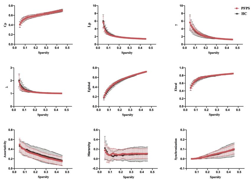
Page 4 of 13 Ma et al. Spatial patterns alteration in facial synkinesis
A
B
C
12
10
D
8
6
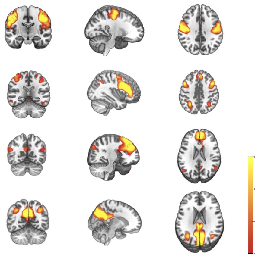
Figure 1 The spatial distribution pattern of four potential sub-networks extracted from resting-data by ICA. Four components (A-D)
resemble the RSNs described in a previous study (18) and consist of regions known to be involved in the sensorimotor network (A), FPN
(B), dDMN (C), and vDMN (D). Images (coronal, sagittal, and axial views) are t-statistics overlaid on the average high-resolution scan
transformed into MNI152. Red to yellow represents t values, ranging from 6.0 to 12.0. The left hemisphere of the brain corresponds to the
right side of the image. ICA, independent component analysis; RSNs, resting-state networks; MNI, Montreal neurological institute.
computational dimension. assortativity r, hierarchy β and synchronization S), were
calculated in this study. All the calculations of network
metrics were completed with GRETNA. Table 2 gives
Network analysis
detailed descriptions of the above metrics (15,22).
Global properties of the whole-brain network and sub- The small-world parameters hold on both modularized/
networks specialized and distributed/integrated information
Graph theory analysis is a suitable method to characterize processing and greatly amplify the efficiency of information
the brain network topology properties (12,21). The transfer at a low-cost. Therefore, small-world networks
commonly used network metrics, including the small- have a shorter Lp than regular networks (high Cp and long
world property (clustering coefficient Cp, characteristic path L p) but greater E local than random networks (low C p and
length Lp, normalized clustering coefficient γ, normalized short Lp). To characterize the small-world properties of
characteristic path length λ and small-worldness σ), network the target network, we calculated its Cp and Lp with the
efficiency (local efficiency Elocal and global efficiency Eglobal), corresponding value, which derive from the average of 100
© Annals of Translational Medicine. All rights reserved. Ann Transl Med 2021;9(3):240 | http://dx.doi.org/10.21037/atm-20-4865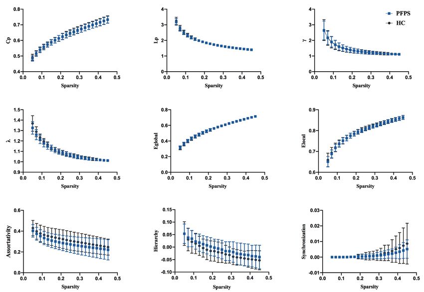
Annals of Translational Medicine, Vol 9, No 3 February 2021                                                                        Page 5 of 13

 Table 2 Descriptions of global network metrics examined in the present study
 Parameter                           Character        Descriptions

 Clustering coefficient              Cp               The extent of local clustering of a network

 Characteristic path length          Lp               The extent of overall routing efficiency of a network

 Gamma                               γ                The deviation of Cp of a network from those of surrogate random networks

 Lambda                              λ                The deviation of Lp of a network from those of surrogate random networks

 Sigma                               σ                The small-worldness indicating the extent of a network between randomness and order

 Local efficiency                    Elocal           How efficient of information propagation over a node’s direct neighbors

 Global efficiency                   Eglobal          How efficient of information propagation through the whole network

 Assortativity                       r                The tendency of nodes to link those nodes with similar number of edges

 Hierarchy                           β                How likely it is that all nodes oscillate with the same wave pattern

 Synchronization                     S                How likely that all nodes fluctuate in the same wave pattern

random networks with the equal scale of nodes and degree                     the second smallest eigenvalue to the largest eigenvalue of
distribution (23). Compared with the random networks,                        the coupling matrix of the network, measures the possibility
small-world networks have a relatively high normalized                       of the fluctuation of all nodes in the same wave pattern.
clustering coefficient γ (C p/C prand) >1 and relatively low
normalized characteristic path length λ (Lp/Lprand) ≈1 (24).                 Regional properties of the whole-brain network
   Global efficiency measures the efficiency of information                  For nodal metrics, we also calculated the regional
transfer at a network level, which is the inverse of the                     properties, including the nodal degree, nodal efficiency, and
harmonic mean of the minimum path length (25). While the                     betweenness centrality of the whole-brain network. The
local efficiency of the network measures the fault tolerance                 nodal characteristics of the brain networks may demonstrate
in a network, showing how the efficient communication                        the importance of specific areas in brain network by
exchanges when the first neighbors of a given node is                        measuring the extent to which a given node connects to all
phased out (25).                                                             other nodes of a network (25,30).
   Additionally, we evaluated the hierarchical nature of
networks using the β parameter (26), which defines the                       Data analysis
size of the power–law relationship between the clustering                    The area under the curve (AUC) over the sparsity range was
coefficient (Cp) and degree (k): Cp ≈ k-β (27). In a network                 used to conduct group comparisons of the metric. Statistical
with hierarchy organizations, some high-degree related                       tests of topological measures between the groups were
nodes form a densely connected cluster. These generated                      performed using permutation testing to assess alterations
clusters act as elements in the next level of the network and                of the topological parameters (10,000 permutations) for
merge into a larger-scale interconnected cluster (28). We                    each topological parameter over a wide range of connection
computed the parameter β for the network through the plot                    densities. For each permutation, participants were
of log(C) versus log(k) applied with regression line fitting.                randomly assigned to patients and HCs with the same size
   In addition, assortativity reflects the tendency of nodes                 as the original two groups. Next, the randomized groups
to link those nodes with a similar number of edges, which                    difference of the metric was measured to obtain a null
measures the correlation between the node degree and                         distribution. Depended on the generated null distribution,
the mean degree of its adjacent neighbor (29). Positive                      a P value was charged to evaluate the extent of alteration
assortativity indicates that the densely connected nodes are                 between groups. A P value of
Page 6 of 13                                                                          Ma et al. Spatial patterns alteration in facial synkinesis

            A                                        B                                       C

            D                                        E                                       F

            G                                       H                                         I

Figure 2 Global topological properties of functional connectome between the groups. The statistical comparison of area under the curve
(AUC) showed no significant difference in Cp, Lp, γ, λ, Eglobal, Elocal, assortativity, hierarchy, or synchronization between groups, and the bin-
width of sparsity was 0.01. PFPS group: red symbols and lines; HC group: gray symbols and lines.

comparisons was performed at a q value of 0.05 (32).                        topological metrics in whole-brain propagation between
                                                                            groups were non-significant. In the nodal analysis, none
                                                                            of the nodes in the regional properties’ analysis (including
Results
                                                                            nodal degree, nodal efficiency, and betweenness centrality)
Demographic and clinical characteristics                                    were significant after FDR correction. That means, there
The patients with PFPS (females; age: 33.61±8.55 years) and                 was no significant difference between the patient group and
19 HC (females; age: 33.21±6.75 years) were included in the                 the HCs on the whole-brain scale.
statistical analysis. We observed non-significant differences
between PFPS and HC concerning age (P=0.86) and gender                      Alterations in sub-network organization of the functional
(P=0.11). Demographic and clinical characteristics of these                 connectome
subjects is provided in Table 1. One patient was excluded
because his head-motion beyond 3 mm or 3°.                                  We identified four subnetworks from the resting-state data
                                                                            after the ICA procedure (Figure 1): SMN (component A),
                                                                            FPN (component B), dDMN (component C), and vDMN
Alterations in whole-brain organization of the functional
                                                                            (component D). A comparison of component A between
connectome
                                                                            groups revealed that γ (10,000 permutations, P=0.048) and
In the initial test of global topological parameters, we found              synchronization (10,000 permutations, P=0.022) within
λ ≈1, γ >1, and σ = γ /λ >1, which indicates the networks are               the SMN progressively decreased in patients with PFPS
of small-worldness property. As displayed by Figure 2, the                  (Figure 3). A comparison of components B and C found a

© Annals of Translational Medicine. All rights reserved.                  Ann Transl Med 2021;9(3):240 | http://dx.doi.org/10.21037/atm-20-4865
Annals of Translational Medicine, Vol 9, No 3 February 2021 Page 7 of 13
A B C J
D E F
G H I K
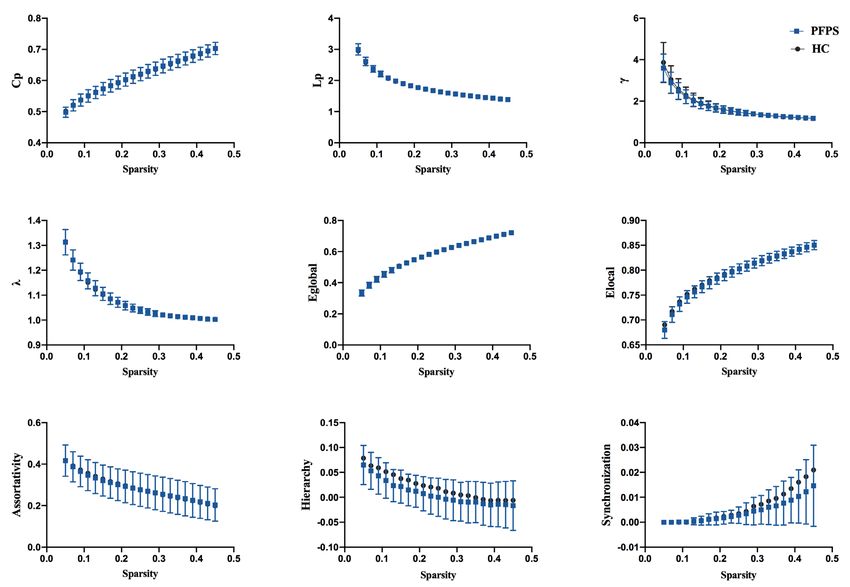
Figure 3 Functional brain network properties of the SMN with a bin-width sparsity of 0.02. The area under the curve (AUC) displayed
no significant differences in Cp, Lp, γ, λ, Eglobal, Elocal, assortativity, hierarchy, or synchronization between the groups. While the index γ was
significantly lower in the PFPS group (P=0.048) and the index of synchronization was significantly decreased in patients (P=0.022). PFPS
group: yellow symbols and lines; HC group: gray symbols and lines. *, indicates a significant difference between the PFPS group and the
HC group.
non-significant difference between the groups. The FPN context, neuro marker candidates will be found to reflect all
and dDMN show analogous patterns in PFPS and HC aspects of involuntary movements in all contexts and help
(Figures 4,5). For component D, significant differences to afford clinical prognostic indicators (36-38). We utilized
were found in γ (10,000 permutations, P=0.019), E local a graph theory analysis to examine the resting-state fMRI
(10,000 permutations, P=0.008), and hierarchy (10,000 data to characterize the global and local features after facial
permutations, P=0.011) between the groups. The PFPS synkinesis, instead of localization information. Beyond
group showed lower γ, Elocal, and hierarchy of the vDMN regional activity, PFPS in our study was found to influence
than did the HCs (Figure 6). intrinsic brain networks (also known as resting-state
networks), mainly the somatosensory networks, and default
mode networks, which subserve homeostatic, attentional,
Discussion
cognitive, executive, and sensory functions together
In recent years, cortical plasticity in PFPS has been and alter their functional connectivity. Therefore, facial
extensively discussed (33,34). To date, most work has synkinesis is a major complication of facial paralysis while
attempted to identify neuro markers that reveal mechanisms it does not induce extensive plasticity of the whole brain.
and neurophysiological processes that are significant in By contrast, it only leads to limited topological changes in
PFPS (9,35). Considering that the experience of synkinesis some functional sub-networks.
has diverse influences, from a deficit in control to social Our study shows that the activity induced by synkinesis
© Annals of Translational Medicine. All rights reserved. Ann Transl Med 2021;9(3):240 | http://dx.doi.org/10.21037/atm-20-4865Page 8 of 13 Ma et al. Spatial patterns alteration in facial synkinesis
A B C
D E F
G H I
Figure 4 Functional brain network properties of the FPN with a bin-width sparsity of 0.02. The area under the curve (AUC) displayed no
significant differences in Cp, Lp, γ, λ, Eglobal, Elocal, assortativity, hierarchy, or synchronization between the groups. PFPS group: blue symbols
and lines; HC group: gray symbols and lines.
is related to many brain regions that belong to different cingulate cortex, medial prefrontal cortex, angular gyrus,
functional brain networks, rather than the dedicated areas hippocampus, parahippocampus, retrosplenial cortex,
within the brain. These components of networks belong to and posterior inferior parietal lobe. Consequently, these
the default mode, and somatosensory networks that control intrinsic networks and dedicated connectomes may be
the cortical and subcortical representations of synkinesis engaged during facial synkinesis. The primary aim of this
movements, which have been clarified in large-scale study was to define the scale affected by facial synkinesis.
resting-state studies, extensively overlap with the regions Although regular networks and random graphs are both
related to PFPS (39). The functional network of the brain useful idealizations, many real networks lie somewhere
is complicated and includes areas associated with coding between order and randomness. Small-world networks have
information regarding muscular control and reactions been extensively researched since the fundamental work by
to muscular control, the recognition and modulation of Watts and Strogatz, who indicated that random mutation of
spatial information, and somatosensory information and a few connections in a regular or lattice network extensively
attention (40). This might be the reason for the deactivated lessened the mean minimum path length between any
SMN, which plays an important role in motor control pair of nodes while holding strong local connectivity
and consists of somatosensory (post-central gyrus) or “cliquishness” (43). The coexistence of integration
and motor (pre-central gyrus) regions, and extends to and separation further reflects the small worldness
the supplementary motor areas. The vDMN, which is characteristics of the networks.
spontaneously active during passive performance and The local efficiency of the network evaluates how
associated with effective processing, memory, and self- efficient communication is among the first neighbors of a
projective thinking (41,42), consists of the posterior given node when it is eliminated. A lower Elocal in SMN and
© Annals of Translational Medicine. All rights reserved. Ann Transl Med 2021;9(3):240 | http://dx.doi.org/10.21037/atm-20-4865Annals of Translational Medicine, Vol 9, No 3 February 2021 Page 9 of 13
A B C
D E F
G H
I
Figure 5 Functional brain network properties of the dDMNs with a bin-width sparsity of 0.02. The area under the curve (AUC) displayed
no significant differences in Cp, Lp, γ, λ, Eglobal, Elocal, assortativity, hierarchy, or synchronization between the groups. PFPS group: blue symbols
and lines; HC group: gray symbols and lines.
vDMN means that the possibility of “off track” increases, seems to be a facial mismatch on neural activity rather
suggesting a trend of “separation” of these functional than deactivation (39,45). We found that facial synkinesis
sub-networks. The decrease in E local also indicates the tends to reduce the cluster efficiency to lower than normal,
randomization of the sub-networks, reflecting the decrease which may illustrate the effects on cortical plasticity from
in overall efficiency and change of the integration-separation the perspective of neural mechanisms. Furthermore,
balance in different neural functional sub-networks. we discovered many measurements, including network
Hierarchy is the tendency of hub regions to connect with efficiency and synchronization, that showed neither
nodes outside the hubs (44). In this study, it was found that significant nor trending differences in any sub-network.
the subjects’ functional complex sub-networks had small- Therefore, we believe that they may not be involved in the
world attributes, and the efficiency of regional information neuroplasticity of facial synkinesis. Such interpretations are
transmitting in SMN and vDMN decreased. It is suggested not difficult as post-facial paralysis synkinesis only causes
that patients with facial synkinesis can cause changes in the changes in topological properties to a limited extent. This
mode of brain information processing. would not be significant if the contrast were executed on
The declined propagation characteristics in the sub- a global scale. These findings should be confirmed with a
network represent less active neural transmitting activity larger sample size in the future.
in facial paralysis. Therefore, the connection of the The present study investigated the topological structure
subnetworks is diminished. The results imply a disruption of functional brain networks between HCs and patients with
of the efficiency of local information in the sub-networks, PFPS. The rs-fMRI images were examined using a graph
which is a manifestation of maladaptive alteration in facial theory analysis to study the brain functional connectomes.
synkinesis. According to previous studies, facial synkinesis No significant differences in the whole-brain scale were
© Annals of Translational Medicine. All rights reserved. Ann Transl Med 2021;9(3):240 | http://dx.doi.org/10.21037/atm-20-4865Page 10 of 13 Ma et al. Spatial patterns alteration in facial synkinesis
A B C J
D E F K
G H I L
Figure 6 Functional brain network properties of the vDMN with a bin-width sparsity of 0.02. The area under the curve (AUC) displayed
no significant differences in Cp, Lp, γ, λ, Eglobal, Elocal, assortativity, hierarchy, or synchronization between the groups. The index γ, Elocal, and
hierarchy were significantly lower in the PFPS group (P values were 0.019, 0.008, and 0.011, respectively). PFPS group: orange symbols and
lines; HC group: gray symbols and lines. *, indicates a significant difference between the PFPS group and the HC group.
observed between patients with PFPS and HCs. However, network. These findings indicate that functional disruption
patients with PFPS had decreased small-worldness in patients with PFPS may be associated with instabilities
and synchronization in the SMN and decreased small- in the local topological organization of the brain functional
worldness, local efficiency, and hierarchy structures in the connectome. In addition, this study provides new insights
vDMN. Moreover, patients with PFPS had altered network into the pathophysiological alterations to further reaching
metrics in the SMN and vDMN brain regions, mainly the mechanism of PFPS in-depth.
involved in information transport, attention, memory, and
emotion.
Acknowledgments
Funding: This work was supported by the Open Project
Conclusions
of Shanghai Key Laboratory of Peripheral Nerve and
The current results provide the first evidence that reduced Microsurgery [Grant Number 17DZ2270500].
intrinsic connectivity represents regionally restricted
information transmission efficiency in the SMNs and
Footnote
vDMNs of patients with PFPS. PFPS produces changes in
neuroplasticity in sub-networks rather than the whole brain Reporting Checklist: The authors have completed the MDAR
© Annals of Translational Medicine. All rights reserved. Ann Transl Med 2021;9(3):240 | http://dx.doi.org/10.21037/atm-20-4865Annals of Translational Medicine, Vol 9, No 3 February 2021 Page 11 of 13
reporting checklist. Available at http://dx.doi.org/10.21037/ and Outcomes in Botulinum Therapy for Patients With
atm-20-4865 Facial Synkinesis. JAMA Facial Plast Surg 2019;21:244-51.
3. Cronin GW. The effectiveness of neuromuscular facial
Data Sharing Statement: Available at http://dx.doi. retraining combined with electromyography in facial
org/10.21037/atm-20-4865 paralysis rehabilitation. Otolaryngol Head Neck Surg
2003;128:534-8.
Peer Review File: Available at http://dx.doi.org/10.21037/ 4. Tenney AP, Livet J, Belton T, et al. Etv1 Controls the
atm-20-4865 Establishment of Non-overlapping Motor Innervation of
Neighboring Facial Muscles during Development. Cell
Conflicts of Interest: All authors have completed the ICMJE Rep 2019;29:437-52.e4.
uniform disclosure form (available at http://dx.doi. 5. Watanabe Y, Akizuki T, Ozawa T, et al. Dual innervation
org/10.21037/atm-20-4865). All authors report grants from method using one-stage reconstruction with free
Open Project of Shanghai Key Laboratory of Peripheral latissimus dorsi muscle transfer for re-animation of
Nerve and Microsurgery [Grant Number 17DZ2270500], established facial paralysis: simultaneous reinnervation of
during the conduct of the study. The other authors have no the ipsilateral masseter motor nerve and the contralateral
conflicts of interest to declare. facial nerve to improve the quality of smile and emotional
facial expressions. J Plast Reconstr Aesthet Surg
Ethical Statement: The authors are accountable for all 2009;62:1589-97.
aspects of the work in ensuring that questions related 6. Battistella G, Simonyan K. Top-down alteration of
to the accuracy or integrity of any part of the work are functional connectivity within the sensorimotor network
appropriately investigated and resolved. The study was in focal dystonia. Neurology 2019;92:e1843-51.
conducted in accordance with the Declaration of Helsinki 7. Smith SM, Vidaurre D, Beckmann CF, et al. Functional
(as revised in 2013), and the Harmonized Tripartite connectomics from resting-state fMRI. Trends Cogn Sci
Guideline for Good Clinical Practice from the International 2013;17:666-82.
Conference on Harmonization. This study was reviewed 8. Wang Y, Wang WW, Hua XY, et al. Patterns of cortical
and approved by the Ethics Committee of the Shanghai Jiao reorganization in facial synkinesis: a task functional
Tong University (approval number 2017-365-T267), and magnetic resonance imaging study. Neural Regen Res
the study protocol was registered in the Chinese Clinical 2018;13:1637.
Trial Registry (ChiCTR1800014630). All patients enrolled 9. Wu JJ, Lu YC, Zheng MX, et al. Motor Control
completed the informed consent form. Deficits in Facial Synkinesis Patients: Neuroimaging
Evidences of Cerebral Cortex Involvement. Neural Plast
Open Access Statement: This is an Open Access article 2019;2019:7235808.
distributed in accordance with the Creative Commons 10. Lee J, Yang J, Li C, et al. Cortical Reorganization in
Attribution-NonCommercial-NoDerivs 4.0 International Patients Recovered from Bell’s Palsy: An Orofacial and
License (CC BY-NC-ND 4.0), which permits the non- Finger Movements Task-State fMRI Study. Neural Plast
commercial replication and distribution of the article with 2016;2016:8231726.
the strict proviso that no changes or edits are made and 11. Song M, Jiang T. A review of functional magnetic
the original work is properly cited (including links to both resonance imaging for Brainnetome. Neurosci Bull
the formal publication through the relevant DOI and the 2012;28:389-98.
license). See: https://creativecommons.org/licenses/by-nc- 12. Bullmore E, Sporns O. Complex brain networks: graph
nd/4.0/. theoretical analysis of structural and functional systems.
Nat Rev Neurosci 2009;10:186-98.
13. VanderWerf F, Reits D, Smit AE, et al. Blink Recovery
References
in Patients with Bell’s Palsy: A Neurophysiological and
1. Markey JD, Loyo M. Latest advances in the management Behavioral Longitudinal Study. Invest Ophthalmol Vis Sci
of facial synkinesis. Curr Opin Otolaryngol Head Neck 2007;48:203.
Surg 2017;25:265-72. 14. Veen MM, Quatela O, Tavares‐Brito J, et al. Patient‐
2. Shinn JR, Nwabueze NN, Du L, et al. Treatment Patterns perceived severity of synkinesis reduces quality of life in
© Annals of Translational Medicine. All rights reserved. Ann Transl Med 2021;9(3):240 | http://dx.doi.org/10.21037/atm-20-4865Page 12 of 13 Ma et al. Spatial patterns alteration in facial synkinesis
facial palsy: A cross‐sectional analysis in 92 patients. Clin Lett [Internet]. 2002. Available online: http://arxiv.org/
Otolaryngol 2019;44:483-6. abs/cond-mat/0205405
15. Wang J, Wang X, Xia M, et al. GRETNA: a graph 30. He Y, Chen Z, Evans A. Structural Insights into Aberrant
theoretical network analysis toolbox for imaging Topological Patterns of Large-Scale Cortical Networks in
connectomics. Front Hum Neurosci 2015;9:386. Alzheimer’s Disease. J Neurosci 2008;28:4756-66.
16. Pasquini L, Scherr M, Tahmasian M, et al. Link between 31. Xu P, Huang R, Wang J, et al. Different topological
hippocampus’ raised local and eased global intrinsic organization of human brain functional networks
connectivity in AD. Alzheimers Dement 2015;11:475-84. with eyes open versus eyes closed. NeuroImage
17. Calhoun VD, Adali T, Pearlson GD, et al. A method 2014;90:246-55.
for making group inferences from functional MRI data 32. Genovese CR, Lazar NA, Nichols T. Thresholding of
using independent component analysis. Hum Brain Mapp Statistical Maps in Functional Neuroimaging Using the
2001;14:140-51. False Discovery Rate1. Neuroimage 2002;15:870-8.
18. Damoiseaux JS, Rombouts SA, Barkhof F, et al. Consistent 33. Hu S, Wu Y, Li C, et al. Increasing functional connectivity
resting-state networks across healthy subjects. Proc Natl of the anterior cingulate cortex during the course of
Acad Sci U S A 2006;103:13848-53. recovery from Bell’s palsy. Neuroreport 2015;26:6-12.
19. Yu Y, Zhou X, Wang H, et al. Small-World Brain Network 34. Klingner CM, Volk GF, Brodoehl S, et al. The effects
and Dynamic Functional Distribution in Patients with of deefferentation without deafferentation on functional
Subcortical Vascular Cognitive Impairment. PLoS One connectivity in patients with facial palsy. Neuroimage Clin
2015;10:e0131893. 2014;6:26-31.
20. Watts DJ, Strogatz SH. Collective dynamics of ‘small- 35. Ma J, Hua XY, Zheng MX, et al. Structural remodeling
world’ networks. Nature 1998;393:440-2. secondary to functional remodeling in advanced-stage
21. He Y, Evans A. Graph theoretical modeling of brain peripheral facial neuritis. Neurol Sci [Internet] 2020.
connectivity. Curr Opin Neurol 2010;23:341-50. Available online: http://link.springer.com/10.1007/s10072-
22. Wang JH, Zuo XN, Gohel S, et al. Graph Theoretical 020-04325-5
Analysis of Functional Brain Networks: Test-Retest 36. Ballesta S, Mosher CP, Szep J, et al. Social determinants of
Evaluation on Short- and Long-Term Resting-State eyeblinks in adult male macaques. Sci Rep 2016;6:38686.
Functional MRI Data. PLoS One 2011;6:e21976. 37. Hall A. The origin and purposes of blinking. Br J
23. Sporns O, Zwi JD. The Small World of the Cerebral Ophthalmol 1945;29:445-67.
Cortex. Neuroinformatics 2004;2:145-62. 38. Smit A, van der Geest J, Metselaar M, et al. Long-term
24. Achard S. A Resilient, Low-Frequency, Small-World changes in cerebellar activation during functional recovery
Human Brain Functional Network with Highly Connected from transient peripheral motor paralysis. Exp Neurol
Association Cortical Hubs. J Neurosci 2006;26:63-72. 2010;226:33-9.
25. Achard S, Bullmore E. Efficiency and Cost of Economical 39. Klingner CM, Brodoehl S, Witte OW, et al. The impact
Brain Functional Networks. PLoS Comput Biol of motor impairment on the processing of sensory
2007;3:e17. information. Behav Brain Res 2019;359:701-8.
26. Ravasz E, Barabási AL. Hierarchical organization 40. van den Heuvel MP, Hulshoff Pol HE. Exploring the
in complex networks. Phys Rev E [Internet]. 2003. brain network: a review on resting-state fMRI functional
Available online: https://link.aps.org/doi/10.1103/ connectivity. Eur Neuropsychopharmacol 2010;20:519-34.
PhysRevE.67.026112 41. Buckner RL. The brain’s default network: origins and
27. Supekar K, Musen M, Menon V. Development of Large- implications for the study of psychosis. Dialogues Clin
Scale Functional Brain Networks in Children. PLoS Biol Neurosci 2013;15:351-8.
2009;7:e1000157. 42. Otti A, Noll-Hussong M. Acupuncture-Induced Pain
28. Barabási AL, Ravasz E, Oltvai Z. Hierarchical Relief and the Human Brains Default Mode Network
Organization of Modularity in Complex Networks. In: - an Extended View of Central Effects of Acupuncture
Pastor-Satorras R, Rubi M, Diaz-Guilera A, editors. Analgesia. Forsch Komplementmed 2012;19:197-201.
Statistical Mechanics of Complex Networks. Berlin, 43. Salvador R, Suckling J, Coleman MR, et al.
Heidelberg: Springer Berlin Heidelberg; 2003:46-65. Neurophysiological Architecture of Functional Magnetic
29. Newman MEJ. Assortative mixing in networks. Phys Rev Resonance Images of Human Brain. Cereb Cortex
© Annals of Translational Medicine. All rights reserved. Ann Transl Med 2021;9(3):240 | http://dx.doi.org/10.21037/atm-20-4865Annals of Translational Medicine, Vol 9, No 3 February 2021 Page 13 of 13
2005;15:1332-42. Schizophrenia. J Neurosci 2008;28:9239-48.
44. Bassett DS, Bullmore E, Verchinski BA, et al. Hierarchical 45. Diedrichsen J. Neural Correlates of Reach Errors. J
Organization of Human Cortical Networks in Health and Neurosci 2005;25:9919-31.
Cite this article as: Ma ZZ, Lu YC, Wu JJ, Li SS, Ding W,
Xu JG. Alteration of spatial patterns at the network-level in
facial synkinesis: an independent component and connectome
analysis. Ann Transl Med 2021;9(3):240. doi: 10.21037/atm-20-
4865
© Annals of Translational Medicine. All rights reserved. Ann Transl Med 2021;9(3):240 | http://dx.doi.org/10.21037/atm-20-4865You can also read