Facial attractiveness of cleft patients: a direct comparison between artificial-intelligencebased scoring and conventional rater groups - ETH ...
←
→
Page content transcription
If your browser does not render page correctly, please read the page content below
European Journal of Orthodontics, 2019, 1–6
                                                                                                                     doi:10.1093/ejo/cjz007

Original article

                                                                                                                                                                Downloaded from https://academic.oup.com/ejo/advance-article-abstract/doi/10.1093/ejo/cjz007/5353220 by ETH Zürich user on 23 July 2019
Facial attractiveness of cleft patients: a direct
comparison between artificial-intelligence-
based scoring and conventional rater groups
Raphael Patcas1, , Radu Timofte2, Anna Volokitin2, Eirikur Agustsson2,
Theodore Eliades1, , Martina Eichenberger1 and Michael Marc Bornstein3,
1
 Clinic of Orthodontics and Pediatric Dentistry, Center of Dental Medicine, University of Zurich, 2Computer Vision
Laboratory, D-ITET, ETH Zurich, Switzerland, and 3Oral and Maxillofacial Radiology, Applied Oral Sciences, Faculty of
Dentistry, The University of Hong Kong, Prince Philip Dental Hospital, Hong Kong SAR, China

Correspondence to: Raphael Patcas, Clinic for Orthodontics and Pediatric Dentistry, Center for Dental Medicine, University
of Zurich, Plattenstrasse 11, 8032 Zürich, Switzerland. E-mail: raphael.patcas@zzm.uzh.ch

Summary
Objectives: To evaluate facial attractiveness of treated cleft patients and controls by artificial
intelligence (AI) and to compare these results with panel ratings performed by laypeople,
orthodontists, and oral surgeons.
Materials and methods: Frontal and profile images of 20 treated left-sided cleft patients (10 males,
mean age: 20.5 years) and 10 controls (5 males, mean age: 22.1 years) were evaluated for facial
attractiveness with dedicated convolutional neural networks trained on >17 million ratings for
attractiveness and compared to the assessments of 15 laypeople, 14 orthodontists, and 10 oral
surgeons performed on a visual analogue scale (n = 2323 scorings).
Results: AI evaluation of cleft patients (mean score: 4.75 ± 1.27) was comparable to human
ratings (laypeople: 4.24 ± 0.81, orthodontists: 4.82 ± 0.94, oral surgeons: 4.74 ± 0.83) and was not
statistically different (all Ps ≥ 0.19). Facial attractiveness of controls was rated significantly higher
by humans than AI (all Ps ≤ 0.02), which yielded lower scores than in cleft subjects. Variance was
considerably large in all human rating groups when considering cases separately, and especially
accentuated in the assessment of cleft patients (coefficient of variance—laypeople: 38.73 ± 9.64,
orthodontists: 32.56 ± 8.21, oral surgeons: 42.19 ± 9.80).
Conclusions: AI-based results were comparable with the average scores of cleft patients seen in
all three rating groups (with especially strong agreement to both professional panels) but overall
lower for control cases. The variance observed in panel ratings revealed a large imprecision based
on a problematic absence of unity.
Implication: Current panel-based evaluations of facial attractiveness suffer from dispersion-related
issues and remain practically unavailable for patients. AI could become a helpful tool to describe
facial attractiveness, but the present results indicate that important adjustments are needed on AI
models, to improve the interpretation of the impact of cleft features on facial attractiveness.

Introduction                                                                            indispensable to counteract possible long-lasting negative effects,
                                                                                        which clefts would leave on function and appearance. A plethora
Non-syndromic clefts of the lip and palate are one of the most
                                                                                        of therapy approaches have been introduced, including different
common congenital defects occurring in approximately 1 in 600
                                                                                        surgical closure techniques of the clefts, orthodontic treatment, and
live births worldwide (1). Prolonged interdisciplinary care is

© The Author(s) 2019. Published by Oxford University Press on behalf of the European Orthodontic Society.
                                                                                                                                                           1
All rights reserved. For permissions, please email: journals.permissions@oup.com
2 European Journal of Orthodontics, 2019
orthognathic surgery, all to improve function and facial appearance. and Paediatric Dentistry of the University of Zurich. The test group
Yet regrettably, therapy does frequently not result in an average consisted of 20 files of randomly selected adult patients (10 males, 10
facial appearance (2), leaving more than often scars from the sur- females; mean age: 20.5 years) previously treated inter-disciplinarily
gical interventions and an asymmetry around the nose and mouth for left-sided cleft lip and palate. The treatment protocol included lip
(3). This reduced facial attractiveness reportedly affects the patients’ repair according to the Millard–Perko protocol, soft palate repair
psychosocial well-being (4), and assessment of facial appearance is according to Widmaier–Perko protocol, alveolar bone grafting with
therefore considered a crucial factor to measure treatment outcome. cancellous bone from the iliac crest, and fixed orthodontic appli-
Currently, no valid objective model exists to study the aesthetic ances. Orthognathic surgery was conducted in most cases (n = 17),
Downloaded from https://academic.oup.com/ejo/advance-article-abstract/doi/10.1093/ejo/cjz007/5353220 by ETH Zürich user on 23 July 2019
treatment outcome in clefts (5), and facial aesthetics is usually estab- as well as rhinoplasty (n = 14) or minor lip revision (n = 9).
lished by single raters or panels (6). This tactic is however ill-fated, Ten post-retention files of orthodontically treated adults (5
as it suffers from weaknesses associated with the inconsistency of the males, 5 females; mean age: 22.1 years) served as control. These were
subjective raters, which mainly depends on their background (3). To randomly selected cases with pretreatment Angle class I, no major
overcome the inhomogeneity seen in subjective ratings, applying a skeletal discrepancies, and treated only for minor dental misalign-
computerized model would seem promising. ment. Owing to slightly different recall protocols, the orthodontic
In computer science, artificial intelligence (AI) is a term widely records consisted of patients with a discreetly higher average age.
used to describe the capability to mimic cognitive functions ordin- Cases were only considered both as tests and as controls when the
arily associated with humans, such as learning and problem-solving. following criteria could be ascertained: no syndromes, no congenital
For these tasks, convolutional neural networks (CNNs) are fre- facial anomalies other than left-sided cleft lip and palate, and no ex-
quently designed, which are biologically inspired models and often ceptional facial features such as tattoos or piercings.
follow the vision processing in living organism (7). Compared with All patients gave their written informed consent for secondary use
other image classifications that depend on hand-engineered algo- of their records including facial photographs. Guidelines in Medical
rithms, CNNs are subject to very little pre-processing, as they are Ethics (15) were strictly obeyed and all data were handled in accord-
capable to learn and optimize their output by changing connection ance with State and Federal Law (Human Research Ordinance, Art. 25,
weights in relation to the expected result, after each piece of data §2). Judicial and ethical conformity of this study were confirmed by the
is processed. CNNs have been applied for various tasks related to governmental ethics committee (BASEC 2016-990; KEK ZH 2012/28).
facial recognition, spanning from the assessment of facial expres-
sions (7) to the evaluation of apparent age (8), at times clearly out- Methods
performing human reference. It is, however, noteworthy that CNNs The images used consisted of standardized frontal and left-side profile
have not been widely applied to evaluate facial attractiveness. One images of each patient, taken 0.5–2 years post-treatment for cleft
of the underlying reasons might be that while assessing aspects such patients and 3–5 years post-treatment for controls. All photographs
as age or gender, objective criteria are used as labels, and datasets (n = 60) were taken with a single-lens reflex camera and a dedicated
are thus relatively easy to collect. Conversely, the attractiveness of flash reflector against a monochrome dark blue background, with
a face is essentially subjective and a robust average attractiveness patients adopting a neutral facial expression in habitual head pos-
label requires the ratings from a large group of observers. Until very ition. Apart from cropping and brightness or contrast adjustment, no
recently (9), the largest face dataset with attractiveness labels con- image processing was done. The images were digitally archived and
tained only 2048 face images (10), and the reported performances printed for the raters, each image individually and in colour, together
were unsatisfying, remaining neither reliable enough nor tuned for with a visual analogue scale (VAS).
medical purposes. The photographs were sent to 20 randomly selected maxillo-
In medicine, the powerful advantages of CNNs in discriminat- facial surgeons (20 males; members of the Swiss Society of Oral and
ing images have been applied recently—with various success—for Maxillofacial Surgery) with an average age of 56.6 years (range:
instance in melanoma identification (11), sickle cell detection in 43–65 years), 20 randomly selected orthodontists (5 females and
blood (12), and the automatic classification of ovarian (13) or gas- 15 males; members of the Swiss Orthodontic Society) with an
tric cancer (14) types. It seems, however, that no CNN has ever been average age of 51.7 years (range: 28–64 years), and 20 laypersons
used in the field of dentistry. We, therefore, propose the use of a (5 females and 15 males) with an average age of 52.2 years (range:
CNN trained on a very large dataset and fine-tuned for medical 34–65 years). Before randomization, care was taken to exclude pro-
assessment, to extract facial features associated with attractiveness. fessional raters affiliated to the local university. Laypeople were
To validate this approach, the present pilot study contains a com- selected based on incidental contacts, who were not trained in den-
parison of CNN to human capabilities. tistry, surgery, or aesthetics. The observers were instructed in writ-
This investigation aims to provide no more than a proof of con- ing to evaluate the attractiveness of the entire face on the VAS (0:
cept, verifying the feasibility and practical potential of AI-based extremely unattractive, 10: extremely attractive) objectively while
ratings in dentistry. To this end, facial attractiveness of treated cleft disregarding external factors such as hairstyle, jewellery, or make-up.
patients and controls was evaluated by AI and compared with the Of the contacted individuals, 15 laypeople (5 females and 10
results of published ratings performed by laypeople, orthodontists, males), 14 orthodontists (5 females and 9 males), and 10 maxillo-
and oral surgeons (3). The hypothesis of this study was that no iden- facial surgeons (all males) returned their evaluation. Of all possible
tifiable differences between the various evaluations exist. VAS assessments (n = 2340), 17 were missing and were disregarded
for subsequent analyses. Thus, the statistical analyses of the human
ratings were based on 2323 independent assessments.
Materials and methods
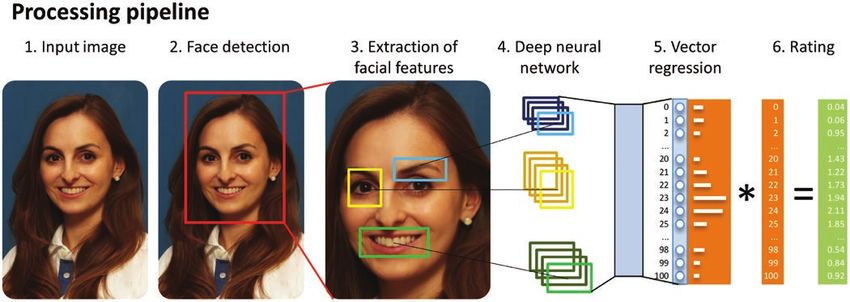
Facial attractiveness was further determined by a computational
Materials and subjects algorithm consisting of a face detector (16) and CNNs trained to
This retrospective analysis was conducted on frontal and left-side pro- extract facial features associated with attractiveness, to provide a
file images retrieved from the archives of the Clinic for Orthodontics prediction of facial attractiveness (9) (Figure 1).R. Patcas et al. 3
Downloaded from https://academic.oup.com/ejo/advance-article-abstract/doi/10.1093/ejo/cjz007/5353220 by ETH Zürich user on 23 July 2019
Figure 1. Processing pipeline of the convolutional neural network (CNN) applied. Note that the person depicted was not part of the assessed cases and is shown
for illustration purposes only (to indicate that the CNN were not solely trained on standard frontal view, this photograph with a slightly tilted head was selected
intentionally).
The Matthias face detector (16) uses a set of learned face tem- by calculating the ratings of each patient separately, for all three
plates at different scales that are evaluated in a sliding window man- rating groups.
ner in the image space and the highest responses are considered face The significance level chosen for all statistical tests was P < 0.05.
detections. The CNN is an artificial neural network inspired by the All analyses were performed in SPSS (version 24.0; IBM Corp.,
visual cortex/vision processing in living organisms. It is defined by Armonk, New York, USA).
a sequence of intermingled layers of neurons, filters, and activation
functions comprising parameters taught to map an input face image
to the corresponding attractiveness class. The learned CNN model Results
extracts facial features by identifying the important regions in the Descriptive statistics are presented in Table 1 for cleft and con-
image, structuring, and combining them. trol patients of each human rating group and the computational
For this task, all face images were brought to an equal size of algorithm (AI) used. Normality tests disclosed that the differences
256 × 256 pixels with a centred face and a 40% background margin between human ratings (all observer groups) and the computational
before being used as input for the CNN models. The CNN mod- algorithms followed normal distribution (all Ps > 0.05) except the
els used VGG-16 architecture (17) and were trained on a dataset differences between AI and laypersons and oral surgeons in the cleft
of a dating site containing >13 000 face images of predominantly subgroup (P = 0.039 and 0.038, respectively).
Caucasians (age range: 18–37 years, mean age: 25 years) with more Overall, control cases were judged more favourably (mean: 6.04–
than 17 million ratings for attractiveness, performed mostly by users 7.15) than clefts (mean: 4.24–4.82) by all three human rater groups
from Switzerland (9). Because images from the dataset were taken in but not by AI. Detailed analysis of the data (Table 2) revealed that
conditions dissimilar to medical assessment, the attractiveness pre- with regard to the evaluation of facial attractiveness for cleft patients
diction network was further fine-tuned, using frontal neutral face no significant differences between human ratings and computational
images taken in a controlled setting, from the Chicago Face Dataset algorithms existed (all Ps ≥ 0.19), with exceptionally strong agree-
(18). The expected attractiveness score was subsequently computed ment between the computational algorithm and both groups of pro-
using the following formula: fessionals raters (i.e. orthodontists and oral surgeons). Conversely,
3 when evaluating the control group, facial attractiveness was consid-
attractiveness = i x probability of class i ered significantly higher across all human raters in comparison to the
i=0 computed results (all Ps ≤ 0.02).
Coefficient of variation as a precision measure on a per case basis
and normalized (scale: 0–10) to enable a direct comparison to VAS
given in Table 1 shows that the variations in human ratings are ra-
scores.
ther large with mean values ranging from 32.56% to 42.19% in cleft
patient images and 23.56% to 29.83% in control patient images.
Statistical analysis The dispersion was thus particularly large in the assessment of cleft
Descriptive statistics were calculated and presented for cleft and cases, demonstrating the absence of unity in all human rater groups.
control patients by different human rating groups and by com-
putational algorithms separately. Data distribution was tested for
normality, and, based on the obtained results, parametric t-tests or Discussion
non-parametric Wilcoxon signed-rank tests were performed accord- This research contribution aimed to tackle the existing obstacles in
ingly, to evaluate whether the difference between human VAS ratings evaluating facial appearance as part of treatment outcome assess-
and computational algorithms varied significantly between the three ment in cleft patients. To this end, facial images of treated cleft
observer groups in cleft patients and control patients separately. patients and controls were fed to a face detector and a previously
To determine the variation in each human rating group, coef- trained CNN to appraise facial attractiveness. The underlying
ficients of variation (i.e. ratio of standard deviation to the mean) hypothesis was that no differences would be observable between the
were also estimated as a measure to describe inter-rater variability, obtained results and previously published panel-based ratings (3).4 European Journal of Orthodontics, 2019
Table 1. Descriptive data for visual analogue scale scores of facial attractiveness of patients treated for clefts and for control patients as
assessed by different rating groups and by using computational algorithms (artificial intelligence [AI])
Layperson (n = 15) Orthodontist (n = 14) Oral surgeon (n = 10) AI#
Cleft (n = 40)
Mean (SD) 4.24 (0.81) 4.82 (0.94) 4.74 (0.83) 4.75 (1.27)
Median (p25, p75) 4.29 (3.73, 4.77) 4.68 (4.29, 5.46) 4.83 (4.21, 5.39) 4.45 (3.81, 5.66)
Minimum 2.20 2.61 2.80 2.74
Maximum 5.90 6.93 6.50 8.44
Downloaded from https://academic.oup.com/ejo/advance-article-abstract/doi/10.1093/ejo/cjz007/5353220 by ETH Zürich user on 23 July 2019
Imprecision, given as coefficient of variance 38.73% (9.64%) 32.56% (8.21%) 42.19% (9.80%) 0% (0%)
(COV) per case (%): mean COV (SD COV)
Control (n = 20)
Mean (SD) 6.04 (0.53) 7.15 (0.62) 6.49 (0.54) 4.16 (1.04)
Median (p25, p75) 6.05 (5.78, 6.40) 7.20 (6.73, 7.74) 6.35 (6.08, 6.87) 3.96 (3.54, 5.00)
Minimum 4.77 6.14 5.35 2.30
Maximum 7.03 7.96 7.56 5.98
Imprecision, given as: COV per case (%): 27.18% (6.56%) 23.56% (7.14%) 29.83% (6.29%) 0% (0%)
mean COV (SD COV)
p25 = 25th percentile; p75 = 75th percentile.
#
Computational algorithm comprising a face detector and convolutional neural networks.
Table 2. Visual analogue scale scores for facial attractiveness of patients treated for clefts and for control patients: differences between
different rating groups to values obtained by trained computational algorithms
Layperson (n = 15) Orthodontist (n = 14) Oral surgeon (n = 10)
Computer
Difference (cleft)
Mean (SD) –0.51 (2.15) 0.07 (2.13) –0.01 (2.45)
Median (p25, p75) –0.49 (–2.00, 0.92) 0.19 (–1.32, 1.38) 0.19 (–1.67, 1.86)
Minimum –8.44 –7.44 –7.94
Maximum 5.79 5.79 5.69
P value .186 .783 .988
Difference (control)
Mean (SD) 1.88 (1.94) 2.97 (1.90) 2.32 (2.17)
Median (p25, p75) 1.84 (0.52, 3.27) 3.13 (1.67, 4.27) 2.26 (0.86, 3.89)
Minimum –3.86 –3.09 –2.52
Maximum 7.20 7.30 7.70
P valueR. Patcas et al. 5
variability, when evaluating the coefficient of variation for each case. represent to some degree ‘social attractiveness’, i.e. the quality to
The panel-based mean values of coefficient of variation, ranging cause ‘social’ interest and desire. On the basis of millions of ratings
from 32.56% to 42.19% in cleft patient images, are considerably retrieved from a dating site, validated and fine-tuned on medical
high and accordingly inherently problematic, as they demonstrate a images, the proposed CNN is unquestionably a fitting tool to mirror
blatant absence of unity within the rater groups. One of the obvious social opinion of treated patients. And perhaps this is what should
benefits of a single AI-based score would therefore doubtlessly be be considered most important for patients as treatment outcomes
the elimination of the evident variability and subjectivity compro- should not be measured by specific panels but by how society views
mising panel-based ratings. Thus, the fact that AI-based results were the aesthetic results.
Downloaded from https://academic.oup.com/ejo/advance-article-abstract/doi/10.1093/ejo/cjz007/5353220 by ETH Zürich user on 23 July 2019
comparable to the average scores in clefts of all three rating groups In this investigation, the applied scoring model rated the facial
(with especially strong agreement to both professional panels) seems attractiveness of the controls similarly to the facial attractiveness
to indicate that AI could replace panel ratings while circumventing of cleft patients. As already mentioned earlier, this finding is very
dispersion-related issues. likely to be a result of the CNN model used and thus warrants
Moreover, panel-based scores are usually unavailable for in- further refinement of the computational algorithm applied. The
dividual case planning. As such, the availability of a dependable CNN tested was apparently unable to detect facial features that
AI-scoring could prove to be a welcome diagnostic tool, enabling render cleft patients less attractive (in the eyes of human raters).
clinicians to analyse objectively the outcome of surgical procedures Great effort was made to train the CNN on more than 13 000
and assisting them in discerning favourable aesthetic results. images of different individuals of a population containing sub-
Analysis of medical images using computers is not entirely of re- jects with clefts, yet the CNN remained clearly unfit to detect and
cent vintage, and early attempts of computerized analysis and diag- estimate the impact of cleft features to the same degree as humans
nostics of medical images were made already in the 1960s (21–23). do. Fine-tuning the algorithm on labelled cleft cases would have
In radiology, data analysed quantitatively by computer as aid to surely help to overcome this shortcoming, but such collections are
make the diagnostic decisions have been common practice already unfortunately unavailable. Because the intention of this pilot study
for some time. Yet in stark contrast to historical advances, the latest was to provide a proof of concept for a novel computational rat-
introduction of trained CNNs producing an automated computer ing system, this must, at this stage, be partially rejected. Although
diagnosis aims not to assist, but rather to replace the radiologist. the potential benefits became evident through direct comparison
Apart from the ethical and moral concerns involved, this approach to over 2000 human ratings, the results clearly underline that
would require a very high-performance level of the computer output, any clinical recommendation of the algorithm tested would be
equal to or better than the performance achieved by radiologists. premature.
Regarding AI-based diagnostics in the field of maxillofacial radi-
ology, scientific literature is scarce and embryonic. Primitive neural
networks were either applied to compare automatic cephalometric
Conclusions
diagnosis for orthodontic treatment to the results obtained from This study introduces a novel method in dental medicine to rate facial
three orthodontic specialists (24) or to evaluate automatic detection attractiveness, by a face detector and a dedicated CNN. Although
of vertical root fractures with intraoral and cone beam computed the introduced method based on AI has the potential to overcome
tomography images using a sample of artificially fractured teeth (25). the shortcomings related to variance and inconsistency prevalent
Although these previous reports attested a high degree of accuracy in panel assessments, this study made it evident that the presented
of AI-based diagnosis, they all rely on rudimentary neural networks AI-based scoring is in need of further perfection and refinement to
operating on task-specific algorithms. This study is the first attempt differentiate cleft features of the face that negatively influence the
to introduce CNNs based on deep learning and constitutes an im- human perception of attractiveness.
portant improvement that has produced—in other fields—results
comparable to and in some cases superior to human experts (26–28). Acknowledgement
The authors are grateful to Ms Kar Yan Li, Centralised Research Laboratories,
Limitations Faculty of Dentistry, The University of Hong Kong, for her valuable assistance
All three human rater groups differed in size. Initially, 20 individuals regarding the statistical analysis.
were contacted for every group to participate, but the response rate
was unequal. Although this in itself did not affect the planned sta- Conflict of Interest
tistical analyses, it limited the amount of data available for analysis.
The authors declare that they have no conflict of interest.
On the basis of the assumption that males have a different aestheti-
cal perception than females, the overrepresentation of males in this
study could potentially have affected the results. References
Caution should be exercised when comparing the computed 1. Mossey, P.A., Little, J., Munger, R.G., Dixon, M.J. and Shaw, W.C. (2009)
results to the different panels assessing facial attractiveness, as com- Cleft lip and palate. Lancet (London, England), 374, 1773–1785.
paring panel ratings to computer-generated results cannot validate 2. Pruzinsky, T. (1992) Social and psychological effects of major craniofacial
the latter. Attractiveness is usually defined as the quality to cause deformity. Cleft Palate-Craniofacial Journal, 29, 578–84; discussion 570.
3. Eichenberger, M., Staudt, C.B., Pandis, N., Gnoinski, W. and Eliades, T.
interest and desire in the observer, and as such, subjectivity is an
(2014) Facial attractiveness of patients with unilateral cleft lip and palate
inherent part of the definition. As every panel is but a representa-
and of controls assessed by laypersons and professionals. European Jour-
tion of its observer, may they be professionals or laypeople, it would
nal of Orthodontics, 36, 284–289.
be unsound to validate one panel through the results of another. 4. Sinko, K., Jagsch, R., Prechtl, V., Watzinger, F., Hollmann, K. and Bau-
Likewise, measurements based on AI are a representation of a par- mann, A. (2005) Evaluation of esthetic, functional, and quality-of-life
ticular opinion that cannot be validated through comparison. The outcome in adult cleft lip and palate patients. Cleft Palate-Craniofacial
CNN-based results thus do not replace other panels but rather Journal, 42, 355–361.6 European Journal of Orthodontics, 2019
5. Gkantidis, N., Papamanou, D.A., Christou, P. and Topouzelis, N. (2013) 17. Simonyan, K. and Zisserman, A. (2014) Very deep convolutional networks
Aesthetic outcome of cleft lip and palate treatment. Perceptions of patients, for large-scale image recognition. arXiv preprint arXiv:1409.1556.
families, and health professionals compared to the general public. Journal 18. Ma, D.S., Correll, J. and Wittenbrink, B. (2015) The Chicago face data-
of Cranio-Maxillo-Facial Surgery, 41, e105–e110. base: a free stimulus set of faces and norming data. Behavior Research
6. Sharma, V.P., Bella, H., Cadier, M.M., Pigott, R.W., Goodacre, T.E. and Methods, 47, 1122–1135.
Richard, B.M. (2012) Outcomes in facial aesthetics in cleft lip and palate 19. Meyer-Marcotty, P., Gerdes, A.B., Reuther, T., Stellzig-Eisenhauer, A. and
surgery: a systematic review. Journal of Plastic, Reconstructive and Aes- Alpers, G.W. (2010) Persons with cleft lip and palate are looked at differ-
thetic Surgery, 65, 1233–1245. ently. Journal of Dental Research, 89, 400–404.
7. Matsugu, M., Mori, K., Mitari, Y. and Kaneda, Y. (2003) Subject inde- 20. Agustsson, E., Timofte, R., Escalera, S., Baro, X., Guyon, I. and Rothe, R.
Downloaded from https://academic.oup.com/ejo/advance-article-abstract/doi/10.1093/ejo/cjz007/5353220 by ETH Zürich user on 23 July 2019
pendent facial expression recognition with robust face detection using a Apparent and real age estimation in still images with deep residual regres-
convolutional neural network. Neural Networks, 16, 555–559. sors on APPA-REAL database. In Automatic Face & Gesture Recognition
8. Rothe, R., Timofte, R. and Van Gool, L. (2015) DEX: Deep EXpectation (FG 2017), 12th IEEE International Conference. 2017. IEEE.
of Apparent Age from a Single Image. 2015 IEEE International Confer- 21. Lodwick, G.S., Haun, C.L., Smith, W.E., Keller, R.F. and Robertson, E.D.
ence on Computer Vision Workshop (ICCVW), pp. 252–257. (1963) Computer diagnosis of primary bone tumors. Radiology, 80, 273–
9. Rothe, R., Timofte, R. and Van Gool, L. (2016) Some like it hot—visual 275.
guidance for preference prediction. 2016 IEEE Conference on Computer 22. Meyers, P.H., Nice, C.M. Jr, Becker, H.C., Nettleton, W.J. Jr, Sweeney, J.W.
Vision and Pattern Recognition (CVPR), pp. 5553–5561. and Meckstroth, G.R. (1964) Automated computer analysis of radio-
10. Gray, D., Yu, K., Xu, W. and Gong, Y. (2010) Predicting Facial Beauty graphic images. Radiology, 83, 1029–1034.
without Landmarks. Springer: Berlin, Heidelberg. 23. Winsberg, F., Elkin, M., Josiah Macy, J., Bordaz, V. and Weymouth, W.
11. Esteva, A., Kuprel, B., Novoa, R.A., Ko, J., Swetter, S.M., Blau, H.M. and (1967) Detection of radiographic abnormalities in mammograms by
Thrun, S. (2017) Dermatologist-level classification of skin cancer with means of optical scanning and computer analysis. Radiology, 89, 211–
deep neural networks. Nature, 542, 115–118. 215.
12. Xu, M., Papageorgiou, D.P., Abidi, S.Z., Dao, M., Zhao, H. and Karni- 24. Mario, M.C., Abe, J.M., Ortega, N.R. and Del Santo, M. Jr. (2010) Para-
adakis, G.E. (2017) A deep convolutional neural network for classification consistent artificial neural network as auxiliary in cephalometric diagno-
of red blood cells in sickle cell anemia. PLoS Computational Biology, 13, sis. Artificial Organs, 34, E215–E221.
e1005746. 25. Johari, M., Esmaeili, F., Andalib, A., Garjani, S. and Saberkari, H. (2017)
13. Wu, M., Yan, C., Liu, H. and Liu, Q. (2018) Automatic classification of Detection of vertical root fractures in intact and endodontically treated
ovarian cancer types from cytological images using deep convolutional premolar teeth by designing a probabilistic neural network: an ex vivo
neural networks. Bioscience Reports, 38, 3. study. Dento Maxillo Facial Radiology, 46, 20160107.
14. Sharma, H., Zerbe, N., Klempert, I., Hellwich, O. and Hufnagl, P. (2017) 26. Ciresan, D., Meier, U., Schmidhuber, J. (2012) Multi-column deep neural
Deep convolutional neural networks for automatic classification of gastric networks for image classification. 2012 IEEE Conference on Computer
carcinoma using whole slide images in digital histopathology. Computer- Vision and Pattern Recognition, pp. 3642–3649.
ized Medical Imaging and Graphics, 61, 2–13. 27. Krizhevsky, A., Sutskever, I., Hinton, G. (2012) ImageNet classification
15. World Medical Association. (2013) World Medical Association Declara- with Deep Convolutional Neural Networks. NIPS’12 Proceedings of the
tion of Helsinki: Ethical Principles for Medical Research Involving Human 25th International Conference on Neural Information Processing Systems,
Subjects. JAMA, 310, 2191–2194. 1, pp. 1097–1105.
16. Mathias, M., Benenson, R., Pedersoli, M. and Van Gool, L. (2014) Face detec- 28. Marblestone, A.H., Wayne, G. and Kording, K.P. (2016) Toward an inte-
tion without bells and whistles. European Conference on Computer Vision gration of deep learning and neuroscience. Frontiers in Computational
8692, 720–735. Neuroscience, 10, 94.You can also read