Acute Kidney Injury: A Guide to Diagnosis and Management
←
→
Page content transcription
If your browser does not render page correctly, please read the page content below
Acute Kidney Injury: A Guide to Diagnosis
and Management
MAHBOOB RAHMAN, MD, MS, Case Western Reserve University School of Medicine, Cleveland, Ohio
FARIHA SHAD, MD, Kaiser Permanente, Cleveland, Ohio
MICHAEL C. SMITH, MD, Case Western Reserve University School of Medicine, Cleveland, Ohio
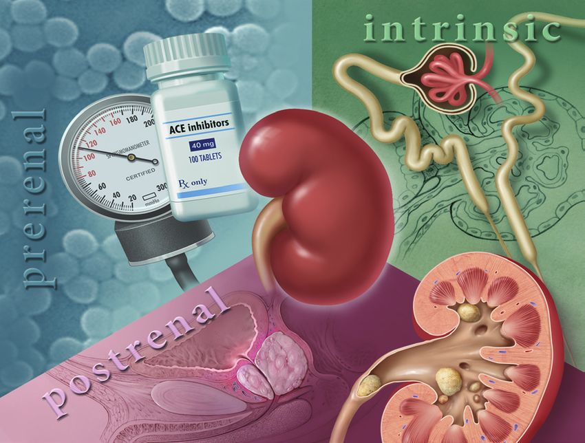
Acute kidney injury is characterized by abrupt deterioration in kidney function, manifested by an increase in serum
creatinine level with or without reduced urine output. The spectrum of injury ranges from mild to advanced, some-
times requiring renal replacement therapy. The diagnostic evaluation can be used to classify acute kidney injury as
prerenal, intrinsic renal, or postrenal. The initial workup includes a patient history to identify the use of nephrotoxic
medications or systemic illnesses that might cause poor renal perfusion or directly impair renal function. Physi-
cal examination should assess intravascular volume status and identify skin rashes indicative of systemic illness.
The initial laboratory evaluation should include measurement of serum creatinine level, complete blood count, uri-
nalysis, and fractional excretion of sodium. Ultrasonography of the
kidneys should be performed in most patients, particularly in older
men, to rule out obstruction. Management of acute kidney injury
involves fluid resuscitation, avoidance of nephrotoxic medications
and contrast media exposure, and correction of electrolyte imbal-
ances. Renal replacement therapy (dialysis) is indicated for refrac-
tory hyperkalemia; volume overload; intractable acidosis; uremic
encephalopathy, pericarditis, or pleuritis; and removal of certain
toxins. Recognition of risk factors (e.g., older age, sepsis, hypovo-
ILLUSTRATION BY CHRISTY KRAMES
lemia/shock, cardiac surgery, infusion of contrast agents, diabetes
mellitus, preexisting chronic kidney disease, cardiac failure, liver
failure) is important. Team-based approaches for prevention, early
diagnosis, and aggressive management are critical for improving
outcomes. (Am Fam Physician. 2012;86(7):631-639. Copyright ©
2012 American Academy of Family Physicians.)
T
he incidence of acute kidney injury Definition
has increased in recent years, both Acute kidney injury is defined as an abrupt
in the community and in hospi- (within 48 hours) reduction in kidney func-
tal settings.1,2 The estimated inci- tion based on an elevation in serum creati-
dence of acute kidney injury is two to three nine level, a reduction in urine output, the
cases per 1,000 persons.3 Seven percent of need for renal replacement therapy (dialy-
hospitalized patients and about two-thirds sis), or a combination of these factors. It is
of patients in intensive care units develop classified in three stages (Table 1).8 The term
acute kidney injury,2 often as part of the acute kidney injury should replace terms
multiple organ dysfunction syndrome.4 such as acute renal failure and acute renal
Acute kidney injury is associated with insufficiency, which previously have been
a high rate of adverse outcomes; mortal- used to describe the same clinical condition.
ity rates range between 25 and 80 percent,
depending on the cause and the clinical sta- Etiology
tus of the patient.5-7 These data highlight the The causes of acute kidney injury can be
importance of recognition and appropriate divided into three categories (Table 2 9):
management, usually in collaboration with prerenal (caused by decreased renal perfu-
nephrologists and other subspecialists. sion, often because of volume depletion),
Downloaded from the American Family Physician Web site at www.aafp.org/afp. Copyright © 2012 American Academy of Family Physicians. For the private, noncommercial
October 1,use
2012
of one Volume
◆
individual86,
userNumber
of the Web7 site. All other rights reserved.
www.aafp.org/afp
Contact copyrights@aafp.org American
for copyright questions and/or permission Physician 631
Familyrequests.Acute Kidney Injury
SORT: KEY RECOMMENDATIONS FOR PRACTICE
Evidence
Clinical recommendation rating References
The diagnosis of acute kidney injury is based on serum creatinine levels, urine output, and C 8
the need for renal replacement therapy.
Renal ultrasonography should be performed in most patients with acute kidney injury to C 17
rule out obstruction.
Adequate fluid balance should be maintained in patients with acute kidney injury by using C 19
isotonic solutions (e.g., normal saline) instead of hyperoncotic solutions (e.g., dextrans,
hydroxyethyl starch, albumin).
Dopamine use is not recommended for the prevention of acute kidney injury. A 21
Diuretics do not improve morbidity, mortality, or renal outcomes, and should not be used A 22
to prevent or treat acute kidney injury in the absence of volume overload.
Consider therapy with immunosuppressive agents (e.g., cyclophosphamide, prednisone) in C 23
patients with rapidly progressive glomerulonephritis.
A = consistent, good-quality patient-oriented evidence; B = inconsistent or limited-quality patient-oriented evidence; C = consensus, disease-
oriented evidence, usual practice, expert opinion, or case series. For information about the SORT evidence rating system, go to http://www.
aafp.org/afpsort.xml.
Table 1. Stages of Acute Kidney Injury
Stage Change in serum creatinine level Urine output Other
1 Increase ≥ 0.3 mg per dL (26.52 µmol per L) or < 0.5 mL per kg per hour for more —
≥ 1.5- to twofold from baseline than six hours
2 Increase > two- to threefold from baseline < 0.5 mL per kg per hour for more —
than 12 hours
3 Increase > threefold from baseline or ≥ 4.0 mg < 0.3 mL per kg per hour for 24 hours Renal replacement
per dL (353.60 µmol per L) with an acute rise or anuria for 12 hours therapy required
of at least 0.5 mg per dL (44.20 µmol per L)
NOTE: Each stage is defined by the change in serum creatinine level, the change in urine output, or the need for renal replacement therapy.
Adapted with permission from Mehta RL, Kellum JA, Shah SV, et al. Acute Kidney Injury Network: report of an initiative to improve outcomes in acute
kidney injury. Crit Care. 2007;11(2):R31.
intrinsic renal (caused by a process within the kidneys), In patients with preexisting chronic kidney disease,
and postrenal (caused by inadequate drainage of urine however, these mechanisms are impaired, and the sus-
distal to the kidneys). In patients who already have ceptibility to develop acute-on-chronic renal failure is
underlying chronic kidney disease, any of these factors, higher.11
but especially volume depletion, may cause acute kidney Several medications can cause prerenal acute kidney
injury in addition to the chronic impairment of renal injury. Notably, angiotensin-converting enzyme inhibi-
function. tors and angiotensin receptor blockers can impair renal
perfusion by causing dilation of the efferent arteriole
PRERENAL CAUSES and reduce intraglomerular pressure. Nonsteroidal anti-
Approximately 70 percent of community-acquired cases inflammatory drugs also can decrease the glomerular
of acute kidney injury are attributed to prerenal causes.10 filtration rate by changing the balance of vasodilatory/
In these cases, underlying kidney function may be nor- vasoconstrictive agents in the renal microcirculation.
mal, but decreased renal perfusion associated with These drugs and others limit the normal homeostatic
intravascular volume depletion (e.g., from vomiting or responses to volume depletion and can be associated
diarrhea) or decreased arterial pressure (e.g., from heart with a decline in renal function. In patients with pre-
failure or sepsis) results in a reduced glomerular filtra- renal acute kidney injury, kidney function typically
tion rate. Autoregulatory mechanisms often can com- returns to baseline after adequate volume status is estab-
pensate for some degree of reduced renal perfusion in lished, the underlying cause is treated, or the offending
an attempt to maintain the glomerular filtration rate. drug is discontinued.
632 American Family Physician www.aafp.org/afp Volume 86, Number 7 ◆ October 1, 2012Acute Kidney Injury
Table 2. Causes of Acute Kidney Injury
Prerenal
Intrarenal vasoconstriction (hemodynamically mediated)
INTRINSIC RENAL CAUSES
Medications: nonsteroidal anti-inflammatory drugs,*
Intrinsic renal causes are also important sources of acute angiotensin-converting enzyme inhibitors,* angiotensin
kidney injury and can be categorized by the component receptor blockers,* cyclosporine (Sandimmune),
of the kidney that is primarily affected (i.e., tubular, glo- tacrolimus (Prograf)
merular, interstitial, or vascular). Cardiorenal syndrome*
Acute tubular necrosis is the most common type of Hepatorenal syndrome
intrinsic acute kidney injury in hospitalized patients. Abdominal compartment syndrome
The cause is usually ischemic (from prolonged hypoten- Hypercalcemia
sion) or nephrotoxic (from an agent that is toxic to the Systemic vasodilation (e.g., sepsis,* neurogenic shock)
tubular cells). In contrast to a prerenal etiology, acute Volume depletion
kidney injury caused by acute tubular necrosis does not Renal loss from diuretic overuse,* osmotic diuresis
improve with adequate repletion of intravascular vol- (e.g., diabetic ketoacidosis*)
ume and blood flow to the kidneys. Both ischemic and Extrarenal loss from vomiting, diarrhea,* burns, sweating,
nephrotoxic acute tubular necrosis can resolve over time, blood loss
although temporary renal replacement therapy may be Intrinsic renal
required, depending on the degree of renal injury and Glomerular (e.g., postinfectious and other
the presence of preexisting chronic kidney disease. glomerulonephritis)
Glomerular causes of acute kidney injury are the Interstitial
result of acute inflammation of blood vessels and glom- Medications: penicillin analogues,* cephalosporins,*
eruli. Glomerulonephritis is usually a manifestation sulfonamides, ciprofloxacin (Cipro), acyclovir (Zovirax),
of a systemic illness (e.g., systemic lupus erythemato- rifampin, phenytoin (Dilantin), interferon, proton pump
inhibitors, nonsteroidal anti-inflammatory drugs
sus) or pulmonary renal syndromes (e.g., Goodpasture
syndrome, Wegener granulomatosis). History, physical Infections (e.g., direct infection of renal parenchyma or
associated with systemic infections)
examination, and urinalysis are crucial for diagnosing
Viruses: Epstein-Barr virus, cytomegalovirus, human
glomerulonephritis (Table 3 9 and Figure 112). Because immunodeficiency virus
management often involves administration of immu-
Bacteria: Streptococcus species, Legionella species
nosuppressive or cytotoxic medications with potentially
Fungi: candidiasis, histoplasmosis
severe adverse effects, renal biopsy is often required to
Systemic disease: sarcoidosis, lupus
confirm the diagnosis before initiating therapy.
Tubular
Acute interstitial nephritis can be secondary to many
Ischemic: prolonged hypotension*
conditions, but most cases are related to medication
use, making patient history the key to diagnosis. In Nephrotoxic: exogenous toxins (e.g., radiographic contrast
agents,* aminoglycosides,* cisplatin, methotrexate,
about one-third of cases, there is a history of macu- ethylene glycol, amphotericin B) and endogenous
lopapular erythematous rash, fever, arthralgias, or a toxins (e.g., hemolysis and rhabdomyolysis [pigment
combination of these symptoms.13 Eosinophiluria may nephropathy], tumor lysis syndrome, myeloma)
be found in patients with acute interstitial nephritis, Vascular
but it is not pathognomonic of this disease. A kidney Renal vein thrombosis, malignant hypertension,
biopsy may be needed to distinguish between allergic scleroderma renal crisis, renal atheroembolic disease,*
interstitial nephritis and other renal causes of acute and renal infarction
kidney injury. In addition to discontinuing offending Postrenal
agents, steroids may be beneficial if given early in the Extrarenal obstruction: prostate hypertrophy*; neurogenic
course of disease.14 bladder; retroperitoneal fibrosis; bladder, prostate, or
Acute events involving renal arteries or veins can also cervical cancer
lead to intrinsic acute kidney injury. Renal atheroem- Intrarenal obstruction: stones,* crystals (acyclovir, indinavir
bolic disease is the most common cause and is suspected [Crixivan]), clots, tumors
with a recent history of arterial catheterization, the pres-
*—Most common causes.
ence of a condition requiring anticoagulation, or after
Adapted with permission from Holley JL. Clinical approach to the
vascular surgery. Physical examination and history pro- diagnosis of acute renal failure. In: Greenberg A, Cheung AK, eds.
vide important clues to the diagnosis (Table 3 9). Vascular Primer on Kidney Diseases. 5th ed. Philadelphia, Pa.: National Kidney
causes of acute kidney injury usually require imaging to Foundation; 2009:278.
confirm the diagnosis.
October 1, 2012 ◆ Volume 86, Number 7 www.aafp.org/afp American Family Physician 633Acute Kidney Injury
POSTRENAL CAUSES Diagnosis
Postrenal causes typically result from obstruction of uri- A patient history and physical examination, with an
nary flow, and prostatic hypertrophy is the most com- emphasis on assessing the patient’s volume status, are
mon cause of obstruction in older men. Prompt diagnosis crucial for determining the cause of acute kidney injury
followed by early relief of obstruction is associated with (Table 39). The history should identify use of nephrotoxic
improvement in renal function in most patients. medications or systemic illnesses that might cause poor
renal perfusion or directly impair renal function. Physi-
Clinical Presentation cal examination should assess intravascular volume status
Clinical presentation varies with the cause and sever- and any skin rashes indicative of systemic illness. The ini-
ity of renal injury, and associated diseases. Most patients tial laboratory evaluation should include urinalysis, com-
with mild to moderate acute kidney injury are asymp- plete blood count, and measurement of serum creatinine
tomatic and are identified on laboratory testing. Patients level and fractional excretion of sodium (FENa). Imaging
with severe cases, however, may be symptomatic and pres- studies can help rule out obstruction. Useful tests are
ent with listlessness, confusion, fatigue, anorexia, nausea, summarized in Table 4.16 Figure 1 presents an overview of
vomiting, weight gain, or edema.15 Patients can also pres- the diagnosis and management of acute kidney injury.12
ent with oliguria (urine output less than 400 mL per day),
SERUM CREATININE LEVEL
anuria (urine output less than 100 mL per day), or normal
volumes of urine (nonoliguric acute kidney injury). Other It is important to compare the patient’s current serum cre-
presentations of acute kidney injury may include develop- atinine level with previous levels to determine the duration
ment of uremic encephalopathy (manifested by a decline and acuity of the disease. The definition of acute kidney
in mental status, asterixis, or other neurologic symptoms), injury indicates that a rise in creatinine has occurred within
anemia, or bleeding caused by uremic platelet dysfunction. 48 hours, although in the outpatient setting, it may be hard
Table 3. History and Physical Examination Findings for Categorizing Acute Kidney Injury
Type of acute
kidney injury History findings Physical examination findings
Prerenal Volume loss (e.g., history of vomiting, diarrhea, diuretic overuse, Weight loss, orthostatic hypotension
hemorrhage, burns) and tachycardia
Thirst and reduced fluid intake Poor skin turgor
Cardiac disease Dilated neck veins, S3 heart sound,
pulmonary rales, peripheral edema
Liver disease Ascites, caput medusae, spider angiomas
Intrinsic renal
Acute tubular History of receiving nephrotoxic medications (including over-the- Muscle tenderness, compartment
necrosis counter, illicit, and herbal), hypotension, trauma or myalgias syndrome, assessment of volume
suggesting rhabdomyolysis, recent exposure to radiographic status
contrast agents
Glomerular Lupus, systemic sclerosis, rash, arthritis, uveitis, weight loss, fatigue, Periorbital, sacral, and lower-extremity
hepatitis C virus infection, human immunodeficiency virus edema; rash; oral/nasal ulcers
infection, hematuria, foamy urine, cough, sinusitis, hemoptysis
Interstitial Medication use (e.g., antibiotics, proton pump inhibitors), rash, Fever, drug-related rash
arthralgias, fever, infectious illness
Vascular Nephrotic syndrome, trauma, flank pain, anticoagulation Livedo reticularis, funduscopic
(atheroembolic disease), vessel catheterization or vascular surgery examination (showing malignant
hypertension), abdominal bruits
Postrenal Urinary urgency or hesitancy, gross hematuria, polyuria, stones, Bladder distention, pelvic mass, prostate
medications, cancer enlargement
Adapted with permission from Holley JL. Clinical approach to the diagnosis of acute renal failure. In: Greenberg A, Cheung AK, eds. Primer on Kidney
Diseases. 5th ed. Philadelphia, Pa.: National Kidney Foundation; 2009:280.
634 American Family Physician www.aafp.org/afp Volume 86, Number 7 ◆ October 1, 2012Acute Kidney Injury
Diagnosis and Treatment of Acute Kidney Injury
Patient presents with acute kidney injury
Progressive increase in blood urea
nitrogen and creatinine levels
Take history and perform physical examination
Obtain serial measurement of blood urea
nitrogen, creatinine, and electrolyte levels
Patient has oliguria?
Yes No
Measure urinary and serum sodium
and creatinine levels and osmolality
Prerenal disease Perform renal ultrasonography
Volume Congestive Normal-size kidneys Hydronephrosis Bilateral small kidneys
depletion heart failure
Renal parenchymal disease Relieve obstruction Chronic renal failure
Administer Administer diuretics
saline and perform
afterload reduction Perform urinalysis
Renal tubular cells, renal tubular Eosinophils, white Red blood cell casts; Orthotolidine positive
cell casts, or pigmented casts blood cell casts proteinuria > 3 g but no red blood cells
Acute tubular necrosis Allergic interstitial nephritis Myoglobinuria or
hemoglobinuria
Glomerulonephritis Multiple myeloma
or vasculitis
Eliminate nephrotoxins; Eliminate offending drug,
treat underlying cause prescribe glucocorticoids Administer fluids,
Serum and urine mannitol; perform
Perform renal biopsy immunoelectrophoresis urine alkalinization
Supportive management
Resolution of Unresponsive volume overload, acidosis, or hyperkalemia
renal insufficiency Signs and symptoms of uremia (blood urea nitrogen
> 100 mg per dL [35.70 mmol per L])
Perform renal replacement therapy (dialysis)
Figure 1. Algorithm for the diagnosis and treatment of acute kidney injury.
Adapted with permission from Smith MC. Acute renal failure. In: Resnick MI, Elder JS, Spirnak JP, eds. Clinical Decisions in Urology. 3rd ed. Hamilton,
Ontario, Canada: BC Decker, Inc.; 2004:61.Acute Kidney Injury
Table 4. Diagnostic Test Results and Corresponding Diseases in Patients with Acute Kidney Injury
Test result When to order Associated diseases/conditions
Elevated antineutrophil cytoplasmic Suspected acute glomerulonephritis, Vasculitis, Goodpasture syndrome
antibody, antiglomerular basement pulmonary renal syndromes
membrane antibody
Elevated antistreptolysin O titer Recent infection and clinical picture Poststreptococcal glomerulonephritis
of acute glomerulonephritis
Elevated creatine kinase level, elevated Recent trauma, muscle injury Rhabdomyolysis
myoglobin level, dipstick positive for
blood but negative for red blood cells
Elevated prostate-specific antigen level Older men with symptoms Prostate hypertrophy, prostate cancer
suggestive of urinary obstruction
Elevated uric acid level History of rapidly proliferating Malignancy, tumor lysis syndrome
tumors, recent chemotherapy
Eosinophiluria Fever, rash Allergic interstitial nephritis
Evidence of hemolysis (schistocytes on Fever, anemia, thrombocytopenia, Hemolytic uremic syndrome, thrombotic
peripheral smear, decreased haptoglobin neurologic signs thrombocytopenic purpura, systemic lupus
level, elevated indirect bilirubin level, erythematosus, other autoimmune diseases
elevated lactate dehydrogenase level)
Hydronephrosis on renal ultrasonography Suspected obstruction Malignancy, prostate hypertrophy, uterine
fibroids, nephrolithiasis, ureterolithiasis
Increased anion gap with increased Suspected poisoning, unresponsive Ethylene glycol or methanol poisoning
osmolar gap* patient
Low complement level Suspected acute glomerulonephritis Systemic lupus erythematosus, endocarditis,
postinfectious glomerulonephritis
Monoclonal spike on serum protein Anemia, proteinuria, acute kidney Multiple myeloma
electrophoresis injury in older patients
Positive antinuclear antibody, double- Proteinuria, skin rash, arthritis Autoimmune diseases, systemic lupus
stranded DNA antibody erythematosus
Positive blood cultures Intravenous drug use, recent Endocarditis
infection, new cardiac murmur
Positive HIV test Risk factors for HIV infection HIV nephropathy
HIV = human immunodeficiency virus.
*—Calculations are as follows:
Anion gap = sodium – (chloride + bicarbonate)
Calculated serum osmolality = 2(sodium [in mEq per L]) + (blood urea nitrogen [in mg per dL] ÷ 2.8) + (glucose [in mg per dL] ÷ 18)
Osmolar gap = measured serum osmolality – calculated serum osmolality.
Adapted with permission from Agrawal M, Swartz R. Acute renal failure [published correction appears in Am Fam Physician. 2001;63(3):445].
Am Fam Physician. 2000;61(7):2081.
to ascertain when the rise actually happened. A high serum urinalysis guide the differential diagnosis and direct fur-
creatinine level in a patient with a previously normal docu- ther workup (Figure 112).
mented level suggests an acute process, whereas a rise over
COMPLETE BLOOD COUNT
weeks to months represents a subacute or chronic process.
The presence of acute hemolytic anemia with the periph-
URINALYSIS eral smear showing schistocytes in the setting of acute kid-
Urinalysis is the most important noninvasive test in ney injury should raise the possibility of hemolytic uremic
the initial workup of acute kidney injury. Findings on syndrome or thrombotic thrombocytopenic purpura.
636 American Family Physician www.aafp.org/afp Volume 86, Number 7 ◆ October 1, 2012Acute Kidney Injury
URINE ELECTROLYTES Management
In patients with oliguria, measurement of FENa is help- Optimal management of acute kidney injury requires
ful in distinguishing prerenal from intrinsic renal causes close collaboration among primary care physicians,
of acute kidney injury. FENa is defined by the following nephrologists, hospitalists, and other subspecialists par-
formula: ticipating in the care of the patient. After acute kidney
injury is established, management is primarily supportive.
(urinary sodium × serum creatinine)
FENa = 100 × Patients with acute kidney injury generally should
(serum sodium × urinary creatinine)
be hospitalized unless the condition is mild and clearly
Online calculators are also available. A value less than resulting from an easily reversible cause. The key to
1 percent indicates a prerenal cause of acute kidney management is assuring adequate renal perfusion by
injury, whereas a value greater than 2 percent indicates achieving and maintaining hemodynamic stability and
an intrinsic renal cause. In patients on diuretic therapy, avoiding hypovolemia. In some patients, clinical assess-
however, a FENa higher than 1 percent may be caused ment of intravascular volume status and avoidance of
by natriuresis induced by the diuretic, and is a less reli- volume overload may be difficult, in which case mea-
able measure of a prerenal state. In such cases, fractional surement of central venous pressures in an intensive care
excretion of urea may be helpful, with values less than setting may be helpful.
35 percent indicating a prerenal cause. FENa values less If fluid resuscitation is required because of intravas-
than 1 percent are not specific for prerenal causes of acute cular volume depletion, isotonic solutions (e.g., normal
kidney injury because these values can occur in other con- saline) are preferred over hyperoncotic solutions (e.g.,
ditions, such as contrast nephropathy, rhabdomyolysis, dextrans, hydroxyethyl starch, albumin).19 A reasonable
acute glomerulonephritis, and urinary tract obstruction. goal is a mean arterial pressure greater than 65 mm Hg,
which may require the use of vasopressors in patients
IMAGING STUDIES with persistent hypotension.20 Renal-dose dopamine is
Renal ultrasonography should be performed in most associated with poorer outcomes in patients with acute
patients with acute kidney injury, particularly in older kidney injury; it is no longer recommended.21 Cardiac
men, to rule out obstruction (i.e., a postrenal cause).17,18 function can be optimized as needed with positive ino-
The presence of postvoid residual urine greater than tropes, or afterload and preload reduction.
100 mL (determined by a bladder scan or via urethral Attention to electrolyte imbalances (e.g., hyperkale-
catheterization if bladder scan is unavailable) suggests mia, hyperphosphatemia, hypermagnesemia, hypona-
postrenal acute kidney injury and requires renal ultra- tremia, hypernatremia, metabolic acidosis) is important.
sonography to detect hydronephrosis or outlet obstruc- Severe hyperkalemia is defined as potassium levels of
tion. To diagnose extrarenal causes of obstruction (e.g., 6.5 mEq per L (6.5 mmol per L) or greater, or less than
pelvic tumors), other imaging modalities, such as com- 6.5 mEq per L with electrocardiographic changes typi-
puted tomography or magnetic resonance imaging, may cal of hyperkalemia (e.g., tall, peaked T waves). In severe
be required. hyperkalemia, 5 to 10 units of regular insulin and dex-
trose 50% given intravenously can shift potassium out of
RENAL BIOPSY circulation and into the cells. Calcium gluconate (10 mL
Renal biopsy is reserved for patients in whom pre of 10% solution infused intravenously over five minutes)
renal and postrenal causes of acute kidney injury have is also used to stabilize the membrane and reduce the
been excluded and the cause of intrinsic renal injury is risk of arrhythmias when there are electrocardiographic
unclear. Renal biopsy is particularly important when changes showing hyperkalemia. In patients without
clinical assessment and laboratory investigations suggest electrocardiographic evidence of hyperkalemia, calcium
a diagnosis that requires confirmation before disease- gluconate is not necessary, but sodium polystyrene sulfo-
specific therapy (e.g., immunosuppressive medications) nate (Kayexalate) can be given to lower potassium levels
is instituted. Renal biopsy may need to be performed gradually, and loop
urgently in patients with oliguria who have rapidly wors- diuretics can be used
ening acute kidney injury, hematuria, and red blood cell in patients who are Patients with acute kidney
casts. In this setting, in addition to indicating a diagno- responsive to diuret- injury are more likely to
sis that requires immunosuppressive therapy, the biopsy ics. Dietary intake of develop chronic kidney
may support the initiation of special therapies, such as potassium should be disease in the future.
plasmapheresis if Goodpasture syndrome is present. restricted.
October 1, 2012 ◆ Volume 86, Number 7 www.aafp.org/afp American Family Physician 637Acute Kidney Injury
Table 5. Preventive Strategies for Patients at High Risk of Acute Kidney Injury
Risk factors Preventive strategies
Cancer chemotherapy Hydration and allopurinol (Zyloprim) administration a few days before chemotherapy initiation in
with risk of tumor lysis patients at high risk of tumor lysis syndrome to prevent uric acid nephropathy
syndrome27
Exposure to nephrotoxic Avoid nephrotoxic medications if possible
medications Measure and follow drug levels if available
Use appropriate dosing, intervals, and duration of therapy
Exposure to radiographic Avoid use of intravenous contrast media when risks outweigh benefits
contrast agents29 If use of contrast media is essential, use iso-osmolar or low-osmolar contrast agent with lowest
volume possible
Optimize volume status before administration of contrast media; use of isotonic normal saline or
sodium bicarbonate may be considered in high-risk patients who are not at risk of volume overload
Use of N-acetylcysteine may be considered
Hemodynamic instability Optimal fluid resuscitation; although there is no consensus, a mean arterial pressure goal of > 65 mm
Hg is widely used; isotonic solutions (e.g., normal saline) are preferred over hyperoncotic solutions
(e.g., albumin)19
Vasopressors are recommended for persistent hypotension (mean arterial pressure < 65 mm Hg)
despite fluid resuscitation; choice of vasoactive agent should be tailored to patients’ needs20
Dopamine is not recommended21
Hepatic failure30 Avoid hypotension and gastrointestinal bleeding
Early recognition and treatment of spontaneous bacterial peritonitis; use albumin, 1.5 g per kg at
diagnosis and 1 g per kg at 48 hours
Early recognition and management of ascites
Albumin infusion during large volume paracentesis
Avoid nephrotoxic medications
Rhabdomyolysis20 Maintain adequate hydration
Alkalinization of the urine with intravenous sodium bicarbonate in select patients (normal calcium,
bicarbonate less than 30 mEq per L [30 mmol per L], and arterial pH less than 7.5)
Undergoing surgery Adequate volume resuscitation/prevention of hypotension, sepsis, optimizing cardiac function
Consider holding renin-angiotensin system antagonists preoperatively 31
Information from references 19 through 21, 27, and 29 through 31.
The main indication for use of diuretics is manage- progressive glomerulonephritis, treatment with pulse ste-
ment of volume overload. Intravenous loop diuretics, as roids, cytotoxic therapy, or a combination may be consid-
a bolus or continuous infusion, can be helpful for this ered, often after confirmation of the diagnosis by kidney
purpose. However, it is important to note that diuretics biopsy.23 In some patients, the metabolic consequences
do not improve morbidity, mortality, or renal outcomes, of acute kidney injury cannot be adequately controlled
and should not be used to prevent or treat acute kidney with conservative management, and renal replacement
injury in the absence of volume overload.22 therapy will be required. The indications for initiation of
All medications that may potentially affect renal func- renal replacement therapy include refractory hyperkale-
tion by direct toxicity or by hemodynamic mechanisms mia, volume overload refractory to medical management,
should be discontinued, if possible. For example, metfor- uremic pericarditis or pleuritis, uremic encephalopathy,
min (Glucophage) should not be given to patients with intractable acidosis, and certain poisonings and intoxica-
diabetes mellitus who develop acute kidney injury. The tions (e.g., ethylene glycol, lithium).24
dosages of essential medications should be adjusted for
the lower level of kidney function. Avoidance of iodinated Prognosis
contrast media and gadolinium is important and, if imag- Patients with acute kidney injury are more likely to
ing is needed, noncontrast studies are recommended. develop chronic kidney disease in the future. They are
Supportive therapies (e.g., antibiotics, maintenance of also at higher risk of end-stage renal disease and prema-
adequate nutrition, mechanical ventilation, glycemic con- ture death.25-27 Patients who have an episode of acute kid-
trol, anemia management) should be pursued based on ney injury should be monitored for the development or
standard management practices. In patients with rapidly worsening of chronic kidney disease.
638 American Family Physician www.aafp.org/afp Volume 86, Number 7 ◆ October 1, 2012Acute Kidney Injury
Prevention report of an initiative to improve outcomes in acute kidney injury. Crit
Care. 2007;11(2):R31.
Because of the morbidity and mortality associated with 9. Holley JL. Clinical approach to the diagnosis of acute renal failure. In:
acute kidney injury, it is important for primary care physi- Greenberg A, Cheung AK, eds. Primer on Kidney Diseases. 5th ed. Phil-
cians to identify patients who are at high risk of developing adelphia, Pa.: National Kidney Foundation; 2009.
this type of injury and to implement preventive strategies. 10. Kaufman J, Dhakal M, Patel B, Hamburger R. Community-acquired
acute renal failure. Am J Kidney Dis. 1991;17(2):191-198.
Those at highest risk include adults older than 75 years; 11. Christensen PK, Hansen HP, Parving HH. Impaired autoregulation of
persons with diabetes or preexisting chronic kidney dis- GFR in hypertensive non-insulin dependent diabetic patients. Kidney
ease; persons with medical problems such as cardiac fail- Int. 1997;52(5):1369-1374.
ure, liver failure, or sepsis; and those who are exposed to 12. Smith MC. Acute renal failure. In: Resnick MI, Elder JS, Spirnak JP, eds.
Clinical Decisions in Urology. 3rd ed. Hamilton, Ontario, Canada: BC
contrast agents or who are undergoing cardiac surgery.28 Decker, Inc.; 2004:60-63.
Preventive strategies can be tailored to the clinical cir- 13. Clarkson MR, Giblin L, O’Connell FP, et al. Acute interstitial nephritis:
cumstances of the individual patient (Table 5).19-21,27,29-31 clinical features and response to corticosteroid therapy. Nephrol Dial
Transplant. 2004;19(11):2778-2783.
Data Sources: We searched PubMed (also with the Clinical Queries 14. González E, Gutiérrez E, Galeano C, et al.; Grupo Madrileño De Nefri-
function), the Cochrane Database of Systematic Reviews, and the tis Intersticiales. Early steroid treatment improves the recovery of renal
National Guidelines Clearinghouse using the key words AKI, acute kidney function in patients with drug-induced acute interstitial nephritis. Kid-
injury, and acute renal failure. Search date: February 2012. ney Int. 2008;73(8):940-946.
15. Meyer TW, Hostetter TH. Uremia. N Engl J Med. 2007;357(13):1316-1325.
16. Agrawal M, Swartz R. Acute renal failure [published correction appears
The Authors in Am Fam Physician. 2001;63(3):445]. Am Fam Physician. 2000;61(7):
MAHBOOB RAHMAN, MD, MS, is an associate professor of medicine at 2077-2088.
Case Western Reserve University School of Medicine in Cleveland, Ohio, 17. Lewington A, Kanagasundaram S. Clinical practice guidelines: acute
and a staff nephrologist at University Hospitals Case Medical Center in kidney injury. 2011. http://www.renal.org/clinical/guidelinessection/
Cleveland and at Louis Stokes Cleveland VA Medical Center. AcuteKidneyInjury.aspx. Accessed September 7, 2012.
18. O’Neill WC. Sonographic evaluation of renal failure. Am J Kidney Dis.
FARIHA SHAD, MD, is a nephrologist at Kaiser Permanente in Cleveland. 2000;35(6):1021-1038.
At the time the article was written, Dr. Shad was a fellow at Case Western 19. Schortgen F, Lacherade JC, Bruneel F, et al. Effects of hydroxyethylstarch
Reserve University School of Medicine. and gelatin on renal function in severe sepsis: a multicentre randomised
study. Lancet. 2001;357(9260):911-916.
MICHAEL C. SMITH, MD, is a professor of medicine at Case Western
Reserve University School of Medicine, and a staff nephrologist at Univer- 20. Brochard L, Abroug F, Brenner M, et al. An Official ATS/ERS/ESICM/
sity Hospitals Case Medical Center. SCCM/SRLF Statement: Prevention and Management of Acute Renal
Failure in the ICU Patient: an international consensus conference in inten-
Address correspondence to Mahboob Rahman, MD, MS, Case Western sive care medicine. Am J Respir Crit Care Med. 2010;181(10):1128-1155.
Reserve University, 11100 Euclid Ave., Cleveland, OH 44106. Reprints 21. Friedrich JO, Adhikari N, Herridge MS, Beyene J. Meta-analysis: low-
are not available from the authors. dose dopamine increases urine output but does not prevent renal dys-
function or death. Ann Intern Med. 2005;142(7):510-524.
Author disclosure: No relevant financial affiliations to disclose. 22. Ho KM, Sheridan DJ. Meta-analysis of frusemide to prevent or treat
acute renal failure. BMJ. 2006;333(7565):420.
REFERENCES 23. Walters G, Willis NS, Craig JC. Interventions for renal vasculitis in adults.
Cochrane Database Syst Rev. 2008;(3):CD003232.
1. Hsu CY, McCulloch CE, Fan D, Ordoñez JD, Chertow GM, Go AS. Com- 24. Mehta RL. Indications for dialysis in the ICU: renal replacement vs. renal
munity-based incidence of acute renal failure. Kidney Int. 2007;72(2): support. Blood Purif. 2001;19(2):227-232.
208-212.
25. Goldberg R, Dennen P. Long-term outcomes of acute kidney injury. Adv
2. Nash K, Hafeez A, Hou S. Hospital-acquired renal insufficiency. Am J Chronic Kidney Dis. 2008;15(3):297-307.
Kidney Dis. 2002;39(5):930-936.
26. Coca SG, Yusuf B, Shlipak MG, Garg AX, Parikh CR. Long-term risk of
3. Hoste EA, Schurgers M. Epidemiology of acute kidney injury: how big is mortality and other adverse outcomes after acute kidney injury: a sys-
the problem? Crit Care Med. 2008;36(4 suppl):S146-S151. tematic review and meta-analysis. Am J Kidney Dis. 2009;53(6):961-973.
4. Hoste EA, Clermont G, Kersten A, et al. RIFLE criteria for acute kidney 27. Pession A, Masetti R, Gaidano G, et al. Risk evaluation, prophylaxis, and
injury are associated with hospital mortality in critically ill patients: a treatment of tumor lysis syndrome: consensus of an Italian expert panel.
cohort analysis. Crit Care. 2006;10(3):R73. Adv Ther. 2011;28(8):684-697.
5. Ympa YP, Sakr Y, Reinhart K, Vincent JL. Has mortality from acute renal 28. Leblanc M, Kellum JA, Gibney RT, Lieberthal W, Tumlin J, Mehta R. Risk
failure decreased? A systematic review of the literature. Am J Med. factors for acute renal failure: inherent and modifiable risks. Curr Opin
2005;118(8):827-832. Crit Care. 2005;11(6):533-536.
6. Gruberg L, Weissman NJ, Pichard AD, et al. Impact of renal function on 29. Rundback JH, Nahl D, Yoo V. Contrast-induced nephropathy. J Vasc
morbidity and mortality after percutaneous aortocoronary saphenous Surg. 2011;54(2):575-579.
vein graft intervention. Am Heart J. 2003;145(3):529-534. 30. Nadim MK, Kellum JA, Davenport A, et al. Hepatorenal syndrome: the
7. Uchino S, Kellum JA, Bellomo R, et al.; Beginning and Ending Supportive 8th international consensus conference of the Acute Dialysis Quality Ini-
Therapy for the Kidney (BEST Kidney) Investigators. Acute renal fail- tiative (ADQI) group. Crit Care. 2012;16(1):R23.
ure in critically ill patients: a multinational, multicenter study. JAMA. 31. Auron M, Harte B, Kumar A, Michota F. Renin-angiotensin system
2005;294(7):813-818. antagonists in the perioperative setting: clinical consequences and rec-
8. Mehta RL, Kellum JA, Shah SV, et al. Acute Kidney Injury Network: ommendations for practice. Postgrad Med J. 2011;87(1029):472-481.
October 1, 2012 ◆ Volume 86, Number 7 www.aafp.org/afp American Family Physician 639You can also read