PROTECTION OF IGE-MEDIATED ALLERGIC SENSITIZATION BY ACTIVE IMMUNIZATION WITH IGE LOOPS CONSTRAINED IN GFP PROTEIN SCAFFOLD
←
→
Page content transcription
If your browser does not render page correctly, please read the page content below
Journal of Immunological Methods 333 (2008) 10 – 23
                                                                                                               www.elsevier.com/locate/jim

                                                            Research paper
     Protection of IgE-mediated allergic sensitization by active
  immunization with IgE loops constrained in GFP protein scaffold
                Swey-Shen Chen a,b,⁎, Teresa Barankiewicz a,b , Yong-Min Yang a,b ,
                                Maurizio Zanetti c , Paul Hill a,b
                       a
                           Department of Allergy and Immunology, The Institute of Genetics, San Diego, CA 92121, USA
                                b
                                  Department of Vaccinology, IgE Therapeutics, Inc. San Diego, CA 92121, USA
                           c
                             Department of Medicine, University of California at San Diego, La Jolla, CA 92093, USA
                                               Received 30 May 2007; accepted 4 October 2007
                                                     Available online 20 November 2007

Abstract

    Green fluorescent protein (GFP) exhibits a rigid central β-barrel, formed by eleven β-strands with floppy loops spanning
between the stands. Herein, we evaluate whether the rigid β-barrel may serve as a scaffold that can constrain the loops of a foreign
protein, and thus its antigenicity. The spanning loops, site 6 of GFP, were engineered with RE cloning sites for inserting
oligonucleotides corresponding to FcɛRI-binding sequence of human IgE. In a high-throughput format, shortened oligonucleotides
encoding eight amino acid residues of the receptor-binding regions were inserted into site 6 of GFP by PCR, followed by enabling
sequences for in vitro transcription and translation at the 5′ end. Antigenized C2-3 linker (C2-3L) was shown by immuno-blots
with polyclonal anti-IgE under native gel electrophoresis and transfer. Recombinant antigenized GFP was expressed and purified to
homogeneity by metal affinity column, followed by Sephacryl S-200 high resolution gel filtration. Hyperimmune sera from mice
immunized with C2-3L antigenized GFP contain anti-IgE reactive with JW8 murine/human chimeric IgE. Further, elevated serum
anti-C2-3L and affinity pure antibodies effectively inhibits binding of JW8 IgE to recombinant FcɛRIα, and desensitizes JW8 to rat
RBL-2H3 transfected with human FcɛRIα. This observation raised the possibility that active IgE vaccine may be employed in
raising active protective anti-IgE in allergic patients as an alternative to passive immunization with MAb-E25 anti-IgE. Taken
together, GFP appears suitable protein scaffold for spanning/constraining the C2-3L of human IgE as active vaccine; and this
technique may be generally employed for eliciting antibodies to specific B-cell epitopes of other proteins.
© 2007 Elsevier B.V. All rights reserved.

Keywords: GFP; Protein scaffold; Constrained B-cell epitope; Human IgE; Protective anti-IgE

   IgE plays a key role in type I-mediated allergic                        by administering monoclonal anti-IgE, MAb-E25,
inflammation. Control of levels of IgE can be achieved                     which neutralizes circulating IgE. The regimen requires
                                                                           repetitive injections of MAb up to 300 to 400 mg per
    Abbreviations: GFP, green fluorescent protein; IgE, immunoglo-         injection on a weekly base. It will be important to
bulin E; FcɛRIα, Type I high affinity IgE Fc receptor alpha subunit.       delineate B-cell epitopes that are directly involved bind-
 ⁎ Corresponding author. Department of Allergy and Immunology,
                                                                           ing to high affinity IgE receptors (FcɛRI). These B-cell
The Institute of Genetics; and Department of Vaccinology, IgE
Therapeutics, Inc. San Diego, CA 92121 USA. Tel.: +1 858 207 7333;
                                                                           epitopes, when appropriately presented are likely to
fax: +1 858 200 2038.                                                      serve as active vaccines that induce antibodies to pre-
    E-mail address: alexchen@igecure.com (S.-S. Chen).                     vent IgE-mediated allergic inflammation. It was recently
0022-1759/$ - see front matter © 2007 Elsevier B.V. All rights reserved.
doi:10.1016/j.jim.2007.10.007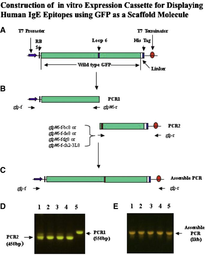
S.-S. Chen et al. / Journal of Immunological Methods 333 (2008) 10–23                                             11

elucidated that four IgE loops around the CHɛ2 and                               formed by 11 β-strands that serve to constrain the elev-
CHɛ3 domains, i.e., BC, DE, FG and C2-3 linker (C2-3L)                           en native loops exposed to the aqueous phase. Native
loops, are intimately involved in binding to FcɛRI as                            loops or peptide library replacing the loops, constrained
shown by co-crystal of IgE and FcɛRI receptor (Garman                            by the β-barrel, are exposed to the aqueous phase.
et al., 2000). It follows that induction of endogenous anti-                     Indeed, members selected from the aptameric peptide
IgE antibodies by active immunization to receptor-binding                        library presented by GFP were shown to block enzymes
IgE B-cell epitopes may protect against IgE-mediated                             regulating signal transduction and cellular growth
immediate hypersensitivity.                                                      (Caponigro et al., 1998; Geyer et al., 1999).
    Previously it was shown that a foreign B-cell an-                               Inserted peptides into Loop 6 (site 6) of GFP are
tigenic loop appropriately constrained in a protein                              known to exhibit constrained conformation similar to
scaffold may exhibit its antigenic conformation similar                          those of peptide loops present in the parent molecules
to that presented in the native loop region of the parent                        (Skerra, 2000). Herein, we test whether such “anti-
molecule, as pioneered by Zanetti and colleagues                                 genized” GFP may therefore serve as an active vaccine
(Billetta et al., 1991; Abedi et al., 1998; Geyer et al.,                        in eliciting neutralizing antibodies, which prevent IgE
1999). The scaffold proteins employed for this purpose                           binding to FcɛRIα. Herein, we showed that antibodies
are reported in the immunoglobulin gene families such                            elicited to C2–C3L antigenized GFP bind native
as complementarity-determining region (CDR2/3) of                                IgE molecules; furthermore this epitope-specific anti-
immunoglobulin (Billetta et al., 1991; Lanza et al.,                             body blocks IgE binding to recombinant FcɛRIα on
1993), loop regions of fibronection type III domain                              solid phase as well as FcɛRIα-transfected mast cells
(Koide et al., 1998), and CTL-A4 (Nuttall et al., 1999)                          (Nechansky et al., 2001).
as well as non-immunoglobulin gene family such as
protease inhibitors (Christmann et al., 1999) and                                1. Materials and methods
lipocalin-related molecules such as β-barrel containing
thioredoxin (Geyer and Brent, 2000; Skerra, 2000).                               1.1. Preparation of antigenized GFP vectors
    Green fluorescent protein (GFP) of the jellyfish
Aequorea victoria encoded by 238 amino acids, (Ormo                                 The general design of the antigenized vector was tested
et al., 1996; Tsien, 1998) exhibits a β-barrel structure                         in Fig. 1. The 5′MCS following the initiation codon was

Fig. 1. Strategy of preparing site 6 and site 7 GFP scaffold vector. Site 6 (Gln 157/Lys 158) or site 7 (Glu 172/Asp 173) of GFP was inserted with unique
RE cloning sites with the primer pairs described in Materials and methods. Diagnostic cut of site 6 and site 7 GFP scaffold vector. Site 6 scaffold vector,
6.5 was digested with Sph I along with either Bgl II or BssH I. Site 7 scaffold vector 7.4 was digested with Sph I along with Nhe I or Pst I.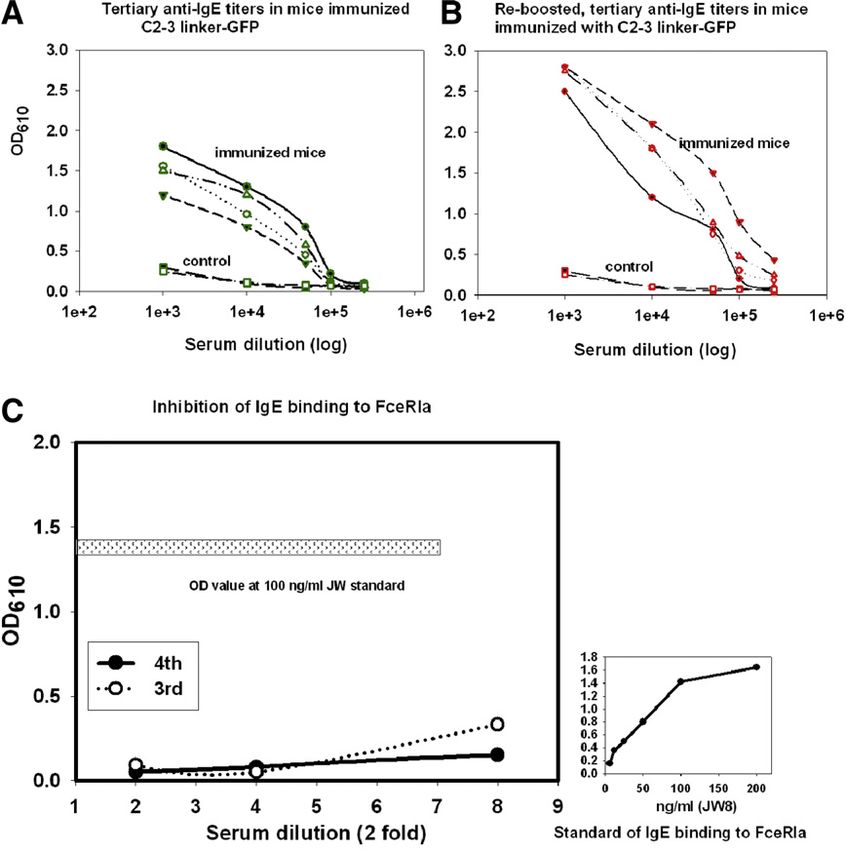
12                              S.-S. Chen et al. / Journal of Immunological Methods 333 (2008) 10–23

shortened to 7 residual amino acids with six out of the eight         10 mM imidazole. The sample was then eluted with
restriction enzyme sites therefore deleted from the 5′MCS             the same buffer containing 250 mM imidazole. The
of GFPUV vector (Clontech, Palo Alto, CA). This permits               flow rate was 2 ml/min, and absorbance was
engineering of RE sites in the loop regions of interest, i.e.,        monitored at A280. Protein eluted from the column
site 6 at Gln 157/Lys 158 in loop 6 (flanked by Ala 155 and           was collected in 1 ml fractions.
Ile 161) and site 7 at Glu 172/Asp 173 in loop 7 (flanked by              Bulk preparation of recombinant protein. Half to 1 l
Ile 171 and Val 176) of the two surface-exposed,                      of DH5α from LB culture containing the recombinant
constrained loops. A three-step PCR strategy and design-              protein were centrifuged at 5000 rpm in a Sorvall GSA
primer sets for the vector were illustrated in Fig. 1. Primers        rotor for 15 min at 4 °C. For each gram of pellet, 2 ml
with restriction enzyme (RE) sites, flanked by the GFP                of cell lysis buffer with 4 μl of 100 mM PMSF and
sequences were used to amplify half GFP molecules. GFP                80 μl of 10 mg/ml lysozyme. The re-suspension was
scaffolds with modified site 6 or 7 were then assembled by            stirred by addition of Triton 100 at a final 1%; the
ligating two pieces of the amplified PCR products, and                viscosity was further relieved by 20 μl of 1 mg/ml
amplified by another round of PCR to yield the scaffolding            DNase per gram starting material for 30 min, and
cloning vector. Thus primers pairs containing the three               finally the lysate with greenish soluble lysates, an
unique efficient end-cutter RE sites, were engineered into            indication without inclusion bodies was clarified by
site 6 (flanked by Gln 157 and Lys 158) or site 7 (flanked by         1 h centrifugation at 20,000 rpm in an SS34 rotor. The
Glu 172 and Asp173), i.e., Bgl II/Kpn I/BssH I for the site 6,        lysate was dialyzed against buffer containing 50mM
and Nhe I/Xba I/Pst I for the site 7, respectively. The               PB with 0.15M NaCl, and kept within 15 ml volume
sequences of the primer pairs were as follow: site 6                  by PEG concentration. The protein yield of half a liter
antisense: TGA GGTAC (Bgl II) AGATCT (Kpn I)                          culture was approximately 1200 mg in the dialysate
TTGTTTGTCTGCCGTGATGTATAC; site 6 sense:                               (80 mg/ml).
TGA GGTACC (KpnI) GCGCGC (BssH I) AA-                                     Protein purification was performed in two steps.
GAATGGAATCAAACGTAACTTC; site 7 antisense: TGA                         Samples were exchanged in PBS with 5 mM imidazole
TCTAGA (XbaI) GCTAGC (Nhe I) TTCAATGTTGTGGC-                          and The IMAC adsorption was performed with 5 ml
GAATTTTGAAG; site 7 sense: TGA TCTAGA (Xba I)                         each batch in five successive runs of 1 ml each on a
CTGCAG (Pst I) GATGGATTCGTTCAACTAGCAG; com-                           10 ml of IMAC column in a upscale enrichment of
mon antisense: TGATGATGATGATGATAGAGCTC (Sac I)                        recombinant GFP, constituting a significant constituent
ATCCATGCCATGTG; common sense: TGATGATGA(GCA-                          in fractions monitored by OD595 in the visible
TGC (SphI)ACCGGT (AgeI) AGAAAAAATGAGTAAAGG.                           spectrum, represented 17 fold enrichment as estimated
    Fig. 1 inset showed diagnostic cuts of non-anti-                  from the bulk of materials measured by OD280 (not
genized site 6 and site 7 expression vector constructs.               shown). The enriched fractions were pooled and
Double digest with Sph I/Bgl II, or Sph I/BssH I                      concentrated in 1 ml PB. GFP enriched fraction was
yielding a small .5 kb fragment and a large 3 kb                      then further fractionated onto a pre-packed Sephacryl
fragment of site 6 construct, while double digest of site 7           S-200 HR (dimension: 26 × 60, Pharmacia, Bridge-
construct with Sph I/Nhe I and Sph I/Pst I yielding a                 water, NJ) with bed volume of approximately 300 ml
similar small fragment and a large 3 kb fragment.                     at the flow rate of 0.2 ml/min at 4 °C with sample
                                                                      volume of in 0.5 ml in a column of bed volume of
1.2. Purification of GFP                                              250 ml packed slurry, and fractions were collected at
                                                                      2.5 ml per tube for one bed volume, and the UV280 was
   Immobilized metal affinity chromatography                          monitored. Peak material was collected and concen-
(IMAC). IMAC was purchased from Biorad (Hercules,                     trated by PEG, and run on Lammeli SDS-PAGE and
CA). IMAC resins containing iminodiacetic acid                        pure material of 28 to 30 kDa was identified as a single
(IDA) is charged with Ni2+ with 60 μm for flow                        band. Throughout the purification, antigenized GFP or
rate. A clarified of E. coli lysate containing C2-3L                  control GFP enriched fraction was sighted by eye or
GFP from DH5α, or GFPUV from JM109 of 100 ml                          visualized by illuminating tubes with a hand UV lamp
culture was exchanged in buffer 50 mM sodium                          at 365 nm.
phosphate, pH 8.0, 0.3 M NaCl containing 5 mM
imidazole, the concentrated sample in 1 ml was loaded                 1.3. Design and expression of the expression cassette
to a 2 ml column pre-equilibrated with the exchange
buffer. The lysate was flowed through the column and                    A complete expression cassette of antigenized GFP
was then washed with the same buffer containing                       was generated using pIVEX 2.3 vector (Roche
S.-S. Chen et al. / Journal of Immunological Methods 333 (2008) 10–23 13
Applied Science, Indianapolis, IN) as a template. The 1.4. Preparation of antibodies against antigenized GFP
two primers used are gfp-f, gatcgagatctcgatcccgcgaaat
(forward25), and gfp-r, gaacctgcagagcaaaaaacccctcaaga For larger scale of protein production, the above PCR
(reverse29). This 1 kb PCR fragment contains, in se- fragments of antigenized GFP (C2-3L and BC loop)
quence from 5′, a T7 promoter, ribosomal binding site were digested with Nco I and Sac I, and the large
(RBS), wide type GFP, a linker, His-tag and T7 ter- fragments were cloned into similar double digest of
minator, which constitutes a complete in vitro expres- pGFPUV. DH5α was then transformed with annealed
sion cassette. PCR1 consists of 5′ regulatory sequence antigenized GFP. Following IPTG induction, the
and upstream of wide type GFP from loop 6, which transformed bacteria were solubilized by 1% Triton,
was produced using pIVEX 2.3 control GFP DNA as 10 mg/ml lysozyme and antigenized GFP was purified
a template with forward primer, gfp-f and the reverse by the IMAC column. BALB/c mice were primed with
primer, gfp#6-r, gtctgccatgatgtatac. PCR2 fragment 10 μg of antigenized GFP in 2 mg alum for 10 days,
incorporating antigenized foreign loop sequences, i.e., boosted three times at the intervals of 14 days, and sera
IgE loop sequences in replacing native loop 6, was pro- were collected 10 days after the last boost. The
duced using the same reverse primer, gfp-r, and differ- purification procedure involved a clarification of 50%
ent forward primers. The four forward primers are gfp#6- ammonia sulfate cut, protein G purification (Pierce),
f-bc8, aatgtatacatcatggcagacGTGGTGGACCTGG followed by purifying on NP-specific chimeric mouse/
CACCCAGCAAG ggaatcaaagttaacttcaaaat, gfp#6-f-de8, human JW8 IgE (clone provided by Dr. Neuberger
aatgtatacatcatggcagacAAGCAGCGCAATGGCACGT- at MRC) coupled on Sepharose 4B, and assayed by
TAACCggaatcaaagttaacttcaaaat, gfp#6-f-fg8, aatgtatacat- human IgE (PS). Anti-immunoglobulin rheumatoid
catggcagacCACCCCCACCTGCCCAGGGCCCTC factor activity was removed by adsorption on mouse
ggaatcaaagttaacttcaaaat, and gfp#6-f-ch2-3L8, aatgtata- and human IgG coupled to Sepharose 4B. Each stage of
catcatggcagacGATTCCAACCCGAGAGGGGT- enrichment was tested by ELISA for immune reactivity
GAGCggaatcaaagttaacttcaaaat. The underlined sequence on JW8 coated 96-well, followed by HRP–MAb rat
is complementary to the primer, gfp#6-r. DNA sequences anti-mouse IgG and substrate. Up to 5% or more of
of IgE inserts were determined with reverse sequencing affinity protein G fraction are specific for human IgE.
primer Rd 4 688r and used.
IgE epitope antigenized GFP chimeric proteins were 1.5. Preparation of recombinant soluble huFcɛRIα
expressed in vitro in a transcription-translation system
based on E. coli lysates for 6 h at 30 °C (Roche, Rapid The total RNA was isolated from KU812 cells
Translation System RTS 100 E. coli HY Kit). Samples (obtained from Mike Robertson, TSRI). RT-PCR
were left overnight at 4 °C to allow for efficient folding performed with appropriate primers and cloned into
of newly synthesized protein molecules. Translated pGEM (Promega). A full-length receptor comprising
protein was precipitated first by acetone to remove the signal sequence, homology domain D1 and D2 and
the interfering material in the lysates, followed by re- membrane anchor was cloned. The first strand cDNA
solubilization in native gel sample treatment buffer was reverse transcribed with both oligo d(T) and ran-
containing 1% NP40 without SDS and 2-ME, and also dom primers. Full length FcɛRIα cDNA was amplified
in the absence of heating during sample solubilization in using forward and reverse primer pairs, gaagaattcgaa-
1% NP-40 alone. The translated proteins were separated gatggctcctgccatgg, and gagtagcaattgctgatgctgga. Secret-
by native gel electrophoresis, followed by transferring ed (sR: transmembrane domain truncated, amino acids
to nitrocellulose in diluted methanol carrier also in from 26 to 204) form was constructed using baculo-
the absence of 0.1% SDS. To diminish background, transfer vector pAcSG2. Another series was made in
blots were incubated and blocked with goat IgG (2 mg/ml) pAcGP67 with GP67 insect signal sequence (sRA), was
followed by detection with anti-GFP antibody (GFP used for production of single soluble receptor with HA
Monoclonal Antibody, purified, mouse clone B34, tag for the convenience of purification.
Covance, Cat. No. MMS-118P, 1/3000, and rat anti- Recombinant monomeric and dimeric receptors were
mouse κ, 1/1000). In parallel, duplicate blots were probed produced in sf9 insect cells. Receptor proteins in super-
with anti-human IgE antibodies of a 1/1 mixture of two natants and cell extracts were pooled. Receptors were
commercial sources of antibodies: goat anti-human IgE– fractionated around 22% NaCl gradient (10 mM to 1 M)
HRP conjugate (Bethyl, Cat. No. A80-108P) and goat on Mono-Q column. Alternatively, monomeric receptor
anti-human IgE–HRP conjugated (Caltag Laboratories, can be enriched by anti-HA column by acidic elution.
Cat. No. H15707). Receptor-based ELISA 96-well were coated overnight14 S.-S. Chen et al. / Journal of Immunological Methods 333 (2008) 10–23
with 10 μg/ml NP-BSA, washed, blocked, and added added to 50 μl of JW8 IgE (200 ng/ml) at the final serum
with NP-specific chimeric mouse/human JW8 IgE for dilutions from 1/2 to 1/8 to 100 ng/ml JW8 IgE. The
2 h at r. t. Plates were washed, followed by addition of mixture were incubated for 30 min at r.t., followed by
dimeric and monomeric receptor at 0.1, 1 and 10 μg/ml addition to receptor-bound 96-well plate for 1 h at r. t.,
for 1 h at r. t. HRP conjugate of rabbit anti-HA was then washed and the presence of receptor-bound IgE on solid
added at 1/3000, followed by substrate. phase was detected by HRP–MAb anti-human IgE.
To determine anti-IgE titers, 96-wells were prepared
1.6. ELISA assays by coating with NP-BSA at 1 μg/ml for 1 h at r. t. followed
by anti-NP chimeric murine/human JW8 IgE at 1 μg/ml
Recombinant huFcɛRIα, prepared from the insect overnight. As standard, MAb anti-human IgE (PharMin-
sf9 cells expressing single or doubly tandem receptor gen) was added from 100 ng to 1.6 ng at two fold
construct, was used for coating the plates at 10 μg/ml dilutions, followed by HRP–rat anti-mouse IgG (HRP–
with similar results. JW8 IgE was titrated on receptor- RaAMG) and substrate. The titers of the antisera were
coated plate from 200 ng to 2.4 ng/ml, washed and determined by adding sera from 1000 to 500,000 dilutions
detected by the HRP–anti-IgE (PharMingen). To per- to the above IgE-coated plate, followed by HRP–
form the inhibition assay, 50 μl of pooled sera were RaAMG, and OD value two fold above the background
Fig. 2. Site 6 antigenized GFP constructs Six sets of 16-mer oligonucleotides corresponding to FcɛRIα binding sites and non-binding site controls
were prepared with Bgl II and Kpn I at 5′ and 3′ ends. The digests of oligonucleotides and scaffold vector were annealed. Diagnostic cut with Bgl II
and Sac I yielded fragments of 250 bp denoted by the star symbol (⁎ Panel A). Lysates prepared from the transformed cells were blotted by polyclonal
rabbit anti-GFP, and expressed antigenized GFP was shown as 25–26 kDa bands (Panel B). Inserted IgE loop sequences at side 6 were verified by
DNA sequencing (Panel C).S.-S. Chen et al. / Journal of Immunological Methods 333 (2008) 10–23 15
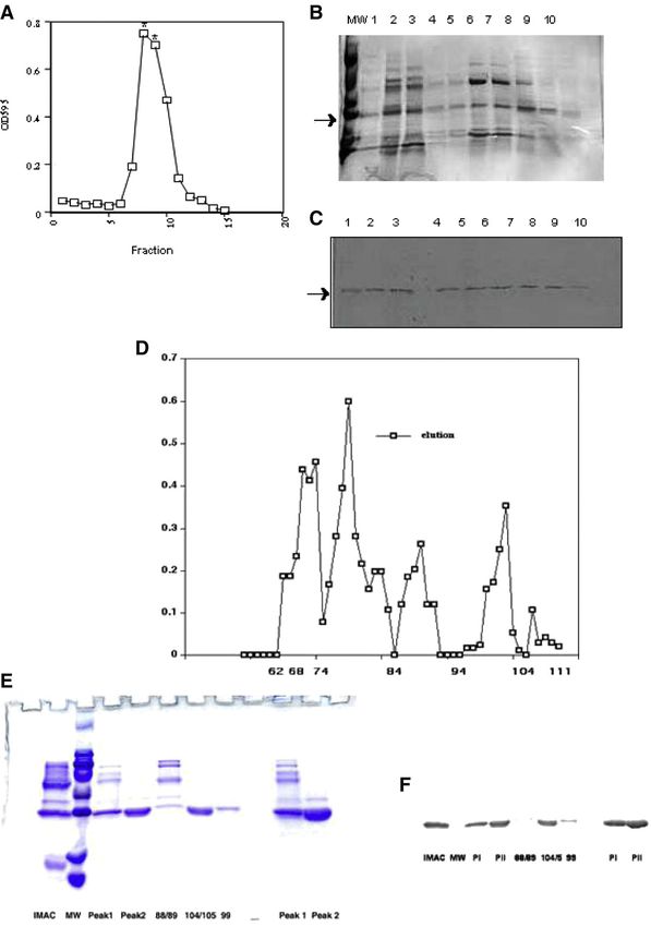
was considered a positive titer, and the concentrations of 2.2. Construction of in vitro expression cassette for
anti-IgE calculated from the standard curve. displaying human IgE epitopes using GFP as a scaffold
molecule
1.7. Mast cell degranulation
The feasibility of expressing a soluble antigenized
Mast cell line, 2H3 stably transfected with huFcɛrIα GFP above prompts development of an in vitro
were pre-treated for 24 h with dexamethasone (10 μM expression platform for studying the immunogenicity
final concentration) to upregulate human FcɛRIα of the inserted sequences. Fig. 3 depicts a strategy to
expression. Mast cells were then sensitized for 30 min bypass the requirement of steps of molecular cloning,
with 1 μg/ml IgE or 1 μg/ml IgE pre-incubated for 1 h and in vivo bacterial transformation and expression.
with anti-C2-3L from 10 to 100 μg/ml, washed and then Four different receptor-binding epitopes of human IgE
challenged with 1 μg/ml NP-BSA. β-hexosaminidase were inserted and replaced the original site 6 residues in
release was then determined. The hexosaminidase GFP. The immunogenicity of the replaced IgE B-cell
release was triggered by NIP23 BSA (obtained from epitopes of antigenized GFP can be expediently
Dr. Garnett Kelsoe, Duke University) added to the evaluated as an in vitro translated product.
cells at the concentrations 1 μg/ml for 45 min at 37 °C. This is achieved via one single tube reaction of
β-hexosaminidase release was calculated as a percen- coupling the in vitro transcription/translation procedure
tage of the secreted versus total available cell hexosa- according to the following three engineering steps: (i)
minidase. The secreted (supernatants) and total (cell PCR1 fragment utilizing a pair of primers that amplify
lysates) enzyme contents were estimated as OD405 after the half molecules spanning the T7 transcription start
incubation with substrate p-nitrophenyl-N-acetyl-β-D- site, Shine/Dargalno sequence (He and Taussig, 1997)
glucosaminide for 90 min at 37 °C. and the beginning of the GFP (delineated by the 5′
primer) up to the upstream GFP sequence to site 6
2. Results (delineated by the 3′ primer). PCR1 consists of 5′ reg-
ulatory sequence and upstream of wide type GFP from
2.1. Expression of antigenized GFP loop 6, which was produced using pIVEX control GFP
DNA as a template with forward primer, gfp-f and the
Site 6 GFP vector scaffold was inserted with reverse primer, gfp#6-r (Fig. 3B and D, lane 5). (ii)
oligonucleotides of human IgE sequences correspond- PCR2 fragment utilizes 5′ primer consisting of GFP
ing to FcɛRIα binding BC loop, FG loop, DE loop and sequences (that pair with the 3′ primer of the first PCR
C2-3 linker loop (C2-3L), respectively (Garman et al., fragment), the eight amino acids corresponding
2000). Sequences from CHɛ4 predicted as B-cell FcɛRIα−binding IgE loop, and 23 oligonucleotides of
epitopes (Mac vector Inc., Cary, NC), were employed gfp sequence downstream and thus deleting the native
as control. Fig. 2A showed diagnostic cuts of site 6 loop 6 sequences. And the 3′ primers include His-tag
antigenized GFP vectors containing the oligonucleotide and T7 terminator (Fig. 3B, D, lanes 1 to 4). (iii) As-
inserts following digestion of Bgl II and Sac I. sembly of the two half molecules of GFP, the stuffer
Expression of antigenized GFP protein was then PCR1 fragment and PCR2 (Fig. 3C). Fig. 3E showed
determined by western blots with anti-GFP antibodies. assembled 1 kb fused products from PCR1 and PCR2
Recombinant native GFP and antigenized GFP from exhibiting antigenized IgE sequences: gfp-6-bc8; gfp-6-
bacterial extracts were electrophoresed and immuno- de8; gfp-6-fg8; gfp-6-ce2-3L8, respectively (Fig. 3E,
blotted, and detected by anti-GFP antibodies (Fig. 2B). lanes 2–5) and GFP control without insert (lane 1).
Antigenized GFP proteins migrated as a specific band Next, the PCR-amplified DNA can then be directly
from 27 to 29 kDa (lanes 2–10; 13–14) in contrast to employed for transcription/translation-coupled reaction
control bacteria transformed with original vector or site for in vitro protein expression. Fig. 4A confirmed the
6 modified vector (lanes 11 and 12), while this material constructs of the assembled PCR products as illustrated
was not found in mock-transformed JM109 (lane 1). in Fig. 3; and base sequences of these newly assem-
The presence of the oligonucleotide inserts in anti- bled products were determined by DNA sequencing
genized GFP constructs was also confirmed by DNA (Fig. 4B). Further, antigenicity of the in vitro expressed
sequencing (Fig. 2C). A parallel set of site 7 oligo- transcribed/translated products (in capital letter): GFP-
nucleotides antigenized constructs was likewise pre- 6-BC8, GFP-6-DE8, GFP-6-FG8, GFP-6-C2-3L8 was
pared, inserts sequenced, and recombinant fusion then evaluated. Fig. 4C showed the detection of trans-
protein migrated as a 27–29 kDa band (not shown). lated antigenized GFP constructs ∼ 27 kDa (Fig. 4C,16 S.-S. Chen et al. / Journal of Immunological Methods 333 (2008) 10–23
Fig. 3. Construction of in vitro expression cassette for IgE B-cell epitopes in GFP scaffold vector. A complete expression cassette of wide type GFP
was generated using pIVEX control vector GFP as a template (Panel A). The primers and designs were described in Materials and methods.
Oligonucleotides encoding different IgE B-cell epitopes prepared with identical 5′ and 3′ of the flanking sequences were inserted in PCR2 fragment
(Panel B). Strategy for reassembled complete antigenized cassette (Panel C), and PCR2, PCR1 fragments and the assembled products were shown
(Panels D and E).
lanes 1 to 4), positive control transcribed/translated wild detectable signal with BC loop antigenized product
type GFP (lane 5), control purified GFPUV protein (lane (Fig. 4D). Furthermore, no reactivities with polyclonal
6), and negative control of translated lysate only without anti-IgE were noted with translated GFP, purified GFP
added template (lane 7). protein and lysates.
To determine the antigenicity of antigenized 8-mer
IgE sequence constrained within GFP scaffold, the trans- 2.3. Preparation of purified C2-3L antigenized GFP
lated products were evaluated under native gel separation
conditions. It is noteworthy that although expression was GFP exhibits 10 histidine residues that interact favor-
observed with four PCR assembled products according to ably with chelated metals such as Ni (II), in particular,
the detection of GFP scaffold (Fig. 4C), FG loop, DE loop with histidines at the top of the β-barrel, His77, His81
antigenized GFP were not detected by polyclonal anti- and His231 as an intrinsic His-tag (Tsien, 1998). Thus,
IgE, distinct reactivities were observed with antigenized immobilized metal affinity chromatography (IMAC)
GFP encompassing C2-3L8 linker loop, and a barely was employed for purification of antigenized GFP (LiS.-S. Chen et al. / Journal of Immunological Methods 333 (2008) 10–23 17 Fig. 4. Evaluation of IgE B-cell epitopes constrained in the GFP scaffold. The amplified DNA fragments:gfp-6-bc8; gfp-6-de8; gfp-6-fg8; gfp-6-ce2- 3L8 and gfp wt were verified by length (Panel A), and by sequencing (Panel B). Translated IgE B-cell epitopes-GFP chimeric proteins: GFP-6-BC8, GFP-6-DE8, GFP-6-FG8, GFP-6-CH2-3L8, and wild type GFP (GFP WT), were prepared and evaluated by western blot with anti-GFP (Panel C) or with polyclonal anti-IgE (Panel D) as described in Materials and methods. et al., 2001). Recombinant protein was solubilized and buffer in fractions 7 to 11 on IMAC column. Fig. 5B prepared as described (Material and methods). Anti- showed that this antigenized GFP was relatively en- genized GFP was observed to distribute homogeneous- riched in fractions 6 to 8; and was highly enriched ly in the cytoplasm and the fluorescence-emitting in tubes 9 and 10, as a 27–29 kDa band away from material was localized in the Triton X-100 clarified the contaminating bands shown by SDS-PAGE. C2-3L lysates and not in the inclusion bodies pellet. C2-3 antigenized GFP was notable across all the fractions by linker-antigenized GFP can be conveniently enriched immuno-blot with a polyclonal anti-GFP. Thus, IMAC via passage of clarified lysates via IMAC column due to affinity adsorption did not yield highly pure antigenized intrinsic His-tag. Fig. 5A showed the fractionation GFP product in our hand. profile of C2-3L antigenized GFP by adsorption to Ni- Next, IMAC-enriched material was further separated based gel bed, followed by elution in 250 mM imidazole by sizing chromatography by Sephacryl S-200 HR due to
18                               S.-S. Chen et al. / Journal of Immunological Methods 333 (2008) 10–23

Fig. 5. Purification of C2-3L antigenized GFP. C2-3 linker transformed DH5α was selected. Procedures of enrichment on IMAC column and gel
filtration on Sephacryl S-200 HR were described in Materials and methods.
S.-S. Chen et al. / Journal of Immunological Methods 333 (2008) 10–23                                        19

Fig. 6. Immune sera were prepared in BALB/c mice, immunized with 10 μg of C2-3L antigenized GFP in 2 mg alum and boosted twice at the
intervals of 14 days. Immune sera were collected day 10 after the third challenge (Panel A) or fourth challenge (Panel B). ELISA for measuring anti-
human IgE was described in Materials and methods. ELISA for measuring IgE bound to recombinant receptor (Panel C) was described in details in
Materials and methods, and the standard curve was presented as an outset of Panel C.

its resolution of separating ∼30 kDa globular proteins                       Assuming no loss of materials during steps of handlings
from neighboring species. As shown in Fig. 5D, five                          and purification, the recombinant antigenized GFP is
distinct peaks were resolved under the current operation                     estimated to be at least 2.1% abundance in the total
conditions. As noted before, the IMAC-enriched, pooled                       clarified protein lysate. In summary, the efficacy of the
starting material exhibited major contaminated molecular                     present scheme of purification is approximately 50 fold of
species (lane IMAC). The peak (tubes 85–90) with                             enrichment following IMAC and Sephacryl-S-200 HR.
approximately 4.5 mg determined according to OD280                           This result indicates the adequacies of expression levels
exhibited a significant amount of antigenized GFP with                       and the suitability of the step-wise procedures for
other contaminated proteins. Importantly, pure C2-3L                         enrichment. To corroborate the purity of the material,
antigenized GFP was indeed isolated in peak 2 (tube 100–                     western blots reacted with antigenized GFP as an ∼27 to
103) with approximately 3.8 mg determined at OD280 and                       29 kDa band, present in IMAC eluted material, peak 1
shown as a single ∼27 to 29 kDa band in SDS-PAGE                             mixture as well as in high intensity in peak 2 pure
(Fig. 5E). Thus the total yield of of peaks 1 and 2                          antigenized GFP (Fig. 5F).
combined was approximately 8.3 mg from the 25 mg of                              To further substantiate the demarcation of the peak 2
IMAC-enriched material following sizing column accord-                       purified antigenized GFP, Fig. 5D to F showed that
ing to the chromatogram (Fig. 5D) monitored by OD280.                        tubes 88/89 delineating the right boundary of peak 1
Thus the sizing step contributed to ~3 fold enrichment.                      contained contaminating materials along with signifi-
Thus, the overall two-step purification yielded 50 fold                      cant presence of antigenized GFP. In contrast, material
enrichment, as calculated from the starting material of                      demarcating the boundaries of peak 2, i.e., material from
~400 mg clarified protein prior to the IMAC column.                          left boundary tube (#99) as well as from the right
20                                  S.-S. Chen et al. / Journal of Immunological Methods 333 (2008) 10–23

Fig. 7. HuFcɛrIα-transfected mast cells were pre-treated for 24 h with dexamethasone. Equal volume of anti-C2-3L antibodies was mixed with JW8 at
the final concentrations from 10 to 100 μg/ml anti-IgE to 1 μg/ml JW8 (to mimic human allergic IgE levels) for 30 min at r.t. Mast cells (0.5 × 10⁶)
were then sensitized to the above mixture for 30 min at 37 °C in a CO2 incubator, washed and then challenged with 1 μg/ml NP-BSA and specific
hexosaminidase release was determined.

boundary tubes (#104 and #105), exhibited only a single                      observations with comparable results were repeated in
band of purified antigenized C2-3L GFP, albeit at a less                     BALB/c and C57BL/6 mice (not shown). The concentra-
concentration than the pooled peak 2 material contain-                       tions of in vivo anti-IgE in these experiments may be
ing the major purified antigenized GFP, i.e., tubes #100                     relevant to the clinical setting of employing passive anti-
to #103). Thus Fig. 5D to F demonstrate unambiguously                        IgE, MAb-E25, i.e., effective blood concentration of
an effective separation of C2-3L antigenized GFP                             MAb-E25 is maintained at a steady state around 30 mg/ml
according to a first enrichment by metal affinity column,                    blood in patients (300 kU/l IgE) that received 300 to
followed by a high resolution gel filtration on Sephacryl                    400 mg bi-weekly injection up to four times.
S-200 HR column. It may be noticed that recombinant                             Next, It is pertinent to determine the protective effect
C2-3L antigenized protein was enriched and purified                          of antisera in neutralizing binding of human IgE to
under native condition throughout preparations from the                      human FceRI. Thus the neutralizing activities against
clarified lysate to final yield.                                             IgE binding to recombinant FceRIa receptors were
                                                                             examined. The outset of Fig. 6C showed the establish-
2.4. Antibodies elicited by the C2-3L antigenized GFP                        ment of a solid phase recombinant receptor capture
block IgE binding to FcεRIα                                                  assay in that high sensitivity of binding of JW8 IgE to
                                                                             solid phase receptor was shown with soluble IgE
    Next, we evaluate whether the purified C2-3L                             (100 ng/ml) captured to plate-bound recombinant
antigenized GFP may be employed as immunogens in                             receptor, exhibiting an OD ∼ 1.4. Importantly, Fig. 6C
eliciting neutralizing, protective anti-IgE antibodies in                    showed that binding of JW8 IgE to recombinant
BALB/c mice. Fig. 6 showed that individual mice that                         receptors was profoundly abrogated by tertiary antisera
received tertiary immunization of C2-3L antigenized GFP                      up to 1/4 dilutions, and re-boosted hyperimmune sera at
exhibited a titer of 1 to 50,000 (Fig. 6A), and upon a re-                   1/8 dilution. This observation suggests that in vivo
boost, serum titers were elevated to a range of 1/50,000 to                  protection of serum IgE from binding to mast cells by
250,000 (Fig. 6B). Concentrations of anti-IgE in immune                      the undiluted, neat sera may be even more effective than
sera were measured at 245 ± 37 μg/ml upon tertiary                           the in vitro correlate.
immunization, and around 784 ± 95 μg/ml following a re-                         Next, we examine the role of anti-C2-3L on protection
boost with ELISA of human IgE-coated plate. Similar                          JW8 IgE to receptors on mast cells. Mast cell
S.-S. Chen et al. / Journal of Immunological Methods 333 (2008) 10–23 21
degranulation via human IgE and human FceRIa was of the GFP scaffold insert and constrain protective IgE
performed with RBL-2H3 was transfected with human sequences into the solvent-exposed loops of GFP as
FceRIa (Robertson, 1993; Nechansky et al., 2001). Thus, antigens (Abedi et al., 1998; Peelle et al., 2001). The
the mast cell-based in vitro protection assay was set with chromophore, consisting of a modified tripeptide, is
serum dilutions as well as with affinity pure anti-C2-3L in buried inside the relatively rigid β-barrel structure.
a range of 10 to 100 mg/ml in neutralizing 1 mg/ml JW8 Insertion of six oligopeptides into site 6 of GFP sig-
IgE. The dosages of protective neutralization assay was nificantly quenched and diminished the intrinsic fluo-
set according to a clinical setting of employing passive rescence. Antigenized GFP with inserts of 16 amino
anti-IgE, maintained around 30 mg/ml blood in treated acids in length were expressed and detected with anti-
patients. As shown in Fig. 7, ∼95% protection was GFP in the immuno-blot (Fig. 2); however, they did not
observed in JW8 incubated with affinity purified anti-IgE react with polyclonal sources of anti-human IgE
at the tested concentrations from 10 to 100 mg/ml. In employed in this study (not shown). This may reflect
contrast, in the absence of anti-C2-3L antibodies, about lack of the constrained conformation of longer inserted
68% of enzyme release was noted in IgE-sensitized, peptides even though GFP is known thermostable and
huFcɛRIα-transfected mast cells followed by NP-BSA can accommodate selectable aptameric peptide library
challenge, and approximately, 5% nonspecific B-hexosa- of shorter length (Peelle et al., 2001). On the other hand,
minidase release was noted in JW8-sensitized mast cells at least one of the four 8-mer cassettes, C2-3 linker can
without allergen challenges. This observation indicates be constrained in site 6 of GFP scaffold, and weak
that anti-IgE elicited by C2-3L antigenized GFP prevent reactivity to BC loop antigenized GFP was detectable. It
IgE sensitization to mast cells; and furthermore, the pre- is possible that the reactivity of BC loop may be im-
incubated immune complexes also did not activate mast proved with shorter core sequence insertion. The ana-
cells, suggesting that elicited antibodies do not react with lyses of the constrained antigenicity can be conducted
non-receptor-binding region of JW8 IgE and may be by the in vitro transcription/translation bypassing steps
safely employed for human use. of laborious cloning and protein production. With the
present in vitro transcription/translation system, future
3. Discussion studies may be expediently conducted with even shorter
oligopeptide inserts, and/or aided with further con-
3.1. Suitability of GFP as antigenic scaffold straining disulfide loops flanking the inserts.
Loop sequences spanning part of the scaffold protein 3.2. Anti-IgE active antibodies protect against IgE
are long known to play a critical role in the biological, binding to FcɛRIα
i.e., enzymatic, ligand/receptor, antibody, and antigenic
specificities. These amino acid residues within the scaf- To test the feasibility of antigenized GFP as ac-
fold molecules are characterized by being hydrophilic in tive vaccine, antibodies elicited to the receptor-binding
nature, surface-exposed, and mobile. Earlier, Zanetti loops of IgE constrained by GFP scaffold may lead to
utilized the immunoglobulin fold as a scaffold to present inhibition of IgE binding to FcɛRI receptors (Garman
a grafted oligopeptide epitope in an immunologically et al., 2000; Wan et al., 2002). The CHɛ2–CHɛ3 linker
accessible and conformationally suitable manner (Bil- region, C2-3L (residue 332 to 337) is critically involved
letta et al., 1991; Lanza et al., 1993). Later, Brent and in the docking of solution-phase or circulating IgE to the
Kamb constrained peptide aptamers on the loop region cognate receptor. Antibodies raised to C2-3L antige-
of thioredoxin and GFP respectively that exhibit a nized GFP, appropriately constrained, indeed prevent
pertinent biological conformation to modulate signal JW8 chimeric murine/human IgE from binding to recom-
transduction to modulate cell growth and differentiation binant FcɛRIα coated on solid phase as well as those
(Abedi et al., 1998; Geyer et al., 1999). Recently, expressed on the cell surface of transfected RBL-2H3.
Bradbury showed that grafting lysozyme binding Formation of immune complexes is essential for
sequence from a camel antibody in GFP loop confer protection by anti-IgE. It is possible that anti-C2-3L
recognition capacity of grafted GFP (Kiss et al., 2006). form effective immune complexes with free circulating
GFP is a stable proteolysis-resistant single chain of IgE by neutralizing each respective determinant on the
238 amino acids exhibiting an 11-stranded β-barrel half molecule of the solution-phase IgE. Indeed, MAb-
wrapped around a single central helix, forming a rigid β- E25 is capable of forming hexameric antigen-antibody
barrel. Herein, we study whether receptor-binding loops complexes via reacting to two identical interaction sites
of IgE may be inserted and protected within the confine of circulating IgE even though the physical binding22 S.-S. Chen et al. / Journal of Immunological Methods 333 (2008) 10–23
sequences to which MAb-E25 reacts are not known References
(Corne et al., 1997; Chang, 2000; Garman et al., 2000;
Wan et al., 2002). Moreover, since antibodies elicited Abedi, M.R., Caponigro, G., Kamb, A., 1998. Green fluorescent
protein as a scaffold for intracellular presentation of peptides.
by C2-3L antigenized GFP recognize conformation
Nucleic Acids Res. 26, 623.
on solution-phase, i.e., circulating but not cell-bound Alexander, J., del Guercio, M.-F., Maewal, A., Qiao, L., Fikes, J.,
IgE, the current construct therefore offers a promise to Chesnut, R.W., Paulson, J., Bundle, D.R., DeFrees, S., Sette, A.,
serve as a safe pan-IgE vaccine for human use. Obser- 2000. Linear PADRE T helper epitope and carbohydrate B cell
vations shown in Figs. 6 and 7 substantiate this notion epitope conjugates induce specific high titer IgG antibody responses.
J. Immunol. 164, 1625.
since actively produced antibodies elicited by antigenized
Billetta, R., Hollingdale, M.R., Zanetti, M., 1991. Immunogenicity of an
C2-C3L did not cross-linked IgE on IgE-sensitized mast engineered internal image antibody. Proc. Natl. Acad. Sci. U.S.A. 88,
cells, while effectively abrogating sensitization of solu- 4713.
tion-phase JW8 IgE to huFcɛRIα-transfected mast cells. Caponigro, G., Abedi, M.R., Hurlburt, A.P., Maxfield, A., Judd, W.,
Kamb, A., 1998. Transdominant genetic analysis of a growth
control pathway. Proc. Natl. Acad. Sci. U.S.A. 95, 7508.
3.3. Relevance of concentrations of active anti-IgE in
Chang, T.W., 2000. The pharmacological basis of anti-IgE therapy.
protecting against IgE-mediated allergy Nat. Biotechnol. 18, 157.
Chen, S.-S., Liu, F.-T., Katz, D.H., 1984. Cellular and molecular
High antibody titers were frequently observed with mechanisms of murine IgE class-restricted tolerance induced by
optimal immunizations in studies conducted in our neonatal administration of soluble or cell-bound IgE. J. Exp. Med.
160, 953.
lab and by others, including anti-peptide antibodies
Christmann, A., Walter, K., Wentzel, A., Kratzner, R., Kolmar, H.,
(Alexander et al., 2000a). Concentrations of anti-IgE 1999. The cystine knot of a squash-type protease inhibitor as a
present in immune sera were in the range from 250 to structural scaffold for Escherichia coli cell surface display of
800 μg/ml according to immunization protocol (Fig. 6), conformationally constrained peptides. Protein Eng. 12, 797.
constituting 1.5 to 4% of IgG fraction (Chen et al., 1984; Corne, J., Djukanovic, R., Thomas, L., Warner, J., Botta, L., Grandordy,
B., Gygax, D., Heusser, C., Patalano, F., Richardson, W., Kilchherr,
Alexander et al., 2000b; Takasuka et al., 2004).
E., Staihelin, T., Davis, F., Gordon, W., Sun, L., Liou, R., Wang, G.,
The data presented herein offers a feasible model for Chang, T.-W., Holgate, S., 1997. The effect of intravenous
the in vivo protection of active immunization against administration of a chimeric anti-IgE antibody on serum IgE levels
IgE-mediated allergy. MAb-E25 recommended for pa- in atopic subjects: efficacy, safety, and pharmacokinetics. J. Clin.
tients higher than 75 kU/l (or 75 IU/ml, i.e., 250 ng/ml). Invest. 99, 879.
Garman, S.C., Wurzburg, B.A., Tarchevskaya, S.S., Kinet, J.P., Jardetzky,
Such patients are administered with MAb-E25 subcut. at
T.S., 2000. Structure of the Fc fragment of human IgE bound to its
300 to 400 mg per injection resulting in a blood level of high-affinity receptor Fc epsilonRI alpha. Nature 406, 259.
anti-IgE of 30 μg/ml, i.e., about 100 M excess of Geyer, C.R., Brent, R., 2000. Selection of genetic agents from random
antibodies to circulating IgE. To ensure sufficient pro- peptide aptamer expression libraries. Methods Enzymol. 328, 171.
tection, patients should receive four bi-weekly or at least Geyer, C.R., Colman_Lerner, A., Brent, R., 1999. “Mutagenesis” by
peptide aptamers identifies genetic network members and pathway
on the monthly of MAb-E25 (half life ∼2 to 3 weeks).
connections. Proc. Natl. Acad. Sci. U.S.A. 96, 8567.
With regard to the active vaccination, the advantage of He, M., Taussig, M.J., 1997. Antibody–ribosome–mRNA (ARM)
undiluted neat sera, extrapolating from the rodent study complexes as efficient selection particles for in vitro display and
herein is likely to prevail over the passive treatment evolution of antibody combining sites. Nucleic Acids Res. 25, 5132.
since 800 to 3,000 fold molar excess of endogenous Kiss, C., Fisher, H., Pesavento, E., Dai, M., Valero, R., Ovecka, M.,
Nolan, R., Phipps, M.L., Velappan, N., Chasteen, L., Martinez, J.S.,
anti-IgE to circulating IgE may be anticipated (Figs. 6
Waldo, G.S., Pavlik, P., Bradbury, A.R.M., 2006. Antibody binding
and 7). loop insertions as diversity elements. Nucleic Acids Res. 34, e132.
In conclusion, this study provides a feasible in vitro Koide, A., Bailey, C.W., Huang, X., Koide, S., 1998. The fibronectin
and in vivo model for studying protection of IgE- type III domain as a scaffold for novel binding proteins. J. Mol.
mediated allergic diseases via active immunization with Biol. 284, 1141.
Lanza, P., Billetta, R., Antonenko, S., Zanetti, M., 1993. Active
appropriately constructed antigenized GFP vaccine.
immunity against the CD4 receptor by using an antibody anti-
Further this study may promise a general method of genized with residues 41–55 of the first extracellular domain. Proc.
molecular engineering for studying conformationally- Natl. Acad. Sci. U.S.A. 90, 11683.
constrained B-cell epitopes of other protein antigens. Li, Y., Agrawal, A., Sakon, J., Beitle, R.R., 2001. Characterization of
metal affinity of green fluorescent protein and its purification
through salt promoted, immobilized metal affinity chromatogra-
Acknowledgements
phy. J. Chromatogr. 909, 183.
Nechansky, A., Robertson, M.W., Albrecht, B.A., Apgar, J.R., Kricek,
This study was supported by the NIH grant R43, and F., 2001. Inhibition of antigen-induced mediator release from IgE-
a grant by IgE Therapeutics, Inc. (IGE-01-101-03). sensitized cells by a monoclonal anti-Fc epsilon RI alpha-chainS.-S. Chen et al. / Journal of Immunological Methods 333 (2008) 10–23 23 receptor antibody: implications for the involvement of the ectodomain. Domain localization of the IgE-binding site. J. Biol. membrane-proximal alpha-chain region in Fc epsilon RImediated Chem. 268, 12736. cell activation. J. Immunol. 166, 5979. Skerra, A., 2000. Engineered protein scaffolds for molecular Nuttall, S.D., Rousch, M.J., Irving, R.A., Hufton, S.E., Hoogenboom, recognition. J. Mol. Recognit. 13, 167. H.R., Hudson, P.J., 1999. Design and expression of soluble CTLA- Takasuka, N., Fujii, H., Takahashi, Y., Kasai, M., Morikawa, S., 4 variable domain as a scaffold for the display of functional Itamura, S., Ishii, K., Sakaguchi, M., Ohnishi, K., Ohshima, M., polypeptides. Proteins 36, 217. Hashimoto, S.-i., Odagiri, T., Tashiro, M., Yoshikura, H., Ormo, M., Cubitt, A.B., Kallio, K., Gross, L.A., Tsien, R.Y., Takemori, T., Tsunetsugu-Yokota, Y., 2004. A subcutaneously Remington, S.J., 1996. Crystal structure of the Aequorea victoria injected UV-inactivated SARS coronavirus vaccine elicits sys- green fluorescent protein. Science 273, 1392. temic humoral immunity in mice. Int. Immunol. 16, 1423. Peelle, B., Lorens, J., Li, W., Bogenberger, J., Payan, D.G., Anderson, Tsien, R.Y., 1998. The green fluorescent protein. Annual Review of D.C., 2001. Intracellular protein scaffold-mediated display of Biochemistry 67, 509. random peptide libraries for phenotypic screens in mammalian Wan, T., Beavil, R.L., Fabiane, S.M., Beavil, A.J., Sohi, M.K., Keown, cells. Chem. Biol. 8, 521. M., Young, R.J., Henry, A.J., Owens, R.J., Gould, H.J., Sutton, B.J., Robertson, M.W., 1993. Phage and Escherichia coli expression of the 2002. The crystal structure of IgE Fc reveals an asymmetrically bent human high affinity immunoglobulin E receptor alpha-subunit conformation. Nat. Immunol. 3, 681.
You can also read