Triceps Brachii Muscle Strength and Architectural Adaptations with Resistance Training Exercises at Short or Long Fascicle Length - MDPI
←
→
Page content transcription
If your browser does not render page correctly, please read the page content below
Journal of
           Functional Morphology
           and Kinesiology

Article
Triceps Brachii Muscle Strength and Architectural
Adaptations with Resistance Training Exercises at
Short or Long Fascicle Length
Angeliki-Nikoletta Stasinaki 1 , Nikolaos Zaras 1 , Spyridon Methenitis 1, * ID ,
Stavroula Tsitkanou 1 ID , Argyro Krase 2 , Angeliki Kavvoura 1 and Gerasimos Terzis 1
 1    Sports Performance Laboratory, School of Physical Education & Sports Science,
      National and Kapodistrian University of Athens, Ethnikis Antistassis 41, Daphne, Athens 17237, Greece;
      agstasin@phed.uoa.gr (A.-N.S.); nikzar@phed.uoa.gr (N.Z.); statsit@phed.uoa.gr (S.T.);
      akavvoura@phed.uoa.gr (A.K.); gterzis@phed.uoa.gr (G.T.)
 2    Department of Physical Education & Sport Science, University of Thessaly, Karies, Trikala 42100, Greece;
      argyrokrase@hotmail.com
 *    Correspondence: smetheni@phed.uoa.gr; Tel.: +30-210-7276173; Fax: +30-210-7276028

 Received: 13 April 2018; Accepted: 16 May 2018; Published: 22 May 2018

 Abstract: The aim of this study was to investigate whether resistance training at short or long triceps
 brachii fascicle length induces different muscular strength and architectural adaptations. Nine young,
 novice, female participants, were trained for 6 weeks (two sessions/week) performing 6 sets × 6-RM
 (repetition maximum) unilateral cable exercises either with push-downs at short fascicle length (S) or
 overhead extensions with the contralateral arm at long fascicle length (L) of triceps brachii. Before and
 after training, 1-RM elbow extension and triceps brachii muscle architecture were evaluated. Muscle
 architecture was analyzed at 50% and 60% of the upper-arm length. Two-dimensional longitudinal
 muscle area of the triceps long head was also analyzed. The results indicated that 1-RM increased
 40.1 ± 21.3% and 44.5 ± 20.1% (p < 0.01) after S and L, respectively. Muscle thickness at 50% length
 was increased 10.7 ± 15.3% (p < 0.05) and 13.7 ± 9.0% (p < 0.01) after S and L, while at 60% it was
 increased 15.5 ± 18.8% (p < 0.05) and 19.4 ± 16.3% (p < 0.01), respectively. Longitudinal muscle area
 increased similarly after S and L (p < 0.01). Fascicle angle and length were not altered with training.
 These results indicate that muscle strength and architecture of elbow extensors adapt similarly during
 the first six weeks of resistance training at either long or short fascicle length.

 Keywords: long head of triceps brachii; panoramic ultrasound imaging; fascicle length

1. Introduction
     Resistance training is popular among training enthusiasts for increasing muscular strength and
mass. Exposure to chronic resistance training induces muscle hypertrophy as well as significant health
benefits both for female and male participants [1]. However, compared to males, females have lower
muscle mass and therefore lower muscle strength [2]. This difference is even more pronounced for the
upper body where the difference may be as large as 50% [2]. This suggests that resistance training for
the upper body musculature is important for females.
     Different resistance exercises have been proposed for the upper body musculature. Some of these
exercises force the protagonist muscles to work at extended lengths while others at short lengths.
Nevertheless, little is known about the effectiveness of each one of these different training positions to
promote muscle mass and hypertrophy. It has been proposed that resistance exercise at different muscle
lengths may force muscles to work at various areas of the length-tension curve [3]. For example, two of
the most popular resistance exercises for strengthening the elbow extensors are the cable push-downs

J. Funct. Morphol. Kinesiol. 2018, 3, 28; doi:10.3390/jfmk3020028                   www.mdpi.com/journal/jfmk
J. Funct. Morphol. Kinesiol. 2018, 3, 28 2 of 10
and the cable overhead extension performed with a pulley. The long head of the triceps brachii, one of
the major elbow extensors, originates from the infraglenoid tubercle of the scapula and it is inserted
to the proximal end of the olecranon of the ulna and the fascia of the forearm [4]. This suggests that
the former of these exercises is performed with the long head of the triceps brachii presumably at a
relatively short muscle length, while the latter exercise is performed with the triceps brachii most likely
at a longer length. However, it remains unexplored whether one of these exercises is more beneficial
for strength and mass development, especially in novice resistance training female participants.
Recent studies aimed to investigate whether training at different muscle lengths of the knee
extensors induces different muscle strength and mass adaptations [5–8]. Recently, it has been reported
that 8 weeks of isometric knee extension training at long muscle length induced greater strength gains
compared to training at short muscle length [5]. However, although muscle thickness was increased
similarly between groups, fascicle length was not altered after the two training interventions [5].
Likewise, isometric training at long muscle length resulted in greater increase in concentric muscle
strength whereas training at short muscle lengths induced increases only in isometric strength, which
was not related to changes in muscle size [6,7]. In another study, dynamic resistance training using
machines and free weights at long muscle length was superior to training at short muscle length [8].
These researchers reported that concentric strength, muscle mass, hypertrophy, and fascicle length of
vastus lateralis increased after both training conditions, but more increases were observed after training
at long muscle length. However, none of these studies examined the fascicle length of contracting
muscles in the respective movement range. To our knowledge, no study has examined triceps brachii
muscle strength and mass development with resistance training at different muscle lengths as defined
by the muscle fascicle length.
The aim of this study was to investigate whether resistance training of elbow extensors at
short or long fascicle length of the triceps brachii long head induces different strength, hypertrophy,
and architectural adaptations. Based on previous research cited above, it was hypothesized that
resistance training at long fascicle length would induce greater increases in muscle strength and
thickness compared to resistance training at short fascicle length.
2. Materials and Methods
2.1. Participants
Ten female first-year physical education students (mean ± SD; age 19.3 ± 0.4 years, height 166.3
± 6.3 cm, body mass 60.1 ± 6.1 kg, one repetition maximum (1-RM) in bench press 36.2 ± 4.8 kg) with
no resistance training experience, participated in the study. All participants had right-arm dominance
and they were healthy with no neuromuscular problems. After detailed oral and written description
of the procedures, they gave their written consent to participate in the study. One participant did not
complete the training program due to personal reasons unrelated to the study. All procedures were
approved by School of Physical Education & Sports Science, National and Kapodistrian University of
Athens Ethics Committee (registration number: 1013, 25 May 2017).
2.2. Experimental Design
To compare the effectiveness of dynamic resistance training of the elbow extensors with two
different resistance exercises that presumably force the triceps long head to work at short (S) and long
(L) fascicle length, respectively, young novice females performed strength training for 6 weeks with
unilateral cable push-downs (S) and cable overhead extensions with the contralateral arm (L). Fascicle
length was determined in a pilot experiment for each one of the two resistance exercises (see description
below). This analysis revealed that during S training, the fascicle length of the long head of the triceps
brachii was significantly shorter than in L training. Elbow extension 1-RM, muscle thickness, and
two-dimensional longitudinal muscle area of the triceps long head, as well as muscle architecture,
were evaluated before and after the training period. Five of the participants were randomly assignedJ. Funct. Morphol. Kinesiol. 2018, 3, 28 3 of 10
to perform S training with the dominant arm and L training with the non-dominant arm. The rest
of the participants performed the S training with the non-dominant arm and the L training with the
dominant arm. This training model was adopted to minimize inter-participant variability in training
adaptations [9]. In addition, cross-education effect (i.e., adaptations in the contralateral arm), seems to
be limited when both limbs are trained with different exercises, due to the fact that cross-education
occurs only if there is an absence of any contralateral repeatable muscle activity [10]. Moreover,
the acute training parameters were the same in the two interventions, except for the fascicle length
during training. However, there is no indication that resistance training at different fascicle lengths
may induce different cross-education effects.
2.3. Training
Before the training period, participants underwent two familiarization sessions separated by
3 days of rest, using the training exercises described below, with various external loads. Training
was performed for 6 weeks, with two training sessions per week, every Monday and Thursday. Each
training session was initiated with a short warm-up with static stretching for the upper extremity- and
shoulder-musculature followed by one set of ten repetitions of standing push-ups against the wall.
Then, participants performed 6 sets × 6-RM unilateral (~85%-1RM; until failure) cable push-downs
(S training) with two minutes rest between sets. Thereafter, participants performed 6 sets × 6-RM
unilateral cable overhead extensions (L training) with the contralateral arm with two minutes rest
between sets. When a participant was able to perform 8 repetitions in the last set of each exercise,
the load was increased in the subsequent session by approximately 5%. Each of these exercises was
performed within a range of 80◦ of elbow movement (ROM). The cable push-downs started with
the elbow at 90◦ and ended at 170◦ (180◦ : full extension). The cable overhead extensions started at
30◦ and ended at 110◦ of the elbow. This range of movement, as well as the exact movement path,
was explained to each participant during the familiarization sessions. To keep the proper technique and
ROM for both the L and S training, a custom made plastic cast was used which limited the movement at
the preferred ROM. Verbal feedback was provided by one of the researchers during training regarding
the desired movement path and range of motion. Participants were also able to monitor their elbow
movement via mirrors during training. The number of sets and repetitions performed for each exercise
was identical throughout the training period. However, the training load was different between the
exercises (higher for the cable push-downs) because of the use of different absolute external loads.
After the initial three training sessions, none of the participants complained about muscle soreness.
2.4. Fascicle Length in Cable Overhead Extension and Cable Push-Down
To determine the actual length of the fascicles during L and S training, fascicle lengths (fLs) were
measured at the middle area (60% of upper arm length; Figure 1) of the triceps long head muscle belly
in six participants during isometric contractions. Subjects were asked to hold the maximum possible
resistance isometrically for 4 s during unilateral elbow extensions with cable push-downs (S) while the
elbow was at 90◦ as well as at 170◦ . Similarly, they were asked to hold the maximum possible external
load isometrically during unilateral cable overhead extensions (L) with the elbow of the contralateral
arm at 30◦ and 110◦ . During contractions, a panoramic sonographic image was obtained with the
method of extended field of view (see details of image capturing and analysis below). Analysis of the
images revealed that there was a significant difference for the fascicle length of the triceps brachii long
head between S and L both at the starting and finishing position (p < 0.05, n = 6). Specifically, fascicle
length ranged between 44.2 ± 5.9 and 51.3 ± 4.8 mm for the S, and between 60.1 ± 7.7 and 66.5 ±
4.8 mm for the L.J. Funct. Morphol. Kinesiol. 2018, 3, 28 4 of 10
J. Funct. Morphol. Kinesiol. 2018, 3, x 4 of 10
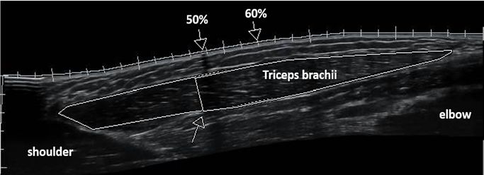
Figure 1. Panoramic sonographic image of triceps brachii muscle. Perimeters of the long head of
Figure 1. Panoramic sonographic image of triceps brachii muscle. Perimeters of the long head of
triceps are shown represented longitudinal muscle area (total, distal, proximal). The arrow on the
triceps are shown represented longitudinal muscle area (total, distal, proximal). The arrow on the
deep aponeurosis of the long head shows the anatomical point where the middle head joins the long
deep aponeurosis of the long head shows the anatomical point where the middle head joins the long
head, which was selected to separate the muscle area as distal (closer to elbow) and proximal (closer
head, which was selected to separate the muscle area as distal (closer to elbow) and proximal (closer
to shoulder). Arrows on the superficial aponeurosis show where muscle thickness, pennation angle,
to shoulder). Arrows on the superficial aponeurosis show where muscle thickness, pennation angle,
and fascicle length were measured at 50% and 60% of the upper‐arm length.
and fascicle length were measured at 50% and 60% of the upper-arm length.
2.5. 1‐RM Elbow Extension
2.5. 1-RM Elbow Extension
Before initiation of the study, all participants followed two familiarization sessions for elbow
Before initiation
extension 1‐RM testingof the study,
of both all extremities.
upper participantsThe followed two familiarization
1‐RM assessment was performed sessions for elbow
according to
extension
previous1-RM testing of
descriptions both
[11] for upper extremities.
each upper extremity The 1-RM
using theassessment was performed
respective exercise according
as used during the to
trainingdescriptions
previous period. After[11]
reporting
for each toupper
the gym, participants
extremity usingperformed a short
the respective warm‐up
exercise (as described
as used during the
previously),
training then
period. onereporting
After set of tento repetitions
the gym, of standing push‐ups
participants performedagainst the warm-up
a short wall and one set of 5
(as described
repetitions then
previously), of submaximal,
one set of unilateral elbow of
ten repetitions extensions.
standingSubsequently, they performed
push-ups against the wall and efforts
onewith
set of
increasing external loads until they were unable to lift a heavier load (6–7 sets
5 repetitions of submaximal, unilateral elbow extensions. Subsequently, they performed efforts within total) for each one
of the two
increasing exercises.
external Two
loads to three
until theyminutes of resttowere
were unable lift aallowed
heavierbetween
load (6–7sets. Oninatotal)
sets different
for occasion,
each one of
the Intraclass Correlation Coefficients (ICC) for elbow extension at S and L was
the two exercises. Two to three minutes of rest were allowed between sets. On a different occasion, measured in our
thelaboratory
Intraclass(ICCs of S = 0.91
Correlation and of L = 0.87,
Coefficients (ICC)n =for
12).elbow extension at S and L was measured in our
laboratory (ICCs of S = 0.91 and of L = 0.87, n = 12).
2.6. Muscle Architecture
2.6. Muscle Architecturemeasurements before and after training were performed during the morning
All ultrasound
hours and after subjects remained supine for 20 min to allow fluid shifts to occur [12]. Pre‐training
All ultrasound measurements before and after training were performed during the morning
ultrasound images were obtained before the familiarization sessions and maximum strength tests,
hours and after subjects remained supine for 20 min to allow fluid shifts to occur [12]. Pre-training
while the post‐training ultrasound images were obtained 5 days after the last training session and 3
ultrasound images were obtained before the familiarization sessions and maximum strength tests,
days after performing the maximum strength tests, in order to avoid osmotic fluid shifts that may
while the post-training ultrasound images were obtained 5 days after the last training session and
confound architectural or morphological measurements. Panoramic B‐mode ultrasound images were
3 days afterwith
obtained performing
a 38‐mmthe maximum
linear strength
probe from tests,head
the long in order
of theto triceps
avoid osmotic fluid the
brachii using shifts that may
“i‐scape”
confound
softwarearchitectural or morphological
of the ultrasound measurements.
device (10.0‐MHz, Product model Panoramic B-mode ultrasound
Z5, Shenzhen, images were
Mindray Bio‐Medical
obtained with a 38-mm linear probe from the long head of the triceps brachii
Electronics Co., Ltd., Shenzhen, China). Initially, participants were placed at a standing position using the “i-scape”
with
software of the
their arms ultrasound
fully extended device (10.0-MHz,
to the side Product
of the body. The model
posterior Z5,surfaces
Shenzhen, of theMindray
acromion Bio-Medical
and the
Electronics Co., Ltd., of
lateral epicondyles Shenzhen,
the humerus China).
were Initially,
markedparticipants
and used as were placed
the total at aofstanding
length the upper position
arm [13].with
their
The upper arm (starting from acromion) was marked at 50% and 60% of the upper‐arm length withthe
arms fully extended to the side of the body. The posterior surfaces of the acromion and
lateral epicondyles
a permanent of the humerus
pen. Self‐adhesive were
paper wasmarked
placed andon theused
skinasatthe
thesetotal
two length
pointsofasthe upper (image
a marker arm [13].
The upper armThen,
shadowing). (starting from acromion)
participants waswith
laid supine markedtheirat 50%rested
arms and 60% on aof the upper-arm
laboratory bed at length with a
90° to their
torso, fully
permanent extended
pen. on elbowpaper
Self-adhesive joint and
wassupinated.
placed onParticipants
the skin at were
theseasked to isometrically
two points as a marker contract
(image
their triceps,Then,
shadowing). so that the researcher
participants laid could
supinelocate
withthe theirinsertion of the on
arms rested triceps long headbed
a laboratory using
at 90 ◦ to
the
ultrasound
their image.
torso, fully Then, the
extended ontransducer
elbow joint was placed
and laterally Participants
supinated. to the arm and oriented
were asked in to
parallel to the
isometrically
muscle fascicles. The transducer’s alignment was considered appropriate when
contract their triceps, so that the researcher could locate the insertion of the triceps long head using several fascicles
thecould be easily
ultrasound outlined
image. Then, without interruption
the transducer wasacross
placed thelaterally
image. Theto thefascicle
arm and pathoriented
was drawn on the to
in parallel
the muscle fascicles. The transducer’s alignment was considered appropriate when several obtain
skin with a pen according to the fascicle path seen from the real‐time ultrasound image. To fascicles
the panoramic image, a continuous single view was taken by moving the probe along the marked
could be easily outlined without interruption across the image. The fascicle path was drawn on the
line [14]. Images were analyzed (as previously aforementioned) for muscle thickness, fascicle angle,
skin with a pen according to the fascicle path seen from the real-time ultrasound image. To obtain the
and fascicle length at 50% and 60% of the upper‐arm length [14]. The two‐dimensional longitudinal
panoramic image, a continuous single view was taken by moving the probe along the marked line [14].
Images were analyzed (as previously aforementioned) for muscle thickness, fascicle angle, and fascicleJ. Funct. Morphol. Kinesiol. 2018, 3, 28 5 of 10
length at 50% and 60% of the upper-arm length [14]. The two-dimensional longitudinal muscle area
was calculated in three regions: total, distal, and proximal with image analysis software (Motic Images
Plus 2.0, Motic, Hong Kong, China). Longitudinal muscle area was measured using the “polygon”
tool of the image analyzing program to analyze the total, proximal, and distal area of the long head of
triceps brachii. In each sonographic image, an anatomical point of the deep aponeurosis was identified
where the mid triceps head joins the long head. This anatomical point was selected in order to separate
the distal (closer to the elbow) and proximal area (closer to shoulder) of the muscle (Figure 1).
Intersession reliability was determined by comparing the analysis of the images obtained by
six participants (both arms) on two separate days when skin markings were completely removed.
The ICC for the measurement of muscle thickness was 0.984 (95% Confidence Intervals (CI): 0.951–0.995,
p = 0.001), ICC for fascicle angle was 0.858 (95% CI: 0.626–0.952, p = 0.001), and for fascicle length it
was 0.794 (95% CI: 0.483–0.928, p = 0.001). The ICC for the measurement of longitudinal muscle area
was 0.963 (95% CI: 0.887–0.988), p = 0.001.
2.7. Statistical Analysis
A post hoc power analysis, was performed according to the study design, the number of the
participants, and the lower partial eta Squared of the significant contrasts or of the Pearson’s r
correlations coefficient that were found, which reveal an actual power of 0.889 for the results of the
present study [15]. Accordingly, the power for the correlation coefficients ranged between 0.817 and 0.900.
All data reports are given as mean ± SD. A series of one-way analyses of variance (ANOVA) were
performed for all baseline data trials to identify any significant differences between training conditions.
As expected, a significant difference was found between the S and the L arms for 1-RM at the initiation
of the training period. Therefore, repeated measures analysis of covariance (ANCOVA; with 1-RM
pre-test values as covariance) was performed to evaluate differences in 1-RM between S and L, whereas
for the rest of the variables separate analyses of variance with repeated measures were performed.
Post-hoc Bonferroni analysis was used to examine pairwise differences whenever a significant F-value
was obtained. Paired-sample t-tests and independent-sample t-tests were performed to evaluate
training-related changes within groups and between training conditions, respectively. Partial eta
squared was also performed; according to Richardson [16], partial eta squared can be used as an
indicator of effect size, and it can be classified as small (0.01 to 0.059), moderate (0.06 to 0.137),
and large (≥0.138). Pearson’s (r) product-moment correlation coefficients were computed to explore
the relationships between variables. The interpretation of the observed correlations was performed
according to Hopkins’ ranking: correlations coefficients between 0.3–0.5 were considered moderate,
between 0.51–0.70 large, between 0.71–0.90 very large, and >0.91 almost perfect [17]. The significance
level was set at p < 0.05. ICCs with 95% CI (two-way random effects with absolute agreement) were
calculated for muscle architecture and longitudinal muscle area. All statistical analyses were performed
using SPSS version 21.0 software (SPSS Inc., Chicago, IL, USA).
3. Results
Elbow extension 1-RM increased significantly by 40.1 ± 21.3% (from 14.86 ± 2.2 kg to 20.83 ±
4.8 kg, p = 0.001) and 44.5 ± 20.1% (from 8.56 ± 2.7 kg to 12.08 ± 3.0 kg, p = 0.000) after S and L training,
respectively, while ANCOVA revealed no difference for these changes between groups (p = 0.640,
η 2 = 0.015).
Results for muscle architecture are presented at Table 1. Muscle thickness at 50% (MT50%) and
60% (MT60%) of the upper arm length increased significantly, while fascicle length (fL50%, fL60%)
and fascicle angle (Angle50%, Angle60%) were not altered significantly either after S or L training,
respectively. There were no differences between groups for any of the muscle architecture variables
at 50% or 60% of the upper-arm length. Analysis of the two-dimensional longitudinal muscle area
revealed a significant increase in total and distal area, while proximal area was not altered significantly
after S and L training, respectively (Table 1). There were no differences between groups for the
longitudinal muscle area (total, distal, or proximal, Table 1).J. Funct. Morphol. Kinesiol. 2018, 3, 28 6 of 10
Table 1. Triceps brachii long head architecture characteristics and longitudinal area (means ± SD) before and after 6 weeks of resistance training at short (S) and
long (L) fascicle length in nine novice physical education female students. p values, independent t-test values, and eta2 for differences between training conditions
are presented.
Time Points/ Short Fascicle Length Long Fascicle Length p Values for the Comparisons
Parameter η2
Percentage Changes Training Group Training Group between the Groups
Before 1.35 ± 0.26 1.29 ± 0.20
0.448 0.036
MT50% (cm) After 1.47 ± 0.21 * 1.46 ± 0.20 §
% change 10.7 ± 15.3 13.7 ± 9.0 0.618
Before 7.95 ± 1.43 8.67 ± 1.36
0.374 0.050
fL50% (cm) After 8.56 ± 1.50 8.72 ± 1.60
% change 9.0 ± 16.8 1.3 ± 15.2 0.323
Before 11.78 ± 2.59 10.89 ± 2.26
0.263 0.078
Angle50% (degrees) After 11.33 ± 2.12 11.78 ± 2.11
% change −1.2 ± 18.8 10.3 ± 21.2 0.241
Before 1.23 ± 0.25 1.22 ± 0.21
0.565 0.021
MT60% (cm) After 1.40 ± 0.25 * 1.43 ± 0.19 §
% change 15.5 ± 18.8 19.4 ± 16.3 0.641
Before 9.68 ± 2.60 9.46 ± 1.71
0.737 0.007
fL60% (cm) After 9.85 ± 1.98 9.28 ± 2.13
% change 6.4 ± 29.4 −1.5 ± 16.3 0.491
Before 11.33 ± 2.45 11.22 ± 1.64
0.359 0.053
Angle60% (degrees) After 11.00 ± 2.12 12.00 ± 2.06
% change 0.3 ± 25.6 7.2 ± 13.5 0.485
Before 16.77 ± 3.71 16.56 ± 3.82
0.917 0.001
Area Total (cm2 ) After 19.24 ± 4.53 * 18.92 ± 3.52 §
% change 15.3 ± 16.4 16.1 ± 14.1 0.912
Before 10.76 ± 2.00 10.43 ± 2.54
0.706 0.009
Area Distal (cm2 ) After 12.59 ± 3.39 * 12.65 ± 2.35 §
% change 16.7 ± 20.3 24.9 ± 25.3 0.460
Before 5.93 ± 1.98 6.34 ± 1.62
0.260 0.079
Area Proximal (cm2 ) After 6.65 ± 2.28 6.27 ± 2.04
% change 13.5 ± 27.8 −0.1 ± 23.0 0.274
* p ≤ 0.05, § p ≤ 0.01 Significant difference after training in each group separately between pre- and post-evaluations. MT50%; MT60%, fL50%; fL60%; Angle50%; Angle60% = muscle
thickness, fascicle length, and pennation angle measured at 50% and 60% of upper arm length, respectively.J. Funct. Morphol. Kinesiol. 2018, 3, 28 7 of 10
J. Funct. Morphol. Kinesiol. 2018, 3, x 7 of 10
Significant
Significant correlations
correlations were
were revealed
revealed between
between thethe percent
percent change
change ofof 1‐RM
1-RM and
and percent
percent change
change
of
of fL50%
fL50% (r (r == −0.
−0.723,
723,pp==0.006)
0.006)and
andpercent
percentchange
changeof ofAngle60%
Angle60%(r(r ==0.827,
0.827,pp ==0.028)
0.028) only
only after
after LL
training (Figure 2). 1‐RM significantly correlated with MT50%, MT60%, and longitudinal
training (Figure 2). 1-RM significantly correlated with MT50%, MT60%, and longitudinal muscle area muscle areaat
at post‐training period (r = 0.706–0.866, p < 0.05, Table 2). No significant correlations
post-training period (r = 0.706–0.866, p < 0.05, Table 2). No significant correlations were found betweenwere found
between
the initialthe
1-RMinitial
and1‐RM
MT50%,andMT60%,
MT50%,andMT60%,
muscleand
areamuscle
before area before
training for training for (p
the S group the
> S0.05),
group (p >
as well
0.05), as well as for the MT60% for the L group (p > 0.05). Finally, in both groups, the
as for the MT60% for the L group (p > 0.05). Finally, in both groups, the 1-RM after the training was 1‐RM after the
training was not correlated with distal muscle
not correlated with distal muscle area (p > 0.05). area (p > 0.05).
Figure 2. Correlation between the percent change in elbow extension 1-RM and the percent change
Figure 2. Correlation between the percent change in elbow extension 1‐RM and the percent change in
in fascicle length (A), and fascicle angle (B) of the long head of the triceps brachii after 6 weeks of
fascicle length (A), and fascicle angle (B) of the long head of the triceps brachii after 6 weeks of
resistance training at long fascicle length in nine novice female physical education students. RM:
resistance training at long fascicle length in nine novice female physical education students. RM:
repetition maximum.
repetition maximum.
Table 2. Correlation coefficients between elbow extension 1-RM and triceps brachii long head muscle
Table 2. Correlation coefficients between elbow extension 1‐RM and triceps brachii long
thickness and two-dimensional longitudinal muscle area, before and after the training period.
head muscle thickness and two‐dimensional longitudinal muscle area, before and after the
training period.Triceps Brachii Long Head Muscle Thickness at
SHORT Muscle Area before Training
50% or 60% of Upper-Arm Length before Training
Triceps Brachii Long Head Muscle Thickness at 50% or 60% of
SHORT 50% 60% Total Distal
Muscle Area before Proximal
Training
1-RM before Upper‐Arm Length before 0.528
0.517 Training 0.426 0.413 0.460
50% 60% Total Distal Proximal
Triceps Brachii Long Head Muscle Thickness at
1‐RM Muscle Area after Training
0.51750% or 60% of Upper-Arm Length0.528
after Training 0.426 0.413 0.460
before
50%
Triceps Brachii 60% at 50% or 60% of
Long Head Muscle Thickness Total Distal Proximal
1-RM after 0.706 * 0.831 § 0.804 § Muscle Area after Training
0.575 0.742 *
Upper‐Arm Length after Training
50%Triceps Brachii Long Head Muscle 60%Thickness at Total Distal Proximal
LONG Muscle Area§ before Training
1‐RM after 50%
0.706 * or 60% of Upper-Arm Length before
0.831 § Training 0.804 0.575 0.742 *
Triceps Brachii
50%Long Head Muscle Thickness
60% at 50% or 60% of
Total Distal Proximal
LONG Muscle Area before Training
1-RM before Upper‐Arm
0.748 * Length before 0.610
Training 0.866 § 0.715 * 0.864 §
50% 60% Total Distal Proximal
Triceps Brachii Long Head Muscle Thickness at
1‐RM Muscle Area after Training
50%
0.748 * or 60% of Upper-Arm Length after
0.610 Training 0.866 § 0.715 * 0.864 §
before
50% 60% Total Distal Proximal
Triceps Brachii Long Head Muscle Thickness at 50% or 60% of
1-RM after 0.804 § 0.779 * 0.756 * Muscle Area after Training
0.586 0.728 *
Upper‐Arm Length after Training
* Significant at p < 60% §
0.05; Significant at p < 0.01.
50% Total Distal Proximal
1‐RM after 0.804 § 0.779 * 0.756 * 0.586 0.728 *
* Significant at p < 0.05; § Significant at p < 0.01.
4. Discussion
The main finding of the present study was that elbow extension strength using either the cable
4. Discussion
push-downs or the cable overhead extensions (i.e., at short or long fascicle length training, respectively)
The main finding of the present study was that elbow extension strength using either the cable push‐
was similarly increased after 6 weeks of resistance training in female novice participants. The former
downs or the cable overhead extensions (i.e., at short or long fascicle length training, respectively) was
exercise is performed at shorter fascicle length compared with the latter exercise, as estimated in
similarly increased after 6 weeks of resistance training in female novice participants. The former exercise
is performed at shorter fascicle length compared with the latter exercise, as estimated in the present study.J. Funct. Morphol. Kinesiol. 2018, 3, 28 8 of 10
the present study. This suggests that during the first weeks of resistance training when muscle
strength development is rapid, the fascicle length during exercise has a relatively low effect on strength
increments. Interestingly, triceps brachii long head muscle thickness as well as longitudinal muscle
area were similarly increased with both exercises. This suggests that triceps brachii long head is
similarly recruited with both exercises. As expected, the increase in elbow extensors’ strength was
much higher (40–44%) than the increase in muscle thickness (10–15%) and longitudinal area (14–25%).
This suggests the presence of neural adaptations, which may be explained by the novice nature of the
participants [18–20]. Longer training periods might have resulted in a greater involvement of muscle
hypertrophy in the strength increases observed, which might have also induced greater triceps brachii
architectural adaptations. However, muscle thickness was similarly increased after the two training
conditions, which is in agreement with a recent study in the lower extremities [5]. This may suggest a
similar recruitment pattern for the long head of triceps brachii between the two exercises used in this
study, despite the different ranges of fascicle lengths during training.
In a recent study [8], it was found that training at long fascicle length induced greater increases
in strength, distal muscle cross sectional area, and fascicle length of vastus lateralis accompanied by
increases in Insulin-Like Growth Factor 1 (IGF-1), as compared to training at short fascicle length.
It is worth noting that the range of fascicle length used during training was not measured in that
previous study [8]. Nevertheless, the apparent discrepancy with the current results might be related to
the longer training duration and/or the training experience of the participants. It may be speculated
that architectural adaptations might have followed the initial 12 training sessions in the current study.
Indeed, the small, however non-significant difference in strength increase (44% vs. 40%), muscle
thickness (15% vs. 10%), and hypertrophy (16–25% vs. 14–17%) increase in favor of the cable overhead
extensions exercise might be an indication for such adaptations, particularly if training was to be
continued for a longer duration. Also, in the study of McMahon et al. (2014), training was performed
for 8 weeks, with three sessions per week, and 4 sets of 8–10 reps for 4–5 exercises per session in
moderately trained males, which also suggests a great variance in the training load in favor of the
earlier study [8]. Although application of a large training load would be inappropriate in the current
study due to the novice nature of the participants, it may not be excluded that the lack of difference
in fascicle length increase between the current study and the study of McMahon et al. (2014) may
be due to the differences in the training volume applied. Moreover, it should be mentioned that the
lower extremities are used more often in everyday life compared to the elbow extensors, which might
imply an elevated initial baseline for vastus lateralis muscle in the study by McMahon et al. (2014),
as compared to the less trained triceps brachii in novice females.
While strength training induced significant increases in strength, the muscle architecture of the
long head of the triceps did not change significantly, which is in accordance with a previous study [21].
Interestingly, in this previous study, there was a uniform architectural adaptation at 50% and 60% of
the triceps brachii length, as found here. Before the initiation of the study it was hypothesized that
training at long muscle length would induce increases in muscle fascicle length, since such a result
has been reported before [8]. However, it seems that the short training duration together with the
novice nature of the participants did not allow for such an adaptation to occur. Similarly, a previous
study reported that 5 weeks of resistance training were inadequate to alter muscle architecture of the
quadriceps, even if strength was increased [22]. The results of the present study revealed that the
fascicle length of the triceps long head was not altered after either S or L training, which is in agreement
with results reported from Alegre et al. (2014) [5]. The current results suggest that performing either
the cable push-downs (short fascicle length) or the cable overhead extensions (long fascicle length)
may not significantly alter the fascicle length of the triceps brachii long head, at least during the first
6 weeks of training, in novice female participants.
Triceps brachii is an important contributor to several sports such as the track and field throws [23],
baseball, and rugby, and it determines most of the muscle volume of the upper arm [24]. There
are a lot of resistance training exercises that are used to develop its mass and strength, however,J. Funct. Morphol. Kinesiol. 2018, 3, 28 9 of 10
little is known about the effectiveness of these exercises. The present study investigated for the first
time the resistance training adaptations using two different exercises where the long head of the
triceps brachii is working at different fascicle lengths. However, there are certain limitations in the
current study. The first limitation was the different training loads between the two experimental
groups. However, this was due to the nature of these exercises. In addition, as mentioned above,
the large increase in muscle strength combined with a much lower increase in muscle thickness suggest
strong neural adaptations after training either with cable push-downs or cable overhead extensions.
Unfortunately, it was not possible to evaluate the electromyographic signals from triceps brachii in this
study, which might have provided a better insight into the training adaptations since such result has
been that occurred. Likewise, a longer training period might have increased the muscular component
of the training adaptations, which would probably have provided a clearer illustration of the triceps
brachii architectural changes. Another possible limitation of this study was the lack of analysis of the
architectural characteristics of the other two heads of the triceps brachii. Given the anatomical points of
origin and insertion of these triceps brachii heads, it may be assumed that their fascicle lengths would
behave similarly as those of the long head during cable push-downs and cable overhead extensions.
However, this question should be addressed in a future study.
The results of the present study are of practical importance. The results of this study suggest that
during the first weeks of resistance training both cable push-downs and cable overhead extensions are
equally effective at inducing elbow extension muscle strength in novice young females. This increase
in strength seems to be partly due to increases in muscle mass of the triceps long head while neural
factors may contribute significantly. This type of resistance training seems not to affect the muscle
architecture (fascicle angle and length) of the triceps long head. Resistance training for six weeks in
novice participants does not seem to be an adequate stimulus for fascicle length increase. In addition,
it seems that when young, novice female participants are involved, just two resistance training sessions
per week with sets of 6-RM provide an adequate stimulus for elbow extension muscle strength increase
and hypertrophy.
5. Conclusions
In conclusion, muscle strength of elbow extensors increased similarly with cable push-downs and
cable overhead extensions during the first six weeks of resistance training in novice female participants.
Muscle thickness also increased similarly. These two different exercises force the muscle to work in
short and long fascicle lengths, respectively. This suggests that during the first weeks of resistance
training, when triceps brachii muscle strength development is rapid, the fascicle length during exercise
has a relatively low effect on strength increments and muscle architecture.
Author Contributions: A.-N.S., S.M., and G.T. conceived and designed the experiments; A.-N.S., N.Z., S.T.,
Ar.K., and An.K. performed the experiments and analyzed the data; A.-N.S., S.M., and G.T. contributed
reagents/materials/analysis tools; A.-N.S., S.M., and G.T. wrote the paper.
Acknowledgments: We wish to express our gratitude to the subjects of the present study. All experimental
procedures used comply with local governmental laws for human subjects. No grant support was received for
this study.
Conflicts of Interest: The authors declare no conflict of interest
References
1. Pedersen, B.K.; Saltin, B. Exercise as medicine–evidence for prescribing exercise as therapy in 26 different
chronic diseases. Scand. J. Med. Sci. Sports 2015, 25, 1–72. [CrossRef] [PubMed]
2. Fleck, J.; Kraemer, W. Designing Resistance Training Programs, 3rd ed.; Human Kinetics: Champaing, IL,
USA, 2009.
3. Vogt, M.; Hoppeler, H. Eccentric exercise: Mechanisms and effects when used as training regime or training
adjunct. J. Appl. Physiol. 2014, 116, 1446–1454. [CrossRef] [PubMed]
4. Agur, A.; Dalley, A. Grant's Atlas of Anatomy; Lippincott Williams & Wilkins: Philadelphia, PA, USA, 2009.J. Funct. Morphol. Kinesiol. 2018, 3, 28 10 of 10
5. Alegre, L.M.; Ferri-Morales, A.; Rodriguez-Casares, R.; Aguado, X. Effects of isometric training on the knee
extensor moment–angle relationship and vastus lateralis muscle architecture. Eur. J. Appl. Physiol. 2014, 114,
2437–2446. [CrossRef] [PubMed]
6. Noorkõiv, M.; Nosaka, K.; Blazevich, A.J. Neuromuscular adaptations associated with knee joint
angle-specific force change. Med. Sci. Sports Exerc. 2014, 46, 1525–1537. [CrossRef] [PubMed]
7. Noorkõiv, M.; Nosaka, K.; Blazevich, A. Effects of isometric quadriceps strength training at different muscle
lengths on dynamic torque production. J. Sports Sci. 2015, 33, 1952–1961. [CrossRef] [PubMed]
8. McMahon, G.; Morse, C.; Burden, A.; Winwood, K.; Onambélé, G.L. Muscular adaptations and insulin-like
growth factor-1 responses to resistance training are stretch-mediated. Muscle Nerve 2014, 49, 108–119.
[CrossRef] [PubMed]
9. MacInnis, M.; McGlory, C.; Gibala, M.; Phillips, S. Investigating human skeletal muscle physiology with
unilateral exercise models: When one limb is more powerful than two. Appl. Physiol. Nutr. Metab. 2017, 42,
563–570. [CrossRef] [PubMed]
10. Lasevicius, T.; Ugrinowitsch, C.; Schoenfeld, B.; Roschel, H.; Tavares, L.D.; De Souza, E.O.; Laurentino, G.;
Tricoli, V. Effects of different intensities of resistance training with equated volume load on muscle strength
and hypertrophy. Eur. J. Sport Sci. 2018, 22, 1–9. [CrossRef] [PubMed]
11. Baechle, T.R.; Earle, R.W.; Wathen, D. Resistance training. In Essentials of Strength Training and Conditioning,
2nd ed.; Baechle, T.R., Earle, R.W., Eds.; Human Kinetics: Champaign, IL, USA, 2000; pp. 395–425.
12. Reeves, N.; Maganaris, C.; Narici, M. Ultrasonographic assessment of human skeletal muscle size. Eur. J.
Appl. Physiol. 2004, 91, 116–118. [CrossRef] [PubMed]
13. Miyatani, M.; Kanehisa, H.; Ito, M.; Kawakami, Y.; Fukunaga, T. The accuracy of volume estimates using
ultrasound muscle thickness measurements in different muscle groups. Eur. J. Appl. Physiol. 2004, 91,
264–272. [PubMed]
14. Noorkoiv, M.; Stavnsbo, A.; Aagaard, P.; Blazevich, A. In vivo assessment of muscle fascicle length by
extended field-of-view ultrasonography. J. Appl. Physiol. 2010, 109, 1974–1979. [CrossRef] [PubMed]
15. Faul, F.; Erdfelder, E.; Lang, A.G.; Buchner, A. G* power 3: A flexible statistical power analysis program for
the social, behavioral, and biomedical sciences. Behav. Res. Methods 2007, 39, 175–191. [CrossRef] [PubMed]
16. Richardson, J.T. Eta squared and partial eta squared as measures of effect size in educational research.
Educ. Res. Rev. 2011, 6, 135–147. [CrossRef]
17. Hopkins, W.G. Measures of reliability in sports medicine and science. Sports Med. 2000, 30, 1–15. [CrossRef]
[PubMed]
18. Narici, M.V.; Roi, G.S.; Landoni, L.; Minetti, A.E.; Cerretelli, P. Changes in force, cross-sectional area and
neural activation during strength training and detraining of the human quadriceps. Eur. J. Appl. Physiol.
1989, 59, 310–319. [CrossRef]
19. Aagaard, P.; Simonsen, E.B.; Andersen, J.L.; Magnusson, P.; Dyhre-Poulsen, P. Neural adaptation to resistance
training: Changes in evoked V-wave and H-reflex responses. J. Appl. Physiol. 2002, 92, 2309–2318. [CrossRef]
[PubMed]
20. Sale, D.G. Neural adaptation to resistance training. Med. Sci. Sports Exerc. 1988, 20, S135–S145. [CrossRef]
[PubMed]
21. Matta, T.; Simão, R.; de Salles, B.F.; Spineti, J.; Oliveira, L.F. Strength training’s chronic effects on muscle
architecture parameters of different arm sites. J. Strength Cond. Res. 2011, 25, 1711–1717. [CrossRef] [PubMed]
22. Blazevich, A.; Gill, N.; Bronks, R.; Newton, R. Training-specific muscle architecture adaptation after 5-wk
training in athletes. Med. Sci. Sports Exerc. 2003, 35, 2013–2022. [CrossRef] [PubMed]
23. Terzis, G.; Karampatsos, G.; Georgiadis, G. Neuromuscular control and performance in shot-put athletes.
J. Sports Med. Phys. Fit. 2007, 47, 284–290.
24. Ogasawara, R.; Thiebaud, R.; Loenneke, J.; Loftin, M.; Abe, T. Time course for arm and chest muscle thickness
changes following bench press training. Interv. Med. Appl. Sci. 2012, 4, 217–220. [CrossRef] [PubMed]
© 2018 by the authors. Licensee MDPI, Basel, Switzerland. This article is an open access
article distributed under the terms and conditions of the Creative Commons Attribution
(CC BY) license (http://creativecommons.org/licenses/by/4.0/).You can also read