Translational research on Myology & Mobility Medicine: 2021 semi-virtual PDM3 from Thermae of Euganean Hills, May 26 - 29, 2021
←
→
Page content transcription
If your browser does not render page correctly, please read the page content below
2021 PDM3 Program and Abstracts, May 26 - 29, 2021
                                     Eur J Transl Myol 2021; 31 (1): 9743. doi: 10.4081/ejtm.2021.9743

                          Translational research on Myology & Mobility Medicine: 2021 semi-
                          virtual PDM3 from Thermae of Euganean Hills, May 26 - 29, 2021
                          Ugo Carraro (1, 2, 3), Zipora Yablonka-Reuveni (4)

                          (1) Department of Biomedical Sciences of the University of Padova, Italy; (2) CIR-Myo -
                          Myology Centre, University of Padova, Italy; (3) A&C Mioni-Carraro Foundation for
                          Translational Myology, Padova, Italy; (4) Department of Biological Structure, University of
                          Washington School of Medicine, Seattle, WA, USA.
                          This article is distributed under the terms of the Creative Commons Attribution Noncommercial License (CC BY-NC 4.0) which permits
                          any noncommercial use, distribution, and reproduction in any medium, provided the original author(s) and source are credited.

                          Abstract

                          On 19-21 November 2020, the meeting of the 30 years of the Padova Muscle Days was virtually
                          held while the SARS-CoV-2 epidemic was hitting the world after a seemingly quiet summer.
                          During the 2020-2021 winter, the epidemic is still active, despite the start of vaccinations. The
                          organizers hope to hold the 2021 Padua Days on Myology and Mobility Medicine in a semi-
                          virtual form (2021 S-V PDM3) from May 26 to May 29 at the Thermae of Euganean Hills,
                          Padova, Italy. Here the program and the Collection of Abstracts are presented. Despite numerous
                          world problems, the number of submitted/selected presentations (lectures and oral presentations)
                          has increased, prompting the organizers to extend the program to four dense days.
                          Key Words: Padua Muscle Days; skeletal muscle; COVID-19; Translational Myology and
                          Mobility Medicine; 2021 PDM3 Program & Abstracts.
                                                                       Eur J Transl Myol 2021; 31 (1): 9743. Doi: 10.4081/ejtm.2021.9743

Meetings on muscle biology, physiology, medicine and                          Program of Sessions and the Collection of Abstracts
rehabilitation, the Padua Muscle Days (PMDs), started                         follow. Despite the debilitating problems, the number of
more than 30 years ago to provide expert advices on                           selected presentations (lectures and oral presentations)
Translational Myology and Mobility Medicine.1-3 The                           has increased to fill in an extend program of four dense
aims were on prevention, managing and rehabilitation of                       days. Beside the traditional sessions, the Program
mobility disorders. Last year the organizers programmed                       presents several new topics: Extracellular Matrix,
for March 2020 an intense 3-day program to be held                            Fasciae & Tendons; Muscle and Sarcomeres in vivo;
either at Padova University or at the Hotel Augustus,                         High density EMG and motor control in health and
Thermae of Euganean Hills (Padova), Italy.4 Abruptly, in                      disease; Mechanisms of Skeletal Muscle Impairment in
January 2020 the SARS CoV-2 outbreak changed the                              Aging and Disease; Muscle Atrophy, Sarcopenia,
world perspectives. In Italy the virus was first detected in                  Cachexia: Physical, Nutritional, and Pharmacological
Lombardy and Veneto. Within a week, the COVID-2019                            Interventions; Light and Electron Microscopy Muscle
affected the first Italian victims in an area south of Milan                  Imaging; Satellite Cells and Muscle Regeneration:
and in a village of the Euganean Hills (Vo Euganeo,                           Biology & Pathology; Therapy of Genetic Muscle
Padova). The village was immediately quarantined, but it                      Dystrophies; Muscle Rehabilitation: Balneology &
was too late. The virus was spreading through the area.                       Balneotherapy; Muscle Rehabilitation: Clinical Aims &
Thus, the PMDs were postponed and successfully held as                        Case Reports; Muscle Rehabilitation: Roles of
Virtual ZOOM Series of Presentations from 19 to 21                            Mitochondria & Excitation-Contraction Coupling.
November, 2020. Luckily, chairs, speakers and attendees                       We hope 2021PDM3 will be as successful as the events
accepted the decisions.5                                                      of the previous 30 years. The Speakers will be double the
In 2021 COVID-19 epidemic is still active, despite                            usual, imposing an extension to four days. It is hoped that
vaccinations. The organizers hope to hold the 2021 Padua                      all Lectures, Speakers and Attendees will patiently wait
Days on Myology & Mobility Medicine (PDM3) semi-                              for the next difficult step: reschedule the time slots to
virtually form May 26 to May 29 at the Thermae of                             allow the interactive presence of people who will
Euganean Hills, Padova, Italy. Here a preliminary                             participate from time zones distributed over 24 hours.
                                                                              It will be a difficult task, but we will succeed.

                                                                       -1-
2021 PDM3 Program and Abstracts, May 26 - 29, 2021
                             Eur J Transl Myol 2021; 31 (1): 9743. doi: 10.4081/ejtm.2021.9743

    TRANSLATIONAL RESEARCH ON MYOLOGY & MOBILITY MEDICINE
     May 26 - 29, 2021 – Thermae of Euganean Hills (Padova), Italy

                                   .

WEDNESDAY May 26, 2021
 9:30 AM     Openings
10:00 AM     Session I:        Extracellular Matrix, Fasciae & Tendons
 3:00 PM     Session II:       Muscle & Sarcomeres in vivo
 5:20 PM     Session III:      High density EMG applied to motor control in health & disease
THURSDAY May 27, 2021
 9:00 AM     Session IV: Poster Presentations
11:00 AM     Session V:   Muscle Atrophy, Sarcopenia, Cachexia: Physical, Nutritional &
             Pharmacological Interventions
 3:00 PM     Session VI: Mechanisms of Skeletal Muscle Impairment in Aging & Disease
5:20 PM      Session VII: Light & Ultrastructural Muscle Imaging
FRIDAY May 28, 2021
 9:00 AM     Session VIII:     Poster Presentations
10:30 AM     Session IX:       Satellite Cells & Muscle Regeneration: Biology & Pathology
 3:00 PM     Session X:        Satellite Cells & Muscle Regeneration: Biology & Pathology
 5:20 PM     Session XI:       Therapy of Genetic Muscle Dystrophies
SATURDAY May 29, 2021
 9:00 AM     Session XII: Muscle Rehabilitation: Balneology & Balneotherapy
11:30 AM     Session XIII: Muscle Rehabilitation: Clinical Aims & Case Reports
 3:00 PM     Session XIV: Muscle Rehabilitation: Roles of Mitochondria & E-C Coupling
 5:00 PM     GENERAL DISCUSSION OF THE CENTER FOR ACTIVE AGING GROUP
7:30 PM      Ugo Carraro:      See you to the 2022 PDM3, March, 2022-Thermae of Euganean Hills & Padova, Italy

Translational Research on Myology & Mobility Medicine
Call for EJTM COMMUNICATIONS - EJTM 31 (2), 2021 – Deadline May 31, 2021
(2500 words, 150-word Abstract, 5 keywords, 4 Figures/Tables, 30 Refs).
EJTM is indexed in PUBMED, ESCI and SCOPUS - CiteScoreTracker 2020 = 2.00
Submit to: pagepressjournals.org/index.php/bam/about/submissions

                                                          -2-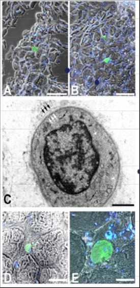
2021 PDM3 Program and Abstracts, May 26 - 29, 2021
                                   Eur J Transl Myol 2021; 31 (1): 9743. doi: 10.4081/ejtm.2021.9743

           2021 Semi-Virtual PDM3 - May 26 to29
                       Thermae of Euganean Hills (Padova), Italy, May 26 - 29, 2021
     Hotel Petrarca, Piazza Roma 23, Montegrotto Terme, Thermae of Euganean Hills, (Padova), 35122 Italy
              Phone +39 049 891 1744 - Email: petrarca@hotelpetrarca.it - https://www.hotelpetrarca.it/
 Organizers: Ugo Carraro, Helmut Kern, Christiaan Leeuwenburgh, Marco Narici, Marco Sandri, Carla Stecco, Zipora Yablonka-Reuveni

                                       Collection of Abstracts

#1. May 26, 2021                                                       #2. May 26, 2021
Invited Lecture: Physical exercise and adaptation of                   Effect of aging on tendon mechanical properties and
connective tissue in tendon and muscle                                 adaptability to chronic loading
Michael Kjaer                                                          Marco Narici
(1) Institute of Sports Medicine, Dept. Orthopedic                     CIR-Myo Myology Centre, Neuromuscular Physiology
Surgery, Copenhagen University Hospital – Bispebjerg,                  Laboratory, Department of Biomedical Sciences,
and Center for Healthy Aging, Faculty of Health and                    University of Padova, Padova, Italy
Medical Sciences, University of Copenhagen, Denmark
                                                                       * Marco Narici: marco.narici@unipd.it
Michael Kjaer: michaelkjaer@sund.ku.dk
                                                                       Tendons play a fundamental role in transmitting forces
The overall turnover of the tendon and cartilage in                    generated by skeletal muscle and in acting as springs
humans seems to be taking place primarily within the                   during locomotion. Because of their anatomical
first 13-17 years of life (markedly lower than turnover of             arrangement, the mechanical properties of tendons affect
contractile protein in muscle), indicating that the major              those of skeletal muscle, above all, the length-force
basic structure remains relatively unchanged through                   relation.1,2 With ageing, human tendon mechanical
adult life. Nevertheless, mechanical loading of adult                  properties (stiffness, Young’s modulus and hysteresis)
human tendon (and intramuscular connective tissue)                     seem to deteriorate,3-6 although reports of no changes also
results in tendon cell responses by producing anabolic                 exist.7 These alterations potentially contribute to the loss
growth factors and loading-induced increase in tendon                  of muscle force with ageing.8 However, tendons retain
collagen synthesis. Comparing tissue turnover in                       considerable plasticity in response to chronic loading in
different tissues simultaneously suggests that a                       old age since their mechanical properties can be
combination of a more basic structure that remains                     substantially improved by high-intensity resistance
relatively unchanged through adult life, and a smaller                 exercise training (RET). Indeed, after 14 weeks of RET
pool of collagen that is more quickly turned over and can              in septuagenarian males, patellar tendon stiffness and
be influenced by mechanical loading. Conversely,                       Young’s modulus and hysteresis were found to improve
physical inactivity down regulates collagen synthesis and              by 65% and 69% respectively, restoring tendon
phenotypic tendon characteristics. Adjustment of the                   properties to values comparable to those of young
tendon mechanical properties in the form of increased                  adults.9,10 These improvements were mostly due to
stiffness and modulus after strength training, and the                 changes in tendon tissue material properties rather than
reverse after period of immobilization occurs relatively               to tendon hypertrophy. Also, the increase in tendon
faster than macroscopic morphological changes of                       stiffness was associated with a faster rate of force
tendon. Age related changes in tendon connective tissue                development, a finding of particular relevance for falls
with reduced stiffness is largely explained by a reduction             prevention. While traditional RET consists of both
in overall physical activity and can thus be at least partly           concentric (CON) and eccentric (ECC) contractions,
counteracted by regular training.                                      ECC contractions are metabolically less demanding and
                                                                       therefore may be more suitable for an older population.
Keywords: Physical exercise; adaptation; connective
                                                                       Thus, we investigated the tendon adaptations and time
tissue; tendon; muscle.
                                                                       course, to 8-week, 3 times/week, submaximal (60%
References                                                             1RM) CON only or ECC only RET in 20 healthy young
1. Agergaard AS, Svensson RB, Malmgaard-Clausen                        (24.5±5.1yrs) and 17 older males (68.1±2.4yrs). Patellar
   NM, Couppé C, Hjortshoej MH, Doessing S, Kjaer M,                   tendon (PT) Young's modulus and stiffness were
   Magnusson SP.Clinical Outcomes, Structure, and                      assessed every 2 weeks. Changes in PT Young's modulus
   Function Improve With Both Heavy and Moderate                       were observed after only 4 weeks in all groups.
   Loads in the Treatment of Patellar Tendinopathy: A                  Importantly, both CON and ECC resulted in similar
   Randomized Clinical Trial. Am J Sports Med. 2021                    changes of PT Young's modulus at week 8, in both young
   Feb 22:363546520988741. doi: 10.1177/03635465                       and older males indicating that tendons are ‘blind’ to the
   20988741. Online ahead of print.                                    direction of muscle loading (11). Notably, changes in
                                                                       tendon tissue seemed to occur in a coordinated fashion

                                                                -3-
2021 PDM3 Program and Abstracts, May 26 - 29, 2021
                                  Eur J Transl Myol 2021; 31 (1): 9743. doi: 10.4081/ejtm.2021.9743

with those of skeletal muscle; presumably in an effort to             #3. May 26, 2021
maintain the efficacy of the muscle-tendon unit. These
                                                                      Effects of Age on Extracellular Matrix of Muscle
findings demonstrate that despite the effects of aging,
                                                                      Spindle in Skeletal Muscle
tendons retain considerable adaptability to chronic
loading, recovering material properties comparable to                 Chenglei Fan (1), Caterina Fede (1), Carmelo Pirri (1),
those of young healthy adults.                                        Diego Guidolin (1), Carlo Biz (2), Lucia Petrelli (1),
                                                                      Andrea Porzionato (1), Veronica Macchi (1), Raffaele De
Keywords: tendon; skeletal muscle; training; aging.
                                                                      Caro (1), Carla Stecco (1)*
References
                                                                      (1) Department of Neuroscience, Institute of Human
1. Lichtwark GA, Wilson AM. Is Achilles tendon                        Anatomy, University of Padua, Padua, Italy; (2)
     compliance optimised for maximum muscle                          Department      of   Surgery,     Oncology     and
     efficiency during locomotion? J Biomech.                         Gastroenterology, Orthopedic Clinic, University of
     2007;40(8):1768-75                                               Padua, Padua, Italy
2. Magnusson SP, Narici MV, Maganaris CN, Kjaer
     M. Human tendon behaviour and adaptation, in                     * Carla Stecco: carla.stecco@unipd.it
     vivo. J Physiol. 2008 Jan 1;586(1):71-81.                        Muscle spindle (MS), as a stretching receptor, plays a
3. Onambele GL, Narici MV, Maganaris CN. Calf                         crucial role in proprioception and locomotor coordination
     muscle-tendon properties and postural balance in                 in musculoskeletal system.1 They are very sensitive to
     old age. J Appl Physiol. 2006 Jun;100(6):2048-56.                stretch, feeling a tension of few grams. For that reason,
4. Stenroth L, Peltonen J, Cronin NJ, Sipilä S, Finni                 the elasticity and viscosity of the extracellular matrix
     T. Age-related differences in Achilles tendon                    (ECM) where MSs are placed could play a key role to
     properties and triceps surae muscle architecture in              define their sensitivity to movements.2 It is well known
     vivo. J Appl Physiol. 2012 Nov;113(10):1537-44.                  that their sensitivity decreases with aging, causing
5. Svensson RB, Heinemeier KM, Couppé C, Kjaer M,                     postural sway and gait stability deterioration,3,4 but the
     Magnusson SP. Effect of aging and exercise on the                relationships, between the extracellular ECM where MSs
     tendon. J Appl Physiol. 2016 Dec 1;121(6):1237-                  are placed and aging, are unclear. Therefore, the aim of
     1246.                                                            this study was to investigate whether the contents of
6. Quinlan JI, Maganaris CN, Franchi MV, Smith K,                     ECM where MSs are placed change with aging, to better
     Atherton PJ, Szewczyk NJ, Greenhaff PL, Phillips                 understand if age-related physiological changes of
     BE, Blackwell JI, Boereboom C, Williams JP, Lund                 muscle spindles could be due also to an alteration of the
     J, Narici MV. Muscle and Tendon Contributions to                 environment where they are located, and not only to
     Reduced Rate of Torque Development in Healthy                    neurodegeneration. Hematoxylin Eosin, Picrosirius-red,
     Older Males. J Gerontol A Biol Sci Med Sci. 2018                 collagen type I (COLI) and III (COLIII) antibodies, and
     Mar 14;73(4):539-545.                                            biotinylated hyaluronan binding protein (HABP)
7. Couppé C, Hansen P, Kongsgaard M, Kovanen V,                       immunohistochemistry staining were used to evaluate
     Suetta C, Aagaard P, Kjaer M, Magnusson SP.                      alterations in intramuscular connective tissue (ICT)
     Mechanical properties and collagen cross-linking                 around the MS and of the ECM of MS itself in 6-weeks
     of the patellar tendon in old and young men. J Appl              puberty (group A), 8-months middle age (group B) and 2
     Physiol. 2009 Sep;107(3):880-6.                                  years old (group C) C57BL/6J male mice hind limb
8. Narici MV, Maganaris CN. Plasticity of the muscle-                 muscles. All the MS capsules are continuous with the
     tendon complex with disuse and aging. Exerc Sport                perimysium of the skeletal muscle. The outer layer of
     Sci Rev. 2007 Jul;35(3):126-34                                   capsule becomes thicker with aging, passing from3.02
9. Reeves ND, Maganaris CN, Narici MV. Effect of                      ±0.26μm in group A to 3.64 ±0.31μm in group B and
     strength training on human patella tendon                        5.81 ±0.85 μm in group C. The capsules of the MSs and
     mechanical properties of older individuals. J                    the ICT mainly comprise COLI and COLIII. The
     Physiol. 2003 May 1;548(Pt 3):971-81.                            collagen content in whole muscle and inside MS was
10. Reeves ND, Narici MV, Maganaris CN. Strength                      significantly higher in the group C, whereas there were
     training alters the viscoelastic properties of tendons           no significantly differences of these parameters between
     in elderly humans. Muscle Nerve. 2003                            group A and group B. The average optical density (AOD)
     Jul;28(1):74-81.                                                 of COLI in the MS was significantly increased with aging
11. Quinlan JI, Franchi MV, Gharahdaghi N, Badiali                    (P0.05). In addition,
     PJ, Smith K, Maganaris CN, Narici MV (2019). The                 HA is present in the ICT and fills the capsule of MS. The
     time course of tendon and muscle adaptations to                  AOD of the HABP immunohistochemistry staining of the
     moderate-load eccentric vs concentric resistance                 MS highlights a decreasing of HA in group C compared
     exercise in young and older males. In Proc. 24th                 to group A (P=0.022), whereas, there were no significant
     Annual ECSS Congress Prague/Czech Republic,                      difference between group A and group B and between
     July 3-6 2019.

                                                               -4-
2021 PDM3 Program and Abstracts, May 26 - 29, 2021
                                 Eur J Transl Myol 2021; 31 (1): 9743. doi: 10.4081/ejtm.2021.9743

group B and group C (P>0.05). The accumulation of                    decline in muscle strength (dynapenia). Several studies
collagen content and decreased HA in ECM where MSs                   suggest that the greater decline of muscle force compared
are placed could change the adaptability of ICT to stretch           to muscle mass is associated with a decreased ability to
and movements, reducing the sensitivity of MS to                     laterally transmit the force generated by muscle fibers2.
dynamic and static lengthening. These alterations of                 This effect, which reduces the efficiency force
ECM where MSs are placed could help to explain the                   transmission at the macroscopic level, is generated, at
peripheral mechanisms of the decline of age-related                  least partially, by alterations of the extra-cellular matrix
proprioception and locomotor coordination.                           (ECM) with aging. ECM is a hierarchical structure that
                                                                     surrounds single fibers (endomysium), fascicles
Keywords: aging, extracellular matrix, muscle spindle,
                                                                     (perimysium) and the whole muscle (epimysium).
collagen, collagen type I, collagen type III, hyaluronan,
                                                                     Therefore, to quantitatively assess the role of this
connective tissue.
                                                                     alteration at any level, multiscale finite element
References                                                           modelling is a powerful tool. However, experimental
1. Windhorst U. Muscle proprioceptive feedback and                   data collection is a crucial step to implement modeling
     spinal networks. Brain Res Bull. 2007 Jul 12;73(4-              and achieve reliable predictions. To this aim we
     6):155-202. doi: 10.1016/j.brainresbull.2007.03.                compared, in a recent work3, the relative contribution of
     010. Epub 2007 Apr 17.                                          the ECM to passive tension and stiffness in small bundles
2. Stecco A, Stecco C., Raghavan P. Peripheral                       in young healthy (Y: 21 years) and older (E: 67 years)
     mechanisms contributing to spasticity and                       males. We showed that the resting passive tension is
     implications for treatment. Current Physical                    significantly higher in the bundles of the older compared
     Medicine and Rehabilitation Reports 2014;2:121-                 to those of the younger males, while no significant
     127.                                                            difference was found in single fibers. The difference was
3.    Miwa T, Miwa Y, Kanda K.. Dynamic and static                   then attributed to the ECM present between the fibers
     sensitivities of muscle spindle primary endings in              composing the bundle. However, our data suggested that
     aged rats to ramp stretch. Neurosci Lett. 1995 Dec              the larger amount of collagen present in older males
     8;201(2):179-82. doi: 10.1016/0304-3940(95)                     (3.3% in the younger and 8.2% in older males) can fully
     12165-x.                                                        account for the observed difference in total tension, and
4. Pavan P, Monti E, Bondí M, Fan C, Stecco C,                       that the intrinsic stiffness of the connective material was
     Narici M, Reggiani C, Marcucci L. Alterations of                almost unchanged or even more compliant in older
     extracellular matrix mechanical properties                      individuals. ECM Young’s modulus was 580 kPa for the
     contribute to age-related functional impairment of              older males and 809kPa for the younger males. To assess
     human skeletal muscles. Int J Mol Sci. 2020 Jun                 how the active force development is affected by the age-
     2;21(11):3992. doi: 10.3390/ijms21113992.                       dependent changes observed in resting conditions, we
                                                                     developed a micromechanical multiscale finite element
                         *****
                                                                     (FE) model. The model simulates a muscle bundle
#4. May 26, 2021                                                     formed by a few fibers connected through the ECM.
                                                                     Importantly, the model uses separated meshes for the
An experimentally-based finite element model for                     connective tissue and the contractile material and specific
estimating the loss of force due to impaired lateral                 assumptions for the links between the meshes at the
force transmission in older humans                                   border between ECM and muscle fibers. Using our
Lorenzo Marcucci,* Silvia Todros (2), Marco Narici                   experimentally-based parameters, we can quantitatively
(1), Carlo Reggiani (1), Piero Pavan (2)                             assess the influence of ECM age-related modifications on
                                                                     lateral force transmission. In detail, we reproduced three
(1) Department of Biomedical Science, University of                  classical experimental evidences4–6 supporting the
Padova, Italy; (2) Department of Industrial                          contribution of lateral force transmission during
Engineering, University of Padova, Italy                             contraction, and analyzed the results using the parameters
* Lorenzo Marcucci: lorenzo.marcucci@unipd.it                        for both younger and older men. The data obtained
                                                                     provide clear evidence of the contribution of changes of
The age-related loss of joint mobility (LJM) sets
                                                                     ECM to the loss of contractile force with aging.
limitations to independent life and increases the risk of
falls and hospitalization, with a strong impact on life              Keywords: aging; skeletal muscle; resting force; active
conditions and social costs1. One of the major causes of             force; extracellular matrix; finite element model.
LJM is muscle weakness, which is associated to
                                                                     References
functional impairment and loss of independence and is
recognized as an independent risk factor for increased               1. Marcucci L, Reggiani C. Increase of resting muscle
mortality of older people. In this respect, a striking                  stiffness, a less considered component of age-related
                                                                        skeletal muscle impairment. Eur J Transl Myol. 2020 Jun
phenomenon is that the decrease of muscle mass with                     17;30(2):8982. doi: 10.4081/ejtm.2019.8982. eCollection
aging (sarcopenia) is accompanied by a much greater                     2020 Jul 13.

                                                              -5-
2021 PDM3 Program and Abstracts, May 26 - 29, 2021
                                   Eur J Transl Myol 2021; 31 (1): 9743. doi: 10.4081/ejtm.2021.9743

2. Csapo R, Gumpenberger M, Wessner B. Skeletal Muscle                 these 11 NTRA features with age and physical activity is
   Extracellular Matrix - What Do We Know About Its                    investigated in the population-based AGES-I dataset (n =
   Composition, Regulation, and Physiological Roles? A                 3,157 elderly volunteers). First, univariate statistical
   Narrative Review. Front Physiol. 2020 Mar 19;11:253. doi:           analyses were performed through IBM SPSS (version 25)
   10.3389/fphys. 2020.00253.
3. Pavan P, Monti E, Bondí M, Fan C, Stecco C, Narici M,
                                                                       using Kruskal Wallis and post-hoc tests, where subjects
   Reggiani C, Marcucci L. Alterations of Extracellular                were grouped by age and self-reported past (youth and
   Matrix Mechanical Properties Contribute to Age-Related              mild-life) and present (within 12 months of the survey)
   Functional Impairment of Human Skeletal Muscles. Int J              physical activity to ascertain which parameters were
   Mol      Sci.    2020     Jun     2;21(11):3992.      doi:          most influent on the grouping variables. Then, machine
   10.3390/ijms21113992.                                               learning (ML) analyses were conducted through Knime
4 Huijing PA. Muscle as a collagen fiber reinforced                    Analytics Platform (version 4.1.1) to classify patients
   composite: a review of force transmission in muscle and             employing NTRA parameters as input features for three
   whole limb. J Biomech. 1999 Apr;32(4):329-45. doi:                  decision tree based ML algorithms (random forests,
   10.1016/s0021-9290(98)00186-9.
5 Ramaswamy KS, Palmer ML, van der Meulen JH, Renoux
                                                                       gradient boosting, and           ADA-boosting). This
   A, Kostrominova TY, Michele DE, Faulkner JA. Lateral                classification analysis yielded poor results (accuracies
   transmission of force is impaired in skeletal muscles of            did not overcome 60%), and regression analyses were not
   dystrophic mice and very old rats. J Physiol. 2011 Mar              particularly reliable or robust. However, univariate
   1;589(Pt 5):1195-208. doi: 10.1113/jphysiol.2010.201921.            statistical models suggested that NTRA parameters may
   Epub 2011 Jan 10.                                                   be strongly sensitive to age and differences between past
6 Street SF. Lateral transmission of tension in frog                   and present physical activity levels. Moreover, for both
   myofibers: a myofibrillar network and transverse                    age and physical activity, lean muscle parameters
   cytoskeletal connections are possible transmitters. J Cell          expressed greater variation, compared with fat and
   Physiol.       1983      Mar;114(3):346-64.           doi:          connective tissues. This paper consists in further
    10.1002/jcp.1041140314.                                            evidence that radiodensitometric distribution values are
                       *****                                           sensitive to changes in physiological parameters.
#5. May 26, 2021                                                       Keywords: Machine learning; CT; soft tissues.
Testing soft tissue radiodensity parameters interplay                  References
with age and self-reported physical activity starting                  1. Edmunds KJ, Árnadóttir Í, Gíslason MK, Carraro U,
from a mid-thigh CT-scan                                                  Gargiulo P. Nonlinear trimodal regression analysis
Carlo Ricciardi (1,2),# Marco Recenti (1),# Kyle J.                       of radiodensitometric distributions to quantify
Edmunds (1,3), Anaïs Monet (1), Deborah Jacob (1)                         sarcopenic and sequelae muscle degeneration.
Jorgelina Ramos (1), Monica Gambacorta (4), Ugo                           Comput. Math. Methods Med. Comput Math Methods
Carraro (5,6), and Paolo Gargiulo (1,7)*                                  Med. 2016;2016:8932950. doi: 10.1155/2016
#
                                                                          /8932950. Epub 2016 Dec 27.
 Marco Recenti and Carlo Ricciardi contributed equally                 2. Edmunds K, Gíslason M, Sigurðsson S, Guðnason V,
as co-first authors                                                       Harris T, Carraro U, Gargiulo P. Advanced
 (1) Institute for Biomedical and Neural Engineering,                     quantitative methods in correlating sarcopenic
Reykjavik University, 102 Reykjavik, Iceland. (2)                         muscle degeneration with lower extremity function
Department of Advanced Biomedical Sciences,                               biometrics and comorbidities. PLoS One. 2018 Mar
University Hospital of Naples ‘Federico II’, Naples,                      7;13(3):e0193241.     doi:    10.1371/journal.pone.
Italy. (3) University of Wisconsin School of Medicine and                 0193241. eCollection 2018.
Public Health, Madison, Wisconsin, USA. (4) Umberto I                                         *****
Hospital, ASL Salerno, Nocera Inferiore, Italy. (5) CIR-
Myo - Myology Centre, Department of Biomedical                         #6. May 26, 2021
Sciences, University of Padova, Italy. (6) A&C Mioni-                  Effects of Aging on Intramuscular Connective Tissue
Carraro Foundation for translational Myology, Padova,
Italy; (7) Department of Science, Landspitali University               Chenglei Fan (1), Caterina Fede (1), Carmelo Pirri (1),
Hospital, 101 Reykjavik, Iceland.                                      Diego Guidolin (1), Carlo Biz (2), Lucia Petrelli (1),
                                                                       Andrea Porzionato (1), Veronica Macchi (1), Raffaele De
* Paolo Gargiulo; paologar@landspitali.is                              Caro (1), Carla Stecco (1)*
Nonlinear trimodal regression analysis (NTRA) is a                     (1) Department of Neurosciences, Institute of Human
recent methodology for modelling the radiodensitometric                Anatomy, University of Padua, Padua, Italy; (2)
distributions of x-ray computed tomography (CT) scans,                 Department      of   Surgery,     Oncology      and
producing 11 patient-specific features which characterize              Gastroenterology, Orthopedic Clinic, University of
qualitatively and quantitatively the lean muscle, fat, and             Padua, Padua, Italy
loose connective tissues. In this paper, the association of
                                                                       * Carla Stecco: carla.stecco@unipd.it

                                                                -6-
2021 PDM3 Program and Abstracts, May 26 - 29, 2021
Eur J Transl Myol 2021; 31 (1): 9743. doi: 10.4081/ejtm.2021.9743
Extracellular matrix (ECM) of intramuscular connective and reduce its’ adaptability, changing gliding of the
tissue (IMCT) play critical role not only in maintaining IMCT and influence the function of muscle fibers. These
integrity of muscle,1 but also in providing mechanical alterations of ECM in IMCT properly can partly explain
properties and local signals to modulate muscle cell the peripheral mechanisms of the decline of age-related
fusion and regeneration. It is known that the muscles locomotor ability.
present many degenerative processes with aging.2,3
Keywords: aging, extracellular matrix, collagen, elastic
However, the effects of aging in the ECM of IMCT are
fiber, collagen type I, collagen type III, hyaluronan,
unclear. Therefore, the aim of this project was to
intramuscular connective tissue
investigate whether the contents of ECM of IMCT
change with aging, to better understand the possible References
effects of age-related ECM alterations of IMCT in the 1. Gillies, AR, Lieber RL. Structure and function of the
peripheral neuro-pathophysiological mechanisms of skeletal muscle extracellular matrix. Muscle Nerve.
locomotor ability deficits. Age-related changes of ECM 2011 Sep;44(3):318-31. doi: 10.1002/mus.22094.
in human quadriceps femoris were compared in 10 young 2. Kragstrup TW, Kjaer M, MackeyA. Structural,
men (37.5±9.0y), 12 elderly men (79.0±12.4y) and 16 biochemical, cellular, and functional changes in
elderly women (80.6±11.4y) patients with traumatic skeletal muscle extracellular matrix with aging.
fracture (Studio 3027P/AO/13). Age-related ECM Scand J Med Sci Sports. 2011 Dec;21(6):749-57. doi:
alterations in mice hind limb were compared in 6-weeks 10.1111/j.1600-0838.2011.01377.x.
puberty (group A); 8-months middle age (group B) and 3. Stearns‐Reider KM, D'Amore A, Beezhold K,
2-years old (group C) C57BL/6J male mice. Hematoxylin Rothrauff B, Cavalli L, Wagner WR, Vorp DA, Tsamis
Eosin, Picrosirius-red, the collagen type I (COLI), III A, Shinde S, Zhang C, Barchowsky A, Rando TA,
(COLIII) antibody, and biotinylated hyaluronan binding Tuan RS, Ambrosio F. Aging of the skeletal muscle
protein (HABP) immunohistochemistry staining were extracellular matrix drives a stem cell fibrogenic
used to evaluate the morphology, collagen content, conversion. Aging Cell. 2017 Jun;16(3):518-528.
COLI, COLIII and HA both in human and mice muscle doi: 10.1111/acel.12578. Epub 2017 Mar 30.
cross-section. Alcian Blue, Weigert Van Gieson staining
were used to evaluate the glycosaminoglycans, elastic *****
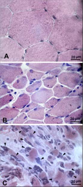
fiber, in human male specimens. Age-related alterations #7. May 26, 2021
of HA concentrations were evaluated both in human and
mice specimens using Purple-Jelley HA assay. Muscle Biomechanical and structural studies of the muscle
loss and fat infiltration were present in most elderly extracellular matrix
samples, especially in the subjects over 70 years old. The Richard L. Lieber
area percentage of collagen contents were significantly
elevated in the elderly group compared to young men Shirley Ryan AbilityLab, Departments of Physical
group (P2021 PDM3 Program and Abstracts, May 26 - 29, 2021
Eur J Transl Myol 2021; 31 (1): 9743. doi: 10.4081/ejtm.2021.9743
be regulated. Finally, the cellular basis of fibrosis in the 8. Meyer GA, Lieber RL. Skeletal muscle fibrosis
ECM was investigated using a transgenic mouse model develops in response to desmin deletion. Am J Physiol
in which Type I collagen producing cells were labeled Cell Physiol. 2012;302(11):C1609-20. doi:
with GFP.9 These studies revealed that muscle satellite 10.1152/ajpcell.00441.2011. PubMed PMID:
cells, fibroblasts and fibroadipogenic progenitor cells all 22442138; PMCID: 3378016.
appear to participate in the fibrotic response. We suggest 9. Chapman MA, Mukund K, Subramaniam S, Brenner
that new high resolution methods are needed at larger D, Lieber RL. Three Distinct Cell Populations
scales as well as a standardized set of rules for the Express Extracellular Matrix Proteins and Increase
reporting of passive mechanics to allow cross-study in Number During Skeletal Muscle Fibrosis. Am J
comparisons. Such studies are required to develop Physiol Cell Physiol. 2017;312(2):C131-C43. Epub
therapies to treat disorders in which passive muscle 2016. doi: 10.1152/ajpcell.00226.2016. PubMed
properties are altered in conditions such as muscular PMID: 27881411
dystrophy, traumatic laceration, and contracture due to *****
upper motor neuron lesion as seen in spinal cord injury,
stroke and cerebral palsy. #8. May 26, 2021
Keywords: fibrosis, modulus, collagen, perimysium, Sarcomere remodeling and function in ageing muscle
extracellular matrix Geoffrey A. Power (1,2), Sean Crooks (2), Jared R.
References Fletcher (2,3), Brian R. Macintosh (2), Walter Herzog (2)
1. Magid A, Law DJ. Myofibrils bear most of the resting (1) Department of Human Health and Nutritional
tension in frog skeletal muscle. Science (Washington Sciences, College of Biological Sciences, University of
DC). 1985;230:1280-2. Guelph, Guelph, Ontario, Canada; (2) Human
2. Meyer G, Lieber RL. Muscle fibers bear a larger Performance Laboratory, Faculty of Kinesiology,
fraction of passive muscle tension in frogs compared University of Calgary, Calgary, Alberta, Canada; (3)
with mice. J Exp Biol. 2018;221(Pt 22). Epub Department of Health and Physical Education, Mount
2018/09/22. doi: 10.1242/jeb.182089. PubMed Royal University, Calgary, Alberta, Canada
PMID: 30237238; PMCID: PMC6262763.
3. Meyer GA, Lieber RL. Elucidation of extracellular * Geoffrey Power: gapower@uoguelph.ca
matrix mechanics from muscle fibers and fiber In old age, there appears to be elevated passive tension
bundles. Journal of Biomechanics. 2011;44(4):771- for a given muscle length1. Aging muscles undergo
3. Epub 2010/11/26. doi: S0021-9290(10)00610-X structural remodeling, whereby muscle fascicle lengths
[pii] 10.1016/j.jbiomech.2010.10.044. PubMed become shorter in old as compared with young2,3. Thus,
PMID: 21092966; PMCID: 3042517. the fascicles and sarcomeres of muscles in older adults
4. Ward SR, Winters TM, O’Connor SM, Lieber RL. may experience greater relative length changes for a
Nonlinear scaling of passive mechanical properties given displacement or joint angular rotation, resulting in
in fibers, bundles, fascicles and whole rabbit muscles. increased passive tension as compared with young. The
Frontiers in Physiology. 2020. doi: purpose of this study was to compare the medial
doi.org/10.3389/fphys.2020.00211. gastrocnemius of young and old rats to determine
5. Gillies AR, Chapman MA, Bushong EA, Deerinck TJ, differences in fascicle length, serial sarcomere numbers
Ellisman MH, Lieber RL. High resolution three- and the sarcomere length at which peak force is obtained
dimensional reconstruction of fibrotic skeletal muscle (optimal reference length; RL), and record passive
extracellular matrix. J Physiol. 2017;595(4):1159- tension from short to long muscle lengths. Two groups of
71. Epub 2016/11/20. doi: 10.1113/JP273376. Fisher344x BN rats were used in this experiment, a
PubMed PMID: 27859324; PMCID: PMC5309386. young cohort (≈9 months ≈20 human years) and old
6. Gillies AR, Bushong EA, Deerinck TJ, Ellisman MH, cohort (~32 months; ≈75-80 human years). The MG from
Lieber RL. Three-dimensional reconstruction of the right leg was surgically isolated, attached to a custom
skeletal muscle extracellular matrix ultrastructure. made muscle puller and force transducer. Passive force
Microsc Microanal. 2014;20(6):1835-40. doi: was recorded at nine muscle lengths relative to the length
10.1017/S1431927614013300. PubMed PMID: producing optimal active force. Following passive force
25275291; PMCID: 4267978. and length measurements, the animals were sacrificed
7. Li Z, Mericskay M, Agbulut O, Butler-Browne G, and the hind limb was fixed in formalin at the muscle
Carlsson L, Thornell LE, Babinet C, Paulin D. length corresponding to optimal force. The muscles were
Desmin is essential for the tensile strength and then dissected into four lengthwise sections medial and
integrity of myofibrils but not for myogenic lateral of the center and sarcomere length measurements4
commitment, differentiation, and fusion of skeletal were obtained. A reduction in muscle fascicle length of
muscle. Journal of Cell Biology. 1997;139(1):129- ~14% was observed in the old rats when compared to
44. Epub 1997/10/06. PubMed PMID: 9314534; young (p2021 PDM3 Program and Abstracts, May 26 - 29, 2021
Eur J Transl Myol 2021; 31 (1): 9743. doi: 10.4081/ejtm.2021.9743
(p0.05). While Queensland, Australia
there was no difference in passive tension at short muscle
* Martino Franchi: martino.franchi@unipd.it
lengths between groups, old rats experienced up to
~125% greater passive tension at long muscle lengths as It is canonically thought that muscle adaptations to
compared with young (p2021 PDM3 Program and Abstracts, May 26 - 29, 2021
Eur J Transl Myol 2021; 31 (1): 9743. doi: 10.4081/ejtm.2021.9743
The influence of training-induced sarcomerogenesis parabolic flight. Gastrocnemius medialis (GM)
on the history dependence of force. J Exp Biol. 2020 electromyographic (EMG) activity and Lf were measured
Aug 13;223(Pt 15):jeb218776. doi: 10.1242/jeb. at rest (bipedal standing), jump-off (JO, with the foot
218776. raised before jumping down), ground contact (GC),
5. Lucas G, Leonard TR, Carvalho A, da Silva ASR, minimum ankle joint (MAJ, end of landing) and PO. GM
Herzog W. Chronic uphill and downhill exercise sarcomere number (Sn) was estimated by dividing Lf
protocols do not lead to sarcomerogenesis in mouse measured by ultrasound at rest by sarcomere length (SL),
skeletal muscle. J Biomech. 2020 Jan 2;98:109469. measured in-vivo at the same joint angle (3.16 µm)1.
doi: 10.1016/j.jbiomech.2019.109469 Changes in SL were estimated by dividing Lf in each
6. Lucas G, Leonard TR, Carvalho A, da Silva ASR, jump phase by Sn calculated individually (Figure 1.). The
Herzog W. Chronic uphill and downhill exercise sarcomere force-generating capacity was estimated from
protocols do not lead to sarcomerogenesis in mouse the sarcomere L-T relation described by Walker and
skeletal muscle. J Biomech. 2020 Jan 2;98:109469. Schrodt.2 At JO in 1g, SL was 2.55µm, close to the L-T
doi: 10.1016/j.jbiomech.2019.109469. relation plateau. At GC and MAJ, SL shifted to the left,
7. Pincheira PA, Boswell MA, Franchi M V, Delp SL, down the ascending limb of the L-T curve. Fascicles and
Lichtwark GA. Biceps femoris long head sarcomere sarcomeres shortened from JO to GC (pre-activation) and
and fascicle length adaptations after three weeks of behaved quasi-isometrically from GC to MAJ, enabling
eccentric exercise training. bioRxiv. January tendon elongation and storage of elastic energy.
2021:2021.01.18.427202. doi:10.1101/2021.01.18. Surprisingly, at PO, SL was extremely short (1.49µm), a
427202. length at which only 40% of maximum sarcomere force
***** is expected to be produced. Notably, this condition is
reached with the ankle in full plantarflexion, causing
#10. May 26, 2021 maximum tendon stretch. From MAJ to PO, a marked
Extreme sarcomere shortening during drop jump in EMG decrease seemed to confirm that most of the energy
variable gravity environment needed for the PO is provided by the tendon recoil. Lf at
JO was shorter in hg and Hg than in 1g, reflecting a
Elena Monti (1), Janice Waldvogel (2); Ramona downward shift of SL along the L-T relationship (range
Ritzmann (2), Kathrin Freyler (2), Kirsten Albracht (3,4), 1.96-2.11µm Hg and 2.23-2.33µm hg). During pre-
Michael Helm (2), Nicola De Cesare (5), Piero Pavan (5), activation, less SL shortening occurred in Hg and hg than
Carlo Reggiani (1), Albert Gollhofer (2), Marco V Narici in 1g, followed by lengthening of Lf and sarcomeres
(1) between GC and MAJ. Remarkably, at MAJ, Lf reached
(1) Department of Biomedical Science, University of values similar to those of 1g, enabling sarcomeres to
Padova, Padova, Italy; (2) Department of Sport and produce about 70% of maximum force. In the last phase,
Sport Science, University of Freiburg, Freiburg, from MAJ to PO, a strong contraction markedly reduced
Germany; (3) Institute of Biomechanics and SL to values of 1.44-1.53 µm. In conclusion, regardless
of the gravity level, muscle activation appears finely
Orthopedics, German Sport University Cologne,
regulated to enable fascicles to operate in the ascending
Cologne, Germany; (4) Department of Medical
Engineering and Technomathematics, Aachen
University of Applied Sciences, Aachen, Germany; (5)
Department of Industrial Engineer, University of
Padova, Padova, Italy
* Elena Monti: elena.monti.1@phd.unipd.it
The regulation of sarcomere length during stretch-
shortening cycle (SSC) activities performed in variable
gravity is unknown. Drop-jumping (DJ), a common SSC
activity, involves three phases: pre-activation of
antigravity muscles, landing and push-off (PO) to
perform a vertical jump. The ability to absorb ground
impact and generate propulsive forces depends on tendon
strain and fascicle length (Lf). Hence, the region of Fig 1. SL in the 4 different DJ phases (JO, GC,
operation of sarcomeres along the muscle length-tension MAJ, PO) in 1g (red dots), hypo-gravity
(L-T) relation is a determinant factor for DJ performance. (blue dots) and hyper-gravity (green dots).
This study investigated changes in Lf and sarcomere The L-T relationship of hypo- and hyper-
operational length throughout the phases of DJs gravity has been shifted on the x axis to the
performed in normo-gravity (1g), hyper-gravity (Hg) and left and right, respectively, to allow for dots
hypo-gravity (hg). Fifteen volunteers performed repeated visualisation.
DJs in 1g, hg (0 to 1g), and Hg (1 to 2g) during a
- 10 -2021 PDM3 Program and Abstracts, May 26 - 29, 2021
Eur J Transl Myol 2021; 31 (1): 9743. doi: 10.4081/ejtm.2021.9743
portion of the L-T relation. Such neuromuscular dramatically influence our ability to rehabilitate these
mechanism seems to limit fascicle elongation during the muscles.8 Recently, we showed that several anti-cancer
breaking phase, enabling sarcomeres to reach, at MAJ, drugs are able to reverse these epigenetic changes, thus
lengths similar to 1 g. This seems a useful mechanism for “rescuing” the satellite cells and promoting myogenesis.
anchoring the tendon, enabling storage of elastic energy Finally, based on the highly unusual transcriptional
and its release in the subsequent PO phase. profile and neural input that these contractures have, we
show that blockage of the neuromuscular junction using
Keywords: sarcomere length, sarcomere operating
neurotoxins can actually assist these muscles in growing.
length, drop-jump, fascicle length, muscle biomechanics,
Taken together, these results support the notion that,
hyper-gravity, hypo-gravity
while contracture formation is multifactorial and neural
References in origin, significant structural alterations in muscle also
1. Sanchez GN, Sinha S, Liske H, Chen X, Nguyen V, occur. An understanding of the specific changes that
Delp SL, Schnitzer MJ. In Vivo Imaging of Human occur in the muscle and extracellular matrix may
Sarcomere Twitch Dynamics in Individual Motor facilitate the development of new conservative or
Units. Neuron. 2015 Dec 16;88(6):1109-1120. doi: surgical therapies for this devastating problem.
10.1016/j.neuron. 2015.11.022. Keywords: satellite cell, cerebral palsy, contracture,
2. Walker SM, Schrodt GR. I segment lengths and thin extracellular matrix
filament periods in skeletal muscle fibers of the
Rhesus monkey and the human. Anat Rec. 1974 References
Jan;178(1):63-81. doi: 10.1002/ar.1091780107. 1. Lieber RL, Fridén J. Spasticity causes a fundamental
***** rearrangement of muscle-joint interaction. Muscle &
Nerve. 2002;25:265-70.
#11. May 26, 2021 2. Lieber RL, Runesson E, Einarsson F, Fridén J.
Intraoperative and Laboratory Studies of Human Inferior mechanical properties of spastic muscle
Skeletal Muscle Contractures bundles due to hypertrophic but compromised
extracellular matrix material. Muscle & Nerve.
Richard L. Lieber 2003;28:464-71.
Shirley Ryan AbilityLab, Departments of Physical 3. Fridén J, Lieber RL. Spastic muscle cells are shorter
Medicine, Rehabilitation and Biomedical Engineering, and stiffer than normal cells. Muscle Nerve.
Northwestern University, Chicago, IL, USA 2003;27(2):157-64. Epub 2003/01/28. doi:
10.1002/mus.10247. PubMed PMID: 12548522.
* Richard L. Lieber: rlieber@sralab.org 4. Smith LR, Ponten E, Hedstrom Y, Ward SR, Chambers
Skeletal muscle is a highly plastic tissue, responding both HG, Subramaniam S, Lieber RL. Novel transcriptional
to level of use and amount of neural input. After cerebral profile in wrist muscles from cerebral palsy patients.
palsy (CP) altered neural input can result in muscle BMC Med Genomics. 2009;2:44. Epub 2009/07/16.
contractures. We have studied the mechanics and biology doi: 10.1186/1755-8794-2-44. PubMed PMID:
of muscle from children with wrist flexion contractures 19602279; PMCID: PMC2722667.
secondary to CP. Dramatic architectural changes are 5. Smith LR, Chambers HG, Subramaniam S, Lieber RL.
observed in these children whereby sarcomere lengths Transcriptional abnormalities of hamstring muscle
are dramatically altered relative to patients without upper contractures in children with cerebral palsy. PLoS
motor neuron lesions.1 This suggests dramatic alterations One. 2012;7(8):e40686. doi: 10.1371/journal.pone.
in the regulation of muscle growth in these children. 0040686. PubMed PMID: 22956992; PMCID:
Biomechanical studies of isolated single muscle cells 3431909.
reveal an increased passive modulus and decreased 6. Smith LR, Chambers HG, Lieber RL. Reduced satellite
resting sarcomere length suggesting alterations in the cell population may lead to contractures in children
cellular cytoskeleton.2,3 Gene expression profiling with cerebral palsy. Dev Med Child Neurol.
reveals a number of “conflicting” biological pathways in 2013;55(3):264-70. doi: 10.1111/dmcn.12027.
spastic muscle. Specifically, this muscle adapts by PubMed PMID: 23210987.
altering processes related to extracellular matrix 7. Dayanidhi S, Dykstra PB, Lyubasyuk V, McKay BR,
production, fiber type determination, fiber hypertrophy Chambers HG, Lieber RL. Reduced satellite cell
and myogenesis.4,5 These transcriptional adaptations are number in situ in muscular contractures from children
not characteristic of muscle adaptations observed in with cerebral palsy. J Orthop Res. 2015;33(7):1039-
Duchenne muscular dystrophy or limb immobilization. 45. doi: 10.1002/jor.22860. PubMed PMID: 25732238.
Superimposed upon the dramatic biological and 8. Domenighetti AA, Mathewson MA, Pichika R, Sibley
structural adaptations is a loss in the number of satellite LA, Zhao L, Chambers HG, Lieber RL. Loss of
cells that are located throughout the muscle.6,7 Even the myogenic potential and fusion capacity of muscle stem
remaining satellite cells have epigenetic changes that can cells isolated from contractured muscle in children
with cerebral palsy. Am J Physiol Cell Physiol.
- 11 -2021 PDM3 Program and Abstracts, May 26 - 29, 2021
Eur J Transl Myol 2021; 31 (1): 9743. doi: 10.4081/ejtm.2021.9743
2018;315(2):C247-C57. Epub 2018/04/26. doi: 2. Sinha U, Malis V, Csapo R, Moghadasi A, Kinugasa
10.1152/ajpcell.00351.2017. PubMed PMID: R, Sinha S. Age-related differences in strain rate
29694232; PMCID: PMC6139501 tensor of the medial gastrocnemius muscle during
***** passive plantarflexion and active isometric
contraction using velocity encoded MR imaging:
#12. May 26, 2021 potential index of lateral force transmission. Magn
3D Fiber aligned strain and strain rate measurements Reson Med. 2015 May;73(5):1852-63. doi: 10.1002
/mrm.25312. Epub 2014 Jul 8.
Shantanu Sinha 3. Malis V, Sinha U, Csapo R, Narici M, Sinha S.
Dept. of Radiology, Muscle Imaging and Modeling Lab, Relationship of changes in strain rate indices
University of California at San Diego, School of estimated from velocity-encoded MR imaging to loss
Medicine, San Diego, CA, USA of muscle force following disuse atrophy. Magn
* Shantanu Sinha: shsinha@ucsd.edu Reson Med. 2018 Feb;79(2):912-922. doi:
10.1002/mrm.26759. Epub 2017 May 30.
We have established velocity encoded MRI as a powerful 4. Lustig M, Donoho D, Pauly JM. Sparse MRI: The
non-invasive imaging technique to monitor skeletal application of compressed sensing for rapid MR
muscle motion.1-3 One of the limitations of this technique imaging. Magn Reson Med. 2007 Dec;58(6):1182-
is that it requires of the order of 70 consistent 95. doi: 10.1002/mrm.21391.
contractions cycles in order to image muscle motion. 5. Malis V, Sinha U, Sinha S. Compressed sensing
Recently, we implemented a ‘compressed sensing’ velocity encoded phase contrast imaging: Monitoring
acquisition,4,5 that enables a reduction in acquisition time skeletal muscle kinematics. Magn Reson Med.
by factors of 4 enabling us to extend the scope of our 2020;84:142-156. Magn Reson Med. 2020
dynamic studies: e.g., imaging multiple slices, different Jul;84(1):142-156. doi: 10.1002/mrm.28100. Epub
and higher %MVCs and ankle positions as well 2019 Dec 11.
accommodate senior and frail subjects. The
implementation of this ‘fast’ technique allowed us to *****
study muscle kinematics in 3D (3 spatial and 3 velocity #13. May 26, 2021
directions) and at three %MVCs in the calf muscle in
young and old subjects. This latter study included Age related structural changes in calf muscle using
diffusion tensor imaging in order to extract the fiber Quantitative Magnetic Resonance Imaging
direction at each voxel. The principal strains and strain John C White, Usha Sinha*
rates were rotated to the fiber basis at each voxel using
the diffusion tensor information at each voxel. The fiber Department of Physics, San Diego State University,
aligned strain and strain rates were not significantly San Diego, CA, USA
different between the young and old age groups even *Usha Sinha: usinha@sdsu.edu
though significant differences were found between the
Structural and functional changes occur in skeletal
two age groups in the strain/ SR indices extracted in the
muscle with age and with disuse resulting in a loss of
principal basis. The Euler angles between the principal
muscle force1,2. Contributors to the loss of muscle
and fiber basis were calculated (represents the rotation of
force with age include decrease in muscle mass,
the strain tensor from the muscle fiber) and we compared
structural changes in the extracellular matrix and
it with the rotation angles derived from multiscale
increase in fat and fibrotic tissue. The extracellular
computational modeling. The multiscale modeling
matrix as well as fibrotic tissue contain a relatively
provides insight into the physiological mechanism
large fraction of macromolecules. Magnetic
responsible for the deviation of the strain from the fiber
Resonance Imaging (MRI) provides a way to map free
axis. Initial results indicate that the rotation of the
protons in tissue while protons bound to
principal strain from the fiber may arise from shear in the
macromolecules (e.g., collagen) are not visible on
endomysium. The implications of these findings with
routine MRI due to their ultra-short T2 values.
respect to muscle force loss with age will be discussed.
Magnetization transfer contrast (MTC) enables an
Keywords: Aging muscle; dynamic MRI; compressed indirect evaluation of the macromolecular content by
sensing; 3D strain imaging; fiber aligned strains. selective saturation of bound protons. It is
hypothesized that collagen is the primary
References
macromolecular pool responsible for the MTC effect
1. Sinha S, Hodgson JA, Finni T, Lai AM, Grinstead J, in skeletal muscle. Collagen content in muscle
Edgerton VR. Muscle kinematics during isometric increases with aging and thus, a non-invasive marker
contraction: Development of phase contrast and spin of collagen in muscle tissue has the potential to
tag techniques to study healthy and atrophied identify aging differences. Further, inflammatory
muscles. J Magn Reson Imaging. 2004 changes are also known to occur in aging muscle and T1
Dec;20(6):1008-19. doi: 10.1002/jmri.20210. mapping may be able to track these changes. Here, we
- 12 -2021 PDM3 Program and Abstracts, May 26 - 29, 2021
Eur J Transl Myol 2021; 31 (1): 9743. doi: 10.4081/ejtm.2021.9743
measured MTsat3 (a semi-quantitative index of MTC) and relaxation obtained from 3D FLASH MRI. Magn
T1 to explore age related changes in calf muscle. We also Reson Med. 2008 Dec;60(6):1396-407. doi:
explored the effects of different fat suppression methods 10.1002/mrm.21732.
on the observed T1 values to identify the effects of 4. Marty B, Carlier PG. Physiological and pathological
incidental magnetization transfer on the measured T1 skeletal muscle T1 changes quantified using a fast
values. A cohort of 10 healthy young subjects (24.5±3 inversion-recovery radial NMR imaging sequence. ci
years) and eight healthy senior subjects (65±8 years) Rep. 2019 May 2;9(1):6852. doi: 10.1038/s41598-
were imaged in a 3T scanner. T1 derived from the fat 019-43398-x.
saturated sequence was significantly lower compared to *****
T1 derived from the other three sequences (no fat
suppression, water excitation -fast and normal). The #14. May 26, 2021
decreased T1 values from the fat saturated sequence is Neural control of movement unraveled by
attributed to the magnetization transfer effects of the off- electromyography
resonance fat saturation pulse. Significant differences
were found between young and old subjects in T1 derived Dario Farina,* Jaime Ibáñez
from the sequences with fat suppression; in all cases T1 Department of Bioengineering, Imperial College
increased with age in fat suppressed sequences4. London, UK
Significant differences in MTsat were found between the
young and old cohorts with MTsat decreasing with age. * Dario Farina: d.farina@imperial.ac.uk
The increase in T1 values with age is hypothesized to
The study of the interactions between the brain, spinal
reflect inflammatory changes in skeletal muscle and cord and muscles during volitional movements is key to
could potentially be an imaging biomarker. That T1 understand the neural basis for motor control. While very
values from the fat unsuppressed sequence did not show important advances are being achieved regarding how
age related changes is attributed to the opposing effects brain's regions and neural populations change their
of increased fat infiltration (shorter T1) and increasing
activity to produce movements, we still know very little
inflammation (longer T1), emphasizing the need for regarding how motor commands are transmitted through
efficient fat suppression in skeletal muscle quantitative descending pathways to the muscles. When this problem
imaging. MTsat decreased significantly with age and this is addressed in animal research, evoked potentials are
decrease cannot be explained by our hypothesis of used to study connectivity in corticospinal tracts. The
increase in collagen with age. The decrease in MTsat may limitation of this approach is that electrical stimulation of
arise from decreases in muscle fibers (atrophy); the
neurons provides biased information as the largest cells
muscle fibers may themselves be the source of the dominate the responses measured. Moreover, evoked
macromolecular pool. In conclusion, fat suppressed T1 potentials are not directly associated to behaviour. We
and MTsat values were significantly different with age in have recently shown that having access to the neural code
skeletal muscle though the direction of the change in driving muscles in combination with brain signals can
MTsat cannot be explained due to a lack of knowledge
provide us with a unique framework to study the
about the macromolecular pool responsible for the MTC
properties of corticomuscular transmission in terms of
effect in skeletal muscle.
the expected populations of cells in the spinal cord that
Keywords: Aging Muscle, macromolecular fraction contribute to the neural information that reaches the
content, T1 mapping muscles. This approach consists in decoding high-density
EMG (HDEMG) signals into the activity of individual
References spinal motoneurons,1 and estimating the sources of
1. Csapo R, Malis V, Sinha U, Du J, Sinha S. Age- synaptic inputs that the motoneurons receive with
associated differences in triceps surae muscle coherence techniques. Using this framework, we have
composition and strength - an MRI-based cross- been able to show that oscillatory activity in the brain
sectional comparison of contractile, adipose and successfully travels to the muscles using the most
connective tissue. BMC Musculoskelet Disord. 2014 straightforward and fast routes available.2 This simple
Jun 17;15:209. doi: 10.1186/1471-2474-15-209.. finding provides a strong rationale towards using
2. Malis V, Sinha U, Csapo R, Narici M, Smitaman E, simplified models of the corticospinal system to
Sinha S. Diffusion tensor imaging and diffusion understand key questions about motor control and also
modeling: Application to monitoring changes in the regarding the mechanisms of actions of different
medial gastrocnemius in disuse atrophy induced by neuromodulation techniques used in the field of neural
unilateral limb suspension. J Magn Reson Imaging. rehabilitation. The talk will focus on the use of HDEMG
2019 Jun;49(6):1655-1664. doi: 10.1002/jmri.26295. for assessing the output of the spinal cord and for
Epub 2018 Dec 19. identifying neural connectivity between the brain and the
3. Helms G, Dathe H, Kallenberg K, Dechent P. High- spinal cord. These methods will be discussed in relation
resolution maps of magnetization transfer with to their applications in neurorehabilitation technologies.
inherent correction for RF inhomogeneity and T1
- 13 -You can also read