Novel Variants of the Sternal Muscle in an Adult and an Anencephalic Infant: Embryological Insights and Clinical Implications
←
→
Page content transcription
If your browser does not render page correctly, please read the page content below
Novel Variants of the Sternal Muscle in an Adult and
an Anencephalic Infant: Embryological Insights and
Clinical Implications
Emilio Farfán Cabello
Pontificia Universidad Católica de Chile https://orcid.org/0000-0002-8819-2945
Marcia Gaete ( mgaets@uc.cl )
Pontificia Universidad Católica de Chile https://orcid.org/0000-0003-1846-2417
Oscar Inzunza H.
Pontificia Universidad Católica de Chile
Mark Echeverría M.
Pontificia Universidad Católica de Chile
Verónica Inostroza R.
Pontificia Universidad Católica de Chile
Research article
Keywords: Sternal Bilateral Muscle, Supernumerary muscle, Muscular variation
Posted Date: September 17th, 2021
DOI: https://doi.org/10.21203/rs.3.rs-798890/v1
License: This work is licensed under a Creative Commons Attribution 4.0 International License.
Read Full License
Page 1/15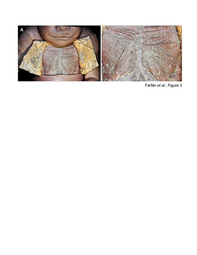
Abstract
Background
The sternal muscle is a supernumerary variant of the thoracic muscles found in 3–8% of the population.
When present, it can be unilateral or bilateral, which can produce confusions during surgeries and
imagenological examinations.

Methods
We report the finding of the sternalis muscle in two human cadavers, one adult and one anencephalic
infant. The muscles were dissected from the fixed bodies and their morphometry analysed.

Results
In the case of the adult, we observed two sternal muscles connected in the superior portion by a central
tendon. In the case of the anencephalic infant, we found a bilateral sternal muscle, in which the bellies
came from the contralateral pectoralis major muscles. The two sternalis muscle variants found here were
impossible to categorise according to the current classifications.

Conclusions
The sternalis muscle displays variants that are still not classified, as observed in the case of the adult
and the infant, in which its presence was correlated with anencephaly. We discuss about this muscular
variation in the clinical, imagenological and surgical context and propose a developmental link with the
occurrence of neural tube closure defects.

Background
The sternal muscle is a supernumerary muscular variant firstly described for Cabrolius in 1604 (Testut,
1884). Throughout history, this muscle has being named “episternalis”, “presternalis”, “rectus thoracicus”
and “rectus sterni” (Jelev et al., 2001), currently, its recognized as “sternal muscle” for the Federative
International Programme for Anatomical Terminology (FIPAT, 2019). Its reported prevalence varies from
4.5 to 8% (Snosek et al., 2014), and between 3–8% with high interracial variability (Arraez-Aybar et al.,
2003).

The localization of the sternal muscle occurs between the superficial fascia and the pectoralis major
muscle, being found unilateral or bilaterally (Hung et al., 2012). In appearance is similar to a band with
two ends: the superior end, which is generally tendinous and in relationship with the sternocleidomastoid
muscle at the level of the sternal manubrium, and the inferior end, which can be muscular or tendinous,

                                                  Page 2/15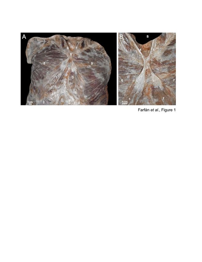
inserted in the ribs or in the aponeurosis of the abdominal external oblique muscle (Testut and Latarjet,
1967). When the sternal muscle contracts, can eventually be visible in people with thin skin (Spalteholz,
1990).

The close relationship between the sternal muscle and the pectoralis major muscle cause diagnostic
dilemmas during surgeries and imagenological examinations: the sternal muscle can be confused with a
tumoral pathology (Standring, 2016). According to this, the sternalis muscle constitutes a structure of
interest for the radiologist, especially when analysing mammograms (Bradley et al., 1996), as well as for
the surgeon, who access to the retromammary pocket when placing mammary implants (Khan, 2008), or
dissect parasternal lymph nodes or internal thoracic blood vessels (Testut and Jacob, 1979).

A connection between anencephaly and the presence of the sternal muscle is described In 1883 Abraham,
studied 11 anencephalic fetuses, of which 6 (55%) presented the sternal muscle (Abraham, 1883).
Variations in the major pectoral muscles were common when the sternal muscle developed, suggesting
an association in the embryological development of both muscles (Abraham, 1883). Shepherd in 1885
backed the studies of Abraham, performing dissections of anencephalic fetuses and investigating the
presence of the sternal muscle in 6 anencephalic infants, in which he found 3 unilateral and 3 bilateral
sternalis muscles, commenting about the origin of the sternalis muscle in the clavicular fibres of the
pectoralis major muscle (Shepherd, 1885).

Methods
Dissection of the anterior chest wall in a cadaver of 65 years of age at the time of death, and in a new-
born anencephalic infant, both white male were performed in the Department of Anatomy, Pontificia
Universidad Católica de Chile, under approval of the institutional ethical committee. They did not had
previous surgeries or interventions in the sternal region and they were intact at the time of dissection.
Fixation was made by perfusion in 10% buffered formaldehyde and samples were kept in that solution
and kept in a cold room at 4ºC until its dissection. After a wash in physiological serum, dissection was
made in planes. First, the skin was lifted after a longitudinal incision from the jugular notch of the
sternum to the infrasternal angle. Then, the cutaneous plane was flapped laterally, mantaining the deep
fascia intact. In the adult cadaver, the presence of an "X" muscle located between the superficial fascia
and the pectoralis major muscle was detected, across the root of the neck to the abdomen. To study this
structure, the deep fascia was removed, leaving a recognizable sternal muscle uncovered. The caudal and
cranial ends were followed to distinguish their insertions, vascularization and innervation together with
their anatomical relationships. Similar procedures were made in the anencephalic infant, determining the
insertions, fascicles and morphological characteristics of the sternal muscle as performed for the adult.

Results
Sternal muscle in the adult
                                                  Page 3/15
During the dissection of a chest in an adult cadaver, we observed an atypical muscular structure shaped
as an "X", located in the anterior side of the thorax, under the skin and the subcutaneous tissue, but
superficial to both pectoralis major muscles. This muscular structure was formed by a right sternal
muscle and a left sternal muscle, joined by a common tendon at the level of sternal manubrium
(Fig. 1A,B). The measurements of this common tendon were: 21 mm in longitudinal diameter, 13 mm in
cross section and 1 mm in anteroposterior diameter.
The right sternal muscle was extended from the sternal head of the right sternocleidomastoid muscle
(RSCM), throughout a cylindrical tendon oriented from cephalic to caudal and lateral to medial, which
descended towards the thorax throughout the anterior side of the sternal manubrium, joining its
homologous tendon from the left side in a common tendon (Fig. 1A,B). This right sternal muscle was
originated from the common tendon, which was running inferiorly and laterally in the thorax passing over
the pectoralis major muscle, then reaching the seventh homolateral costal cartilage. From this point, the
tendinous fibers were curved laterally and oriented from cephalic to caudal and from medial to lateral.
Those tendinous fibers were confused with the fibers of the pectoral fascia and the aponeurosis of the
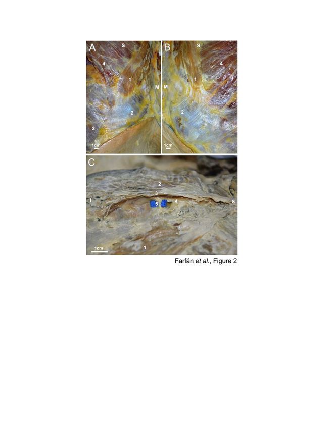
abdominal external oblique muscle (Fig. 2A). The muscular belly of the sternal muscle was formed by
two muscular planes separated by a loose tissue, while its deep side had its own fascia that allowed it to
be separated from the pectoralis major muscle. The total length of the right sternal muscle was 222 mm
measured from the common tendon to the caudal tendon; the length of the muscular belly was 124 mm;
its transverse diameter was 48 mm and its anteroposterior diameter was 1 mm. Its vascularization was
given by thin perforating branches of the internal thoracic vessels, while its innervation was given by
nerves of the anterior cutaneous branches of the intercostal nerves. The blood vessels and nerves of this
muscle came from the intercostal spaces 2, 3, 4 and 5 (Fig. 2C).
The left sternal muscle was extended through the head of the left sternocleidomastoid muscle, by a
cylindrical tendon from cephalic to caudal and from lateral to medial that ran towards the thorax on the
sternal manubrium, joining in this level to its homologous of the right side. This left sternal muscle was
originated from the previously described common tendon, which pointed downwards and laterally,
passing over the superficial side of the pectoralis major muscle, then reaching the sixth costal cartilage of
the same side (Fig. 2B). In this area, it originated some tendinous fibres that were curved medially, getting
a cephalic to caudal and lateral to medial orientation, and were confused with the fibers of the fascia
pectoralis and the aponeurosis of the abdominal external oblique muscle (Fig. 2B). Similar to the right
side, the muscular belly was formed by two muscular planes separated by a loose tissue, while its deep
side had its own fascia that separated it from the pectoralis major. The total length of the right sternalis
muscle was 184 mm, measured from the common tendon to the caudal tendon. The length of the
muscular belly was 85 mm, the transversal diameter was 42 mm and the anteroposterior diameter was 1
mm. Its vascularization was given by thin perforating branches coming from the internal thoracic vessels,
and its innervation was from the cutaneous nerves coming from the intercostal nerves. The vessels and
nerves of this muscular fascicle came from the intercostal spaces 2, 3 and 4 (Fig. 2C).
Page 4/15The continuity of the common tendon with the sternal muscles of both antimeres, together with the
sternal heads of the sternocleidomastoid muscles, forms, at the level of the sternal angle, a structure
reminiscent to a large tendinous chiasm (Fig. 1A).
Sternal muscle in the anencephalic infant
In the anencephalic infant, the right sternal muscle was originated from a muscular fascicle of the
sternocostal head of the left pectoralis major muscle, crossing the midline from the left to the right going
down and lateral, passing over the pectoralis major muscle and extending up to the costal arch. The deep
side of the muscular belly had a fascia that separated the sternalis muscle from the pectoralis major
muscle (Fig. 3A,B). The total length of the right sternal muscle was 39 mm and its transversal diameter
was 8 mm (Fig. 3A,B). It was not possible to determine its vascularization and innervation.
The left sternal muscle was originated from a muscular fascicle of the sternocostal portion of the right
major pectoral muscle, by a fascicle that crossed deeply to the right sternal muscle and then gave rise to
two muscle fascicles located superficially on the left major pectoral muscle, remaining one superior and
the other inferior (Fig. 3A,B). The upper fascicle was positioned horizontally, its length was 26 mm and its
transversal diameter was 9 mm, this fascicle was subdivided into 2 minor fascicles. The inferior fascicle,
which was positioned obliquely had a major axis of 28 mm and a transverse axis of 7 mm (Fig. 3A,B). It
was not possible to determine its vascularization and innervation.
Discussion
The occasional presence of sternal muscle in humans has been of great interest to anatomists. Since the
sternal muscle was firstly described by Cunningham (Cunningham, 1884), a high number of anatomical
publications report the presence of the sternalis muscle, which have a prevalence of about 3–8% and a
variable presentation (Arraez-Aybar et al., 2003; Orts Llorca, 1970; Snosek et al., 2014). There are
differences by gender: the sternal muscle is slightly more frequent in women (8.7%) than in men (6.4%)
(Scott-Conner and Al-Jurf, 2002) and differences by ethnic groups: in white population its incidence is
approximately 4–7%, while in the black population 8.4%, and in the Asian population 11.5% (Bergman et
al., 1988). The cases described in this work correspond to white male, meaning they belong to the less
probable group of occurrence.
Embryology of the sternal muscle
The embryological origin of the sternalis muscle is still on discussion. It has been postulated that it
comes from adjacent muscles, which may be the panniculus carnosus, sternocleidomastoid, pectoralis
major or rectus abdominis muscles (Jelev et al., 2001; Kida et al., 2000; Orts Llorca, 1970; Raikos et al.,
2011). Turner (1867) considered it as an atavic form of the pectoralis cutaneous of lower animals (Turner,
1867). It is described that the somatic origin of the sternalis muscle, is a part of the ventral longitudinal
muscular column that arises from the ventral portion of the thoracic hypomeres, whose equivalent in the
abdomen gives rise to the rectus abdominis muscle while its persistence in the thorax gives rise to the
Page 5/15sternal muscle (Kumar et al., 2003; Saeed et al., 2002). Other work indicate that is derived from the
pectoralis muscle group including the subcutaneous trunci muscle (Kida and Kudoh, 1991). The
innervation of the muscle give us some hints about its origin: most of the sternalis muscles are
innervated by branches of the internal or external thoracic nerves (55%), or by branches of the intercostal
nerves (43%), or both (2%) (O'Neill and Folan-Curran, 1998). Some cases describe innervation from the
pectoral nerves, even if the muscle is in direct contact with the thoracic wall (Kida and Kudoh, 1991),
however it is also described that the innervation comes from the intercostal or extramural nerves
(Yamada & Mannen, 1985; Kodama et al., 1986). It is also suggested that the sternalis muscle could be
arising from pectoralis major with innervations from pectoral nerve or from rectus abdominis with
innervations from intercostal nerves (Saeed et al., 2002; Vaithianathan et al., 2011).
A high correlation between anencephaly and the presence of the sternal muscle is described together with
some variations and origins in the pectoralis major muscle, implicating similar developmental pathways
(Abraham, 1883; Shepherd, 1885). These results motivated us to examine an anencephalic infant, finding
it bilaterally and corroborating the variations of the major pectoral muscles mentioned by Abraham
(Abraham, 1883): both sternal muscles of the infant were originated from the larger pectoralis on the
opposite side, a unique characteristic that was not seen in the case of the adult.
The relationship between anencephaly and the presence of alteration on the chest muscles is not clear. At
the light of the current knowledge in developmental biology, this can be caused by the effect of a
common molecular control, in which a common signal can be involved in the generation of both
conditions. Probably, if this signal is altered at early stages. it can affect to the neural tube and somites.
However, if the signal is altered later in time and in a downstream component, it can affect just the
muscles, as we saw in the case of the adult. Other possibility is that two separate aetiologies are acting,
however, same molecular control can be shared in same phenotype. Several genes are involved in the
differentiation of the neck and chest muscles, such as Mef2c-AHF, Islet1, Mesp1and Pax3. Chest muscles,
such as pectoralis, are controlled by the most posterior somite myogenic program, which depends on
Pax3 (Heude et al., 2018). Importantly, mice homozygous for Pax3 mutation develop significantly higher
cranial neural tube defects (Burren et al., 2008; Epstein et al., 1991), making Pax3 a candidate gene to be
analysed for this muscular variation. Relation with anencephaly can be also linked with a positional
effect, however the anencephaly is mainly caused in anterior regions in which paraxial mesoderm and the
muscles of the chest are related to the more posterior somites, making this unlikely.
Classification and innervation
Given the different forms of presentation of the sternal muscle, Jelev developed a classification (Jelev et
al., 2001) that was later modified by Snosek (Snosek et al., 2014) trying to accommodate varieties not
previously considered. The former classification of Jelev (Jelev et al., 2001) established four
morphological criteria to denominate a muscle as "sternal muscle" and propose a numerical
nomenclature to define the different types. The modified classification of Snosek (Snosek et al., 2014)
includes the four criteria indicated, but redefines the types and the subtypes of the sternal muscle. The
sternal muscles of the adult and the infant presented in this study meet all the four above mentioned
Page 6/15criteria to be considered as a sternal muscle: 1) to be placed on the fascia of the pectoralis major muscle;
2) to be originated from the sternum or from the infraclavicular region; 3) to be inserted in the lower ribs,
costal cartilages, aponeurosis of the external oblique muscle of the abdomen or the sheath of the rectus
abdominis muscle; and 4) to be innervated by the medial or intercostal pectoral nerves (Jelev et al.,
2001). Applying the classification modified by Snosek (Snosek et al., 2014), the variety found in the adult
cadaver seems to be similar to the type "others" subtype "cross-linked", although with some differences
with respect to the muscle described in this work. The "cross-linked" variety as schematically represented
in this classification, corresponds to a muscular arrangement in an "X", similar to the observed in this
study (Fig. 1); but with two muscular bellies fused in the midline in front of the sternum, in contrast to the
case we are describing here, in which the muscular bellies are not fused, rather they share a common
superior tendon (Fig. 2). The "X" aspect in our case is given by the extension of their superior tendons with
the sternal head of the sternocleidomastoid muscle. Therefore, the muscle described in this work does
not match the classification of Snosek (Snosek et al., 2014).
In relation to the classification of the sternal muscles of the infant, the right sternal muscle could be close
to a muscle of a simple type and of a cross-linked subtype, while the muscle of the left side could be
similar to a mixed type and divergent bicipital subtype crisscrossed (Snosek et al., 2014), nevertheless,
they do not match exactly the current classification due to its origin is in the larger pectoralis muscles.
Regarding the innervation of the sternal muscle, Shepherd (Shepherd, 1885) indicated that it was given by
the medial pectoral nerve, similar to what was found by Kida (Kida et al., 2000). Other cadaveric and
surgical explorations have reported that the sternal muscle is innervated by the pectoral nerves or anterior
branches of the intercostal nerves (Snosek et al., 2014), or a combination of both (Hung et al., 2012). In
the adult case described here, the nervous branches from the anterior cutaneous nerves of the
neighbouring intercostal nerves reached the sternalis muscle (Fig. 2B), but no innervation of the medial
pectoral nerve was observed in none of the two sternalis bellies. In our current report was not possible to
identify the vascularization and innervation in the sternal muscles of the infant, probably due to its small
size.
Clinical implications
The clinical interest of the sternalis muscle relates to its imagenological and surgical implications: it is
necessary for professionals to become familiar with this muscle to improve their medical practice. Here
we confirmed the high variability in the presentation of this muscle that can be found and how numerous
classifications are still not covering all types of variants that can be founded. The sternal muscle
presence and variability is extremely important in the field of imagenology, in which this muscle has been
studied due to its accidental finding during mammograms. To reduce the possibility of omitting a
neoplasic condition in the image, it has been emphasized the correct positioning of the patient to cover
the largest amount of breast tissue on the detector. Nowadays this, together with the improvements of the
technique, results in a greater detection of the sternal muscle. In the imagenological studies, the sternalis
muscle is observed as an irregular mass of medial situation in the craniocaudal projections of the
mammary gland, and its shape varies according to the position of the patient, resembling a band when in
Page 7/15supine position and making a bulge in a prone position (Nuthakki et al., 2007). Reports of the sternal
muscle in imagenological tests increased in the last decades. The study by Bradley et al., (Bradley et al.,
1996), reported finding 4 cases with sternal muscles in the review of mammograms corresponding to
more than 32,000 women. In contrast, the prevalence reported in more recent studies that have used
multidetector computed tomography is similar to the values of the cadaveric reports: reaching 5.8%
according to the study by Ge et al., (Ge et al., 2014), which involved the revision of 6000 images of
Chinese adults; 6.2% according to the study by Young et al., (Young Lee et al., 2006) that involved the
review of 1,387 images of Korean patients; and 10.5% according to the study by Shiotani et al., (Shiotani
et al., 2012), that considered 948 exams taken consecutively in Japan. Undoubtedly, the soft tissue image
quality that this test provides, the intention to spot the muscle, and the study of the Asian population,
explain the higher prevalence on these studies compared with the older ones.
The prevalence of the sternalis muscle reported in surgical studies is also low. The study by Bailey &
Tzarnas (Bailey and Tzarnas, 1999) identified the sternal muscle in the mastectomies of only 3 patients
in a period of 15 years, whereas in the study by Harish & Gopinath (Harish and Gopinath, 2003), which
reviewed 1151 operative mastectomy records, the prevalence was 0.7%. According to Snosek et al.
(Snosek et al., 2014), this low prevalence can be related to the lack of awareness of the surgeon regarding
the existence of the sternal muscle, together with its high variability. It can also be unnoticed during
mastectomies or breast implant surgeries (Salval et al., 2012), despite of breast implant is the most
frequent cosmetic surgery in the United States (Alderman et al., 2014).
Numerous contributions from the surgical field take into account the sternal muscle. For example,
Schulman & Chun (Schulman and Chun, 2005) reported a modified technique of tissue expander
placement in breast reconstructive surgery in the presence of a sternal muscle. Kabay et al., (Kabay et al.,
2005) reported that they include the sternalis muscle when removing a breast during a radical
mastectomy in cancer surgery and according to Khan (Khan, 2008) the sternal muscle can be used to
give more coverage of the breast implant. Other authors also suggest that it could be used as a flap in
reconstructive surgery of the thorax or neck (Raikos et al., 2011; Salval et al., 2012).
Conclusion
The sternalis muscle has been more frequently identified the last years due to modern anatomical,
surgical and radiological technics, however general practitioners and medical specialist are not always
aware of the sternalis muscle variant, which has a moderate prevalence and its presence need to be
considered during diagnosis and surgeries of the anterior thoracic wall. Here we reported new
morphological presentations in this muscles, making them highly variable in their anatomical
presentation and difficult to classify. We also confirmed the correlation between its formation and the
anencephalic condition, which is consistent with previous reports, and that could link the appearance of
this muscle with neural tube defects. All of these considerations contribute to the general understanding
and awareness of this muscle during development, diagnosis and therapies.
Page 8/15Declarations
ETHICS APPROVAL AND CONSENT FOR PUBLICATION
This study was approved by CEC (Comité Ético Científico) MED-UC of the Pontificia Universidad Católica
de Chile (No: 190115002). Consent for publication is not applicable as cadaveric material was used in
this research.
AVAILABILITY OF DATA AND MATERIALS
All data generated or analyzed during this study are included in this published article.
COMPETING INTERESTS
The authors declare that they have no competing interests.
FUNDING
This worked was supported by the Pontificia Universidad Católica de Chile’s Department of Anatomy.
AUTHOR’S CONTRIBUTIONS
EF, OI, ME and VI acquired the data and dissected the cadavers. EF and MG wrote the manuscript. MG
make the figures. All authors contributed to the data analysis, interpretation, read and approval of the
final manuscript.
ACKNOWLEDGEMENTS
As researchers and teachers, we thank the people who, in an enormous act of generosity and greatness,
donate their bodies to the science to contribute to the morphological research and the training of new and
better professionals. Thanks to Jose Miguel Masman for the photographies.
References
1. Abraham, P.S., 1883. Notes on the occurrence of the musculus sternalis in human anencephalic
foetuses. Transactions of the Academy of Medicine in Ireland 1, 301–304.
2. Alderman, A.K., Bauer, J., Fardo, D., Abrahamse, P. and Pusic, A., 2014. Understanding the effect of
breast augmentation on quality of life: prospective analysis using the BREAST-Q. Plast Reconstr
Surg. 133, 787-95.
3. Arraez-Aybar, L.A., Sobrado-Perez, J. and Merida-Velasco, J.R., 2003. Left musculus sternalis. Clin
Anat. 16, 350-4.
4. Bailey, P.M. and Tzarnas, C.D., 1999. The sternalis muscle: a normal finding encountered during
breast surgery. Plast Reconstr Surg. 103, 1189-90.
Page 9/155. Bergman, R., Thompson, S., Afifi, A. and Saadeh, F., 1988. Compendium of human anatomic
variations. Urban & Schwarzenberg, Baltimore-Munich. .
6. Bradley, F.M., Hoover, H.C., Jr., Hulka, C.A., Whitman, G.J., McCarthy, K.A., Hall, D.A., Moore, R. and
Kopans, D.B., 1996. The sternalis muscle: an unusual normal finding seen on mammography. AJR
Am J Roentgenol. 166, 33-6.
7. Burren, K.A., Savery, D., Massa, V., Kok, R.M., Scott, J.M., Blom, H.J., Copp, A.J. and Greene, N.D., 2008.
Gene-environment interactions in the causation of neural tube defects: folate deficiency increases
susceptibility conferred by loss of Pax3 function. Hum Mol Genet. 17, 3675-85.
8. Cunningham, D., 1884. The musculus sternalis. J Anat Physiol 18: 208–210.
9. Epstein, D.J., Vekemans, M. and Gros, P., 1991. Splotch (Sp2H), a mutation affecting development of
the mouse neural tube, shows a deletion within the paired homeodomain of Pax-3. Cell. 67, 767-74.
10. FIPAT, 2019. Federative International Programme for Anatomical Terminology. Terminologia
Anatomica. Halifax, Dalhousie University, 2019. Disponible en: https://fipat.library.dal.ca
11. Ge, Z., Tong, Y., Zhu, S., Fang, X., Zhuo, L. and Gong, X., 2014. Prevalence and variance of the
sternalis muscle: a study in the Chinese population using multi-detector CT. Surg Radiol Anat. 36,
219-24.
12. Harish, K. and Gopinath, K.S., 2003. Sternalis muscle: importance in surgery of the breast. Surg
Radiol Anat. 25, 311-4.
13. Heude, E., Tesarova, M., Sefton, E.M., Jullian, E., Adachi, N., Grimaldi, A., Zikmund, T., Kaiser, J.,
Kardon, G., Kelly, R.G. and Tajbakhsh, S., 2018. Unique morphogenetic signatures define mammalian
neck muscles and associated connective tissues. Elife. 7.
14. Hung, L.Y., Lucaciu, O.C. and Wong, J.J., 2012. Back to the Debate: Sternalis Muscle. Int. J. Morphol.
30(1), 330-336.
15. Jelev, L., Georgiev, G. and Surchev, L., 2001. The sternalis muscle in the Bulgarian population:
classification of sternales. J Anat. 199, 359-63.
16. Kabay, B., Akdogan, I., Ozdemir, B. and Adiguzel, E., 2005. The left sternalis muscle variation detected
during mastectomy. Folia Morphol (Warsz). 64, 338-40.
17. Khan, U.D., 2008. Use of the rectus sternalis in augmentation mammoplasty: case report and
literature search. Aesthetic Plast Surg. 32, 21-4.
18. Kida, M.Y., Izumi, A. and Tanaka, S., 2000. Sternalis muscle: topic for debate. Clin Anat. 13, 138-40.
19. Kida, M.Y. and Kudoh, H., 1991. Innervation of the sternalis muscle accompanied by congenital
partial absence of the pectoralis major muscle. Okajimas Folia Anat Jpn. 67, 449-55.
20. Kumar, H., Rath, G., Sharma, M., Kohli, M. and Rani, B., 2003. Bilateral sternalis with unusual left-
sided presentation: a clinical perspective. Yonsei Med J. 44, 719-22.
21. Nuthakki, S., Gross, M. and Fessell, D., 2007. Sonography and helical computed tomography of the
sternalis muscle. J Ultrasound Med. 26, 247-50.
Page 10/1522. O'Neill, M.N. and Folan-Curran, J., 1998. Case report: bilateral sternalis muscles with a bilateral
pectoralis major anomaly. J Anat. 193 ( Pt 2), 289-92.
23. Orts Llorca, F., 1970. Anatomía Humana. Tomo Primero. Aparato Locomotor. Tronco. Cabeza y
Cuello. 4° ed. Barcelona, Editorial Científico-Médica.
24. Raikos, A., Paraskevas, G.K., Tzika, M., Faustmann, P., Triaridis, S., Kordali, P., Kitsoulis, P. and Brand-
Saberi, B., 2011. Sternalis muscle: an underestimated anterior chest wall anatomical variant. J
Cardiothorac Surg. 6, 73.
25. Saeed, M., Murshid, K.R., Rufai, A.A., Elsayed, S.E. and Sadiq, M.S., 2002. Sternalis. An anatomic
variant of chest wall musculature. Saudi Med J. 23, 1214-21.
26. Salval, A., Scevola, A. and Baruffaldi Preis, F.W., 2012. Sternalis muscle: an uncommon finding during
aesthetic breast surgery. Aesthet Surg J. 32, 903-5.
27. Scott-Conner, C.E. and Al-Jurf, A.S., 2002. The sternalis muscle. Clin Anat. 15, 67-9.
28. Schulman, M.R. and Chun, J.K., 2005. The conjoined sternalis-pectoralis muscle flap in immediate
tissue expander reconstruction after mastectomy. Ann Plast Surg. 55, 672-5.
29. Shepherd, F.J., 1885. The Musculus Sternalis and its occurrence in (Human) Anencephalous
Monsters. J. Anat. Physiol., 19(3):310.2-319.
30. Shiotani, M., Higuchi, T., Yoshimura, N., Kiguchi, T., Takahashi, N., Maeda, H. and Aoyama, H., 2012.
The sternalis muscle: radiologic findings on MDCT. Jpn J Radiol. 30, 729-34.
31. Snosek, M., Tubbs, R.S. and Loukas, M., 2014. Sternalis muscle, what every anatomist and clinician
should know. Clin Anat. 27, 866-84.
32. Spalteholz, W., 1990. Atlas de Anatomía Humana, Tomo Segundo. Regiones, músculos aponeurosis,
corazón y vasos sanguíneos. 14 ed, editorial Labor, Barcelona.
33. Standring, S., 2016. Gray’s Anatomy. The anatomical basis of clinical practice. 41 st edition. Ed.
Elseiver. U.K.
34. Testut, L., 1884. Anomalies Musculaires, Chez L´Homme.Editorial G. Masson. Paris.
35. Testut, L. and Jacob, O., 1979. Tratado de Anatomía Topográfica, Tomo Segundo. Editorial Salvat,
Barcelona.
36. Testut, L. and Latarjet, A., 1967. Anatomía Humana, Tomo Primero. Osteología-Artrología-Miología.,
9° edición, Ed. Salvat. Barcelona.
37. Turner, W., 1867. On the musculus sternalis. J Anat Physiol. 1: 246–253.
38. Vaithianathan, G., Aruna, S., Rajila, R.H. and Balaji, T., 2011. Sternalis "mystery" muscle and its
clinical implications. Ital J Anat Embryol. 116, 139-43.
39. Young Lee, B., Young Byun, J., Hee Kim, H., Sook Kim, H., Mee Cho, S., Hoon Lee, K., Sup Song, K., Soo
Kim, B. and Mun Lee, J., 2006. The sternalis muscles: incidence and imaging findings on MDCT. J
Thorac Imaging. 21, 179-83.
Figures
Page 11/15Figure 1
A. Sternalis muscle in an adult, dissection of the previous wall of the thorax. 1. Rigth sternalis muscle 2.
Lower tendon; 3. Left sternalis muscle; 4. Lower tendon; 5. Common tendon; 6. Sternal hea of the SCMR;
7. Sternal head of the SCML; 8. M. Right Pectoralis major muscle; 9. Left Pectoralis major muscle. 1B
Sternalis muscle in infant, dissection of the previous wall of the thorax . 1. Rigth sternalis muscle; 2. Left
Page 12/15sternalis muscle; 3. Common tendon; 4. Sternal head of the SCMR; 5. Sternal head of the SCML; 6. Right
Pectoralis major muscle; 7. Left Pectoralis major muscle. Superior; I. Lower.
Figure 2
A. Dissection of the anterior chest wall, on A on the right side and on B on the left side. A. 1. M. Sternalis
right; 2. Lower tendon; 3. M. External oblique of the abdomen right; 4. M. Pectoral major right; 2B. 1. M.
Sternalis left; 2. Lower tendon; 3. M. External oblique of the abdomen left; 4. M. Pectoral major left; S.
Page 13/15Superior; M. Medial. 2C. Dissection of the anterior chest wall. 1. M. Sternal Left; 2. M. Sternal right; 3.
Surface layer of sternal muscle right; 4. Deep layer of sternal muscle right; 5. Anterior cutaneous nerve
and perforating vessels; S. Superior; I. Lower.
Figure 3
A. Dissection of the anterior wall of the newborn anencephalic thorax. B. Magnification of the area of
dissection. 1. M. Sternal left, upper fascicle; 2. M. Sternal left, lower fasciculus; 3. M. Pectoral major left 4.
Page 14/15M. Sternal right; 5. M. Pectoral greater right; * Muscle fascicles originating from the left upper fascicle.
Page 15/15You can also read