Diverse midbrain dopaminergic neuron subtypes and implications for complex clinical symptoms of Parkinson's disease - NET
←
→
Page content transcription
If your browser does not render page correctly, please read the page content below
Carmichael et al. Ageing Neur Dis 2021;1:4
DOI: 10.20517/and.2021.07
                                                                                                               Ageing and
                                                                                                         Neurodegenerative
                                                                                                                 Diseases

Review                                                                                                                        Open Access

Diverse midbrain dopaminergic neuron subtypes
and implications for complex clinical symptoms of
Parkinson’s disease
Kathleen Carmichael1,2, Breanna Sullivan1, Elena Lopez1, Lixin Sun1, Huaibin Cai1
1
 Transgenic Section, Laboratory of Neurogenetics, National Institute on Aging, National Institutes of Health, Bethesda, MD
20892, USA.
2
  The Graduate Partnership Program of NIH and Brown University, National Institutes of Health, Bethesda, MD 20892, USA.

Correspondence to: Dr. Huaibin Cai, Transgenics Section, Laboratory of Neurogenetics, National Institute on Aging, National
Institutes of Health, Building 35, Room 1A112, MSC 3707, 35 Convent Drive, Bethesda, MD 20892-3707, USA.
E-mail: caih@mail.nih.gov

How to cite this article: Carmichael K, Sullivan B, Lopez E, Sun L, Cai H. Diverse midbrain dopaminergic neuron subtypes and
implications for complex clinical symptoms of Parkinson’s disease. Ageing Neur Dis 2021;1:4.
https://dx.doi.org/10.20517/and.2021.07

Received: 16 Jun 2021 First Decision: 9 Jul 2021 Revised: 12 Jul 2021 Accepted: 14 Jul 2021 First online: 15 Jul 2021

Academic Editor: Weidong Le Copy Editor: Xi-Jun Chen Production Editor: Xi-Jun Chen

Abstract
Parkinson’s disease (PD), the most common degenerative movement disorder, is clinically manifested with various
motor and non-motor symptoms. Degeneration of midbrain substantia nigra pas compacta (SNc) dopaminergic
neurons (DANs) is generally attributed to the motor syndrome. The underlying neuronal mechanisms of non-motor
syndrome are largely unexplored. Besides SNc, midbrain ventral tegmental area (VTA) DANs also produce and
release dopamine and modulate movement, reward, motivation, and memory. Degeneration of VTA DANs also
occurs in postmortem brains of PD patients, implying an involvement of VTA DANs in PD-associated non-motor
symptoms. However, it remains to be established that there is a distinct segregation of different SNc and VTA
DAN subtypes in regulating different motor and non-motor functions, and that different DAN subpopulations are
differentially affected by normal ageing or PD. Traditionally, the distinction among different DAN subtypes was
mainly based on the location of cell bodies and axon terminals. With the recent advance of single cell RNA
sequencing technology, DANs can be readily classified based on unique gene expression profiles. A combination of
specific anatomic and molecular markers shows great promise to facilitate the identification of DAN
subpopulations corresponding to different behavior modules under normal and disease conditions. In this review,

                   © The Author(s) 2021. Open Access This article is licensed under a Creative Commons Attribution 4.0
                   International License (https://creativecommons.org/licenses/by/4.0/), which permits unrestricted use, sharing,
                   adaptation, distribution and reproduction in any medium or format, for any purpose, even commercially, as
long as you give appropriate credit to the original author(s) and the source, provide a link to the Creative Commons license, and
indicate if changes were made.

                                                                                                                  www.ageneudisjournal.com
Page 2 of 26                        Carmichael et al. Ageing Neur Dis 2021;1:4   https://dx.doi.org/10.20517/and.2021.07

we first summarize the recent progress in characterizing genetically, anatomically, and functionally diverse
midbrain DAN subtypes. Then, we provide perspectives on how the preclinical research on the connectivity and
functionality of DAN subpopulations improves our current understanding of cell-type and circuit specific
mechanisms of the disease, which could be critically informative for designing new mechanistic treatments.

Keywords: Parkinson’s disease, ageing, dopaminergic neurons, dopamine, SNc, VTA, ALDH1A1, RNA sequencing,
subpopulation

INTRODUCTION
Parkinson’s disease (PD) is the second most common degenerative neurological disorder affecting millions
of elderly individuals worldwide. PD patients display canonical motor symptoms, including resting tremor,
slowed movement, impaired posture and balance, and rigid muscles[1]. The motor syndrome is generally
regarded as the result of extensive loss of nigrostriatal dopaminergic neurons (DANs) in the substantia
nigra pars compacta (SNc) of the midbrain[2,3]. Dopamine replacement medications and deep brain
stimulation surgery can improve some of the patient’s motor conditions. However, no cure is available.
Moreover, long-term medication can cause severe side effects, such as dyskinesia and impulsive control
disorders[4]. New mechanistic insights and therapeutic agents are still needed to improve patients’ treatment
and life quality.

In addition to motor symptoms, PD patients often suffer from depression, dementia, and other
neuropsychiatric symptoms[5]. For example, PD patients often develop cognitive dysfunctions, which leads
to Parkinson’s disease dementia (PDD)[6,7]. Approximately 75% of PD patients develop dementia within 10
years of diagnosis, and the prevalence of PDD is 0.3%-0.5% in the general population older than 65 years[6].
The exact pathogenic mechanisms of PDD and other PD-related non-motor symptoms are largely
unknown. The cognitive dysfunctions are not improved by levodopa, the most effective drug to treat the
motor symptoms in PD[6]. Therefore, an important step in intervening a complex neurological disorder such
as PD is to fully elucidate the functional roles of different neural circuits responsible for specific behavioral
phenotypes.

It has been generally accepted that ageing, environmental toxins, and genetic mutations contribute to the
etiopathogenesis of PD. Ageing is the most significant risk factor in the development of PD and other
neurodegenerative diseases. Genomic instability, epigenetic alterations, loss of proteostasis, mitochondrial
dysfunction, and altered intracellular communication are among the key ageing hallmarks[8]. Various
environmental toxins and genetic risk factors have been linked to PD, which affect largely overlapping
molecular and cellular pathways as those implicated in ageing[9-11]. However, the molecular genetic studies
often fall short in pinpointing any specific cell-types or neural circuits critical for the alterations and
impairments of motor and non-motor behaviors. To better understand how different brain cells and neural
circuits control diverse behaviors will therefore provide the structural framework to better appreciate the
impacts of ageing and environmental and genetic factors on the cause and progression of the disease-related
behavioral abnormalities.

The midbrain DANs are composed of diverse neuron subpopulations based on the location of cell bodies,
projection patterns, morphology, gene expression profiles, electrophysiological properties, physiological
functions, and vulnerabilities to diseases[12-17]. Since a preferential degeneration of midbrain DANs
represents the most significant neuropathological feature of PD, in this review, we focus our discussion
mainly on the diversity of midbrain DANs in their distinct genetic makeups, connectivity, and functionality.

Carmichael et al. Ageing Neur Dis 2021;1:4   https://dx.doi.org/10.20517/and.2021.07             Page 3 of 26

Because animal research has been instrumental in understanding the pathophysiological mechanisms and
developing the treatments of PD, such as the current dopamine replacement therapy and deep brain
stimulation surgery[18], we mainly summarize the research findings from preclinical animal models.

MOLECULAR GENETIC DIVERSITY OF MIDBRAIN DOPAMINERGIC NEURONS
Midbrain DANs, residing in the ventral region of the midbrain, are grouped together based on their ability
to synthesize and release dopamine, a key neuromodulator involved in motor control and learning,
motivation, cognition, and reward[19,20]. Dysfunction of midbrain DAN-mediated dopamine transmission
has been associated with PD, schizophrenia, addiction, and other neurological and psychological
disorders[5,21,22]. Traditionally, midbrain DANs can be divided into three main subgroups, retrorubral field
(RRF, A8), SNc (A9), and ventral tegmental area (VTA, A10), in humans and rodents[23,24]. The midbrain
DANs also differ in their axon projections to different brain regions and physiological functions. In
addition, the SNc DANs are relatively more vulnerable to neuronal toxins in rodent PD models and
preferentially degenerated in PD patients[25,26]. However, molecular genetic makers are needed to better
characterize different DAN subtypes, as well as their distinctive connectivity and functionality.

The midbrain DANs selectively express genes critical for the dopamine synthesis, transport, and
degradation, such as tyrosine hydrolase (TH), vesicular monoamine transporter 2 (VMAT2), dopamine
transporter (DAT), and aldehyde dehydrogenase 1a1 (ALDH1A1)[27]. The midbrain DANs also express
transcription factors critical for the DAN differentiation and survival, including nuclear receptor related 1
protein (NURR1), pituitary homeobox 3 (PITX3), and forkhead box protein A1/2 (FOXA1/2)[28,29]. The
molecular genetic difference between SNc and VTA DANs was initially studied by immunostaining and in
situ hybridization using preselected genetic makers. Those markers included G-protein activated inwardly
rectifying potassium channel 2 (GIRK2/KCNJ6), which is more abundant in SNc DANs[30,31], and calbindin
(D-28K, CALB1), which is more enriched in VTA DANs[32-34]. However, those genetic markers are not
exclusively expressed by the SNc or VTA DANs. Additional genetic markers are required to specifically
investigate the functions of different SNc and VTA DAN subtypes. The whole-genome gene expression
studies using microarray and RNA sequencing (RNAseq) technology, especially the latest single cell
RNAseq (scRNAseq) techniques, in combination with Laser Capture Microdissection (LCM) and
Fluorescence-Activated Cell Sorting (FACS) procedures, provide the means to systematically identify
distinct genetic identifiers for diverse DAN subpopulations[35].
Laser capture microdissection, fluorescence-activated cell sorting, and microarray studies of
differential gene expression between SNc and VTA DANs
The TH-positive DANs in SNc and VTA were visually marked with a so-called rapid immunostaining
procedure, and then collected separately by LCM. RNAs were extracted from the LCM-isolated SNc and
VTA samples and subjected to microarray analyses. In both rat[36] and mouse[37] studies, numerous
differentially expressed genes were found between SNc and VTA DANs. Some of those differentially
expressed genes were reported by both studies. For example, the expression of Igf1, Gad1, Drd2, and Sncg is
higher in the SNc, while the expression of Otx2, Tacr3, and Lpl is higher in the VTA. However, very few
genetic markers can be used to distinguish between the SNc and VTA DANs, indicating that the
molecularly defined DANs may not always be confined within the anatomical boundaries. In our studies,
while we found that the majority of ALDH1A1-positive DANs are distributed in the ventral tier of SNc, a
minority population is scattered in a broad region in the VTA[38,39]. A combination of two or more genetic
markers would be required to identify a distinct subtype of DANs in SNc or VTA. On the other hand, since
there were non-dopaminergic cells within the LCM samples, additional immunostaining and in situ
hybridization experiments are needed to verify the expression of any gene of interest in the DANs.
Furthermore, because of difficulty in physically isolating small DAN clusters by LCM, the distinct gene
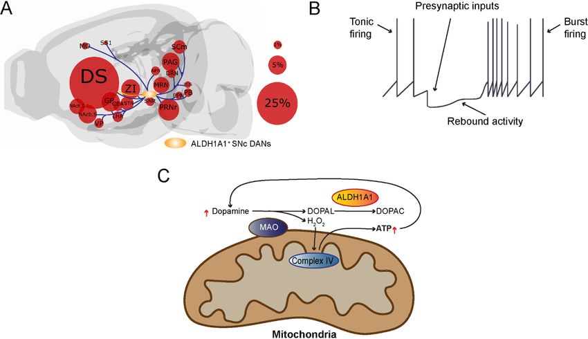
Page 4 of 26                        Carmichael et al. Ageing Neur Dis 2021;1:4   https://dx.doi.org/10.20517/and.2021.07

expression profile of DAN subtypes can only be elucidated by the later FACS and scRNAseq techniques.
Multiple subtypes of midbrain DAN identified by single-cell RNA-sequencing
By employing the newly available scRNAseq technology, recently multiple studies have been performed to
reveal the diverse gene expression profiles of midbrain DANs at single cell level[35]. These high-resolution
gene expression studies demonstrate more complex gene expression patterns in individual midbrain DANs,
identify more DAN subtypes with additional genetic markers, and improve our understanding of the
genetic diversity of midbrain DANs[14,40-43]. Based on distinct gene expression patterns, midbrain DANs may
constitute about 10 or even more subtypes. However, different numbers of DAN subtypes were reported
from different studies, which often used different subtype names and proposed different genetic markers for
clustering the subtypes. These differences reflect different technical approaches and classification criteria in
their studies, while presenting a challenge to other researchers to consistently define the major subtypes of
midbrain DANs[44]. We highlighted seven major midbrain DAN subtypes and listed their unique
classification criteria based on the published mouse scRNAseq studies [Figure 1]. The heterogenous gene
expression profiles of midbrain DANs revealed by scRNAseq and verified by the follow-up RNA scope in
situ hybridization demonstrate the distribution of distinct subgroups of DANs in the developing and adult
mouse midbrains. For example, the ALDH1A1-positive SNc DANs, which are mainly located in the ventral
tier of SNc, express high levels of Sox6, Th, Dat, Aldh1a7, Lmo3, Anxa1, and Sncg, but not Otx2 and
CALB1[40,43]. By contrast, the ALDH1A1-positive VTA DANs, which are found to be intermingled with
other cell types in the VTA, express high levels of CALB1 and Vglut2, but not Sox6[14,17,40,43-46]. Therefore,
based on the co-expression of Sox6 or CALB1, ALDH1A1-positive DANs can be assigned to SNc or VTA
subregions in the midbrain [Figure 1]. Further in-depth analyses of distinct gene expression in ALDH1A1-
positive SNc or VTA DANs may reveal additional molecularly and anatomically defined subtypes
responsible for distinct physiological functions. To reliably interpret scRNAseq data, future studies need to
improve the sensitivity in detecting low-level gene expression, as well as increase the numbers of cells
collected in each experiment.

DIVERSE SNC DOPAMINERGIC NEURON SUBPOPULATIONS
The midbrain DAN system is the largest and most complex in primates compared to rodent species, with
up to 600,000 TH-positive cells in humans compared to ~25,000 TH-positive cells in mice[47]. In rodents,
about half of the TH-positive cells across A8, A9, and A10 are found within the substantia nigra (SN)[47]. In
monkeys and humans, there is both a large increase in the number of TH-positive cells and an even larger
percentage (> 70%) of the number of TH-positive cells that are located in the SN[47]. Beyond simply noting
the number and percentage of SNc dopamine neurons across species, studies have also gone on to
document the anatomic, molecular, and functional diversity of SNc DANs.
Anatomic and molecular diversity of SNc dopaminergic neuron subtypes
The DANs in the midbrain of rodents and primates can also be classified into a dorsal tier and a ventral
tier[48-50]. The SN itself can be further subdivided into the SNc and substantia nigra pars reticulata (SNr).
DANs are very densely packed within the SNc, while neurons are more sparse and diffuse within the SNr[51].
The DANs in the SNc are well known for their projections to the dorsal striatum (which consists of the
caudate nucleus and the putamen) in what is known as the nigrostriatal dopamine pathway[52]. It has been
well established that the striatum can be divided into two neurochemically distinct compartments, each with
differential inputs, gene expression, connectivity, and distributions of neurotransmitters and
neuromodulators[53-61]. These two compartments are referred to as striosomes (also known as patches) and
matrix. When considering striatal input from midbrain DANs, unique sets of DANs produce projections to
the patch and matrix compartments in both rodents and primates[62,63]. This organization of SNc DAN
projection begins during development. Early on, dopamine input to postnatal striatum is organized into
Carmichael et al. Ageing Neur Dis 2021;1:4      https://dx.doi.org/10.20517/and.2021.07                                  Page 5 of 26

Figure 1. We outline the regional distribution of seven molecularly defined DAN subtypes in the SNc and VTA: (A) a list of molecularly
defined DAN subtypes in the SNc and VTA; (B) a sagittal view of adult mouse brain where the three vertical black lines mark the
positions of three cross-sections depicted in (C); and (C) regional distribution of molecularly defined DAN subtypes in the midbrain at
Bregma-2.92, -3.16, and -3.64 mm. SNcD: SNc dorsal; SNcM: SNc medial; SNcL: SNc lateral; PBP: parabrachial pigmented nucleus;
Cli: caudal linear nucleus of the raphe; PN: paranigral nucleus; PIF: parainterfascicular nucleus; SNc: substantia nigra pars compacta;
VTA: ventral tegmental area.

patch compartments. As development progresses, the matrix is eventually innervated by its own DA
afferents[50,52,64,65].

Recent work has demonstrated that even within a specific target region, DAN subtypes have partially
overlapping yet distinct projections[66]. With respect to SNc DANs, three projection patterns were identified.
These projections segregate along different axes of the caudate putamen. One of these projection patterns
projects for the most part to locomotor areas of the striatum and consists of neurons positive for
ALDH1A1, Sox6, and Ndnf that mainly project their fibers to rostral, intermediate, and caudal caudate
putamen[66]. Another projection group originating in the dorsal SNc consists of neurons positive for CALB1
and Sox6 but negative for ALDH1A1 that project to the medial rostral caudate putamen and ventromedial
regions of the intermediate and caudal caudate putamen. A third group consists of neurons positive for
Vglut2 and CALB1 but negative for Sox6 that are located in the lateral SNc and project to the tail of the
caudate putamen more so in response to novel cues and salience than error prediction[67]. DANs that project
to cortical and limbic areas in both rat and monkey, while mostly derived from the VTA, are also present in
lower numbers in the dorsal tier of the SNc[47,68-70].

In addition to projections, research has also investigated the inputs to nigrostriatal DANs and supports an
integration of inputs from the autonomic, somatosensory, and motor areas. The greatest source of input is
from the dorsal striatum, with a lot of input coming from the globus pallidus as well[71]. There are also
notable projections from the central nucleus of the amygdala, entopeduncular nucleus, bed nucleus of stria
terminalis, paraventricular hypothalamus nucleus, parasubthalamic nucleus, zona incerta, superior
colliculus, supraoculomotor periaqueductal gray, dorsal raphe nucleus, pendunculotegmental nucleus,
cuneiform nucleus, and parabrachial nucleus[71]. Additionally, strong excitatory inputs from the subthalamic
nucleus and somatosensory and motor cortices may contribute to quick responses in the SNc DANs during
salient events.
Page 6 of 26                         Carmichael et al. Ageing Neur Dis 2021;1:4   https://dx.doi.org/10.20517/and.2021.07

Functional diversity of SNc DAN subtypes
Various genetic, behavioral, and pharmacological studies have established a role of the nigrostriatal
dopamine pathway in motor function, with reward largely associated with the mesolimbic dopamine
pathway, which consists of dopaminergic connections that project from the VTA to the ventral striatum[49].
However, research also supports the importance of the nigrostriatal dopamine pathway in reward,
suggesting that both SNc and VTA midbrain DANs have altered firing in response to reward prediction and
prediction errors[72]. Studies show that reward-predicting stimuli result in SN activation, highlighting the
fact that changes in midbrain DAN activity are not limited to the VTA[73]. When considering SNc DANs
specifically, there is again support for neuronal activation in response to reward or sensory stimuli that
predict reward and inhibition by aversive stimuli[74]. These DANs are located within the ventromedial part
of the SNc adjacent to VTA, which raises the need to better define those two DAN subpopulations with
more definitive genetic makers. On the other hand, there are also some SNc DANs that are activated by
aversive stimuli or cues that predict aversive stimuli, which are located in the dorsolateral part of the SNc[74].
The role of dopamine is expanding further to include response to novel, salient, and even aversive
stimuli[75-81].

The entire reward circuitry within a brain is a complex neuronal network with different aspects of reward-
and incentive-based learning associated with pathways and connections within the larger reward
network[49]. Reward systems are strongly associated with not only reward processing but also cognitive
planning and motor control pathways that together all contribute to implementing an action plan for goal-
directed behavior in response to reward and motivation[49]. For a long time, the basal ganglia were well
known mostly for their role in motor behavior[49]. The nigrostriatal pathway has commonly been recognized
as being involved in the facilitation and control of voluntary movement[82]. Nigrostriatal DANs allow
information regarding movement to be sent from the SN to the striatum and are critical for normal
movement capabilities. When information is transmitted to the striatum, desired movements can be
initiated[83]. Nigrostriatal dopamine input to the striatum also plays a role in the initial learning and memory
of sequential motor tasks and motor skill learning[84,85]. We now know that the basal ganglia are involved not
just in motor function, but more widely in a range of emotional, cognitive, and motivation functions that
allow for goal-directed behaviors[49].

In terms of where in the SNc motivational value or salience signals, reward value coding, activity in
response to aversive stimuli, and signals for trial start of unexpected time cues occur, each specific signal
type is most strongly associated with some part within the SNc[75]. Ventromedial SNc DANs are strongly
associated with motivational value signals, dorsolateral SNc DANs are strongly associated with motivational
salience signals, ventromedial SNc DANs are strongly associated with standard reward value coding, lateral
SNc DANs are strongly associated with aversive cues that lead to excitation, and DANs throughout the SNc
are associated with trial start cues and unexpected time cues[74,86,87]. Adding to what we know about reward
prediction error of dopamine signaling, recent research has worked on investigating the role of dopamine in
impulsivity[88-91]. Different SNc groups and their corresponding projections are suggested to be able to
distinguish between decisional impulsivity and motor impulsivity. Research suggests that the two types of
impulsivity are regulated by different dopamine systems[88]. Specifically, the medial SNc dopamine group
plays a role in value-coding, or the difference in response between reward and punishment, and is
associated with impulsive choice. On the other hand, the ventral and lateral SNc DAN groups play a role in
salience-coding, or the difference in response between either reward or punishment and aversive stimuli,
which are associated with response inhibition.
Carmichael et al. Ageing Neur Dis 2021;1:4   https://dx.doi.org/10.20517/and.2021.07                Page 7 of 26

Differences in function between ventrally and laterally positioned SNc DANs have also been demonstrated
for other behaviors. ATP-sensitive potassium (K-ATP) channel activity in medial SN DANs, but not lateral
SN DANs, allows for in vivo burst firing that is critical for novelty-dependent exploratory behavior but not
standard locomotion[92]. Research also suggests nigrostriatal DAN activity signals the start or stop of action
sequences and is involved in action selection, in addition to its role in reward-based learning[93-96]. Pathways
that involve the SNc are also critical for learning to orient to food cues and for increasing motivation to
perform reward-seeking actions[75,97-101].
Distinct characteristics of ALDH1A1-positive SNc DAN subpopulation
Although the classification of midbrain DANs into the A8, A9, and A10 cell groups is still commonly used,
recent work suggests the existence and importance of functional and gene expression heterogeneity of
subgroups within each of these cell groups[12,14,66,75,102-105]. One subtype of SNc DANs that is of particular
interest, especially in the context of PD, is the ALDH1A1-positive subtype of SNc DANs[106]. ALDH1A1
oxidizes the highly reactive dopamine catabolite 3,4-dihydroxyphenylacetaldehyde (DOPAL)[107], and
neurons that express ALDH1A1 correspond to ventral tier nigrostriatal DANs, which are preferentially
degenerated in PD[15,38,108]. The ALDH1A1-positive SNc DAN subtype accounts for about 70% of SNc
DANs[15,39]. The ALDH1A1-positive subtype has its own pattern of projections and inputs that is distinct
from the connectivity patterns of other SNc DAN subtypes, in addition to playing an important role in the
acquisition of skilled movements in rodent models of PD[39,106].

ALDH1A1-positive DANs in the SNc project primarily to the dorsal striatum[39,109]. The projections to the
striatum appear to be arranged along a medial to lateral axis based on the position of their cell bodies. The
more caudal ALDH1A1-positive SNc DANs project to more rostral areas in the striatum[39]. However, only a
small fraction of ALDH1A1-positive SNc DAN axons converge to the striosomes[39,66,110]. In terms of inputs,
ALDH1A1-positive SNc DANs receive most of their input from the caudate putamen, but they also receive
substantial input from other areas in the striatum, pallidum, hypothalamus, and midbrain [Figure 2A][39].
While there is less input from cortical areas, most of the input that is derived from there is coming from
primary and secondary motor cortices and the somatosensory cortex, implicating a role of the regulation of
ALDH1A1-positive SNc DANs in sensorimotor activity[39]. Relative to ALDH1A1-positive DANs in the
VTA, those in the SNc receive more inputs from the caudate putamen, particularly the lateral caudate
putamen which is heavily innervated by the ALDH1A1-positive SNc DANs, supporting a strong reciprocal
innervation[39]. Compared to inputs to all SNc DANs, more inputs to ALDH1A1-positive SNc DANs come
from the ventral striatum and the hypothalamus while fewer inputs originate from neurons in the cerebral
cortex, pallidum, amygdala, and midbrain[39,71].

With respect to its distinct functional role, ALDH1A1-positive SNc DANs are critical for the acquisition of
motor skill learning in the rotarod task in a mouse model that is not alleviated by dopamine replacement
therapy[39]. More generally, it seems that timely and dynamic regulation of dopamine release by ALDH1A1-
positive SNc DANs plays an important role in mediating goal-oriented actions requiring high levels of
motor motivation. Compared to the ALDH1A1-negative SNc DANs, the ALDH1A1-positive SNc DANs
possess a distinct rebound activity after hyperpolarization [Figure 2B], resulting in alteration of firing
pattern from evenly paced tonic firing to high frequency burst firing[16,106,111]. The burst firing may lead to
increase of dopamine release, an indicator for engagement of certain actions. The presynaptic inhibitory
inputs from striosome direct pathway spiny neurons play a major role in regulating the transition from
tonic firing to burst firing of ALDH1A1-positive SNc DANs[111]. During the burst firing, the cytosolic
dopamine can be oxidized by mitochondria-attached monoamine oxidase (MAO) to produce H2O2 and
DOPAL [Figure 2C][112]. The H2O2 can then be utilized for mitochondrial Complex IV-mediated ATP
production[112], while the increase of ATP production may lead to increased dopamine release and reuptake,
Page 8 of 26                                Carmichael et al. Ageing Neur Dis 2021;1:4        https://dx.doi.org/10.20517/and.2021.07

Figure 2. Presynaptic inputs alter the firing pattern of ALDH1A1-positive SNc DANs, in which the burst firing recruits dopamine for ATP
production: (A) We outline the locations of major input neurons in the brain that directly innervate ALDH1A1-positive SNc DANs. While
the ALDH1A1-positive SNc DANs integrate diverse synaptic inputs from different brain regions, they provide the output mainly to the
dorsal striatum. The size of the circles represents the percentage of overall inputs. (B) Presynaptic inputs regulate the transition from
tonic firing to burst firing of ALDH1A1-positive SNc DANs. (C) Cytosolic dopamine can be oxidized by mitochondria-attached MAO to
produce H2O2 and DOPAL. H2O2 can be used for Complex IV-mediated ATP production, while the cytotoxic byproduct DOPAL can be
neutralized by ALDH1A1. The increase of ATP production may lead to increased dopamine release and reuptake, resulting in further
increase of ATP production and dopamine release during the burst firing. DS: Dorsal striatum; ZI: zona incerta; NAcb.C: nucleus
accumbens core; NAcb.S: nucleus accumbens shell; MO: motor cortex; SS1: somatosensory cortex; VP: ventral pallidum; GPe: globus
pallidus; STN: subthalamic nucleus; PAG: periaqueductal gray; APN: anterior pretectal nucleus; MRN: medial raphe nucleus;
PRNr: pontine reticular nucleus roastral; PPN: posterior pretectal nucleus; PCG: pontine central gray; SCm: superior colliculus medial;
DRN: dorsal raphe nucleus; CEA: central nucleus of the amygdala; LHA: lateral hypothalamus; PB: pontine parabrachial nucleus.

resulting in further increase of ATP production and dopamine release during the burst firing [Figure 2C].
The presence of ALDH1A1 neutralizes the cytotoxic byproduct DOPAL and maintains the normal function
and survival of this distinct DAN subtype[106]. However, there is still more research needed regarding the
integration of specific excitatory and/or inhibitory inputs in functionally regulating ALDH1A1-positive SNc
DANs before we fully understand the role of ALDH1A1-positive SNc DANs in motor learning.

DIVERSE VTA DOPAMINERGIC NEURON SUBPOPULATIONS
VTA holds an intriguing and diverse population of DANs in the midbrain[104]. The VTA neurons synthesize
several major neurotransmitters, including dopamine, GABA, and glutamate[113]. While most neurons in the
VTA are dopaminergic, the exact percentage can vary between subregions. Overall, only around 50%
exclusively secrete dopamine, while others co-secrete glutamate and GABA or do not secrete dopamine at
all[114,115]. In contrast to the SNc, which is greatly associated with movement, the VTA is more related to
emotion and cognition[116,117]. All of these functions are impaired in PD[118], making both regions of great
translational interest. The anatomical separation of the VTA is not clear, hence its name ending in “area”,
not “nucleus”. Its separation from the SNc is best described based on both its functional projections and
molecular markers[104]. While the SNc tends to project to the striatum via the nigrostriatal pathway, which is
critical for motor movement, the VTA largely mediates dopamine secretion through limbic and cortical
Carmichael et al. Ageing Neur Dis 2021;1:4   https://dx.doi.org/10.20517/and.2021.07                 Page 9 of 26

projections[119]. These two, divergent VTA pathways are known as the mesolimbic and mesocortical,
respectively[120]. They are especially important for incentive-based behavior, motivation, and
cognition[121-123]. The mesocortical pathway projects to the prefrontal cortex and is related to the attention to
reward experience, interpretation of motivation, and the cognitive appraisal to seek out reward again[122]. On
the other hand, the mesolimbic pathway projects to limbic structures, such as the amygdala, nucleus
accumbens, and hippocampus[124]. Different stimuli can lead to different degrees of dopamine secretion or
firing patterns[125-127], helping to explain why some drugs may be more addictive than others.
Molecular, anatomical, and functional subclassification of VTA components
The VTA DANs were originally classified into five subgroups but are presently further segregated into seven
due to analyzing the differential expression of GIRK2, calbindin, DAT, and TH[128]: interfascicular nucleus
(IF), rostral linear nucleus (RL), caudal linear nucleus of the raphe (CLi), paranigral nucleus (PN),
parabrachial pigmented nucleus (PBP), parainterfascicular nucleus (PIF), and ventral tegmental area rostral
(VTAR) [Figure 1]. The VTA has a greater diversity of DAN subtypes when compared to RRF and SNc
groups. Despite these classifications, several studies have detected heterogeneity, even within the clusters,
both molecularly and functionally[129]. Some of the main differences found were between the medial and
lateral VTA DANs[128,130]. These regions tended to have more calbindin-positive DANs and less DAT- and
GIRK2-positive expression; in addition, the IF subregion had the smallest size DANs out of all VTA
nuclei[128]. Moreover, in the lateral VTA, PBP and VTAR are on the edge of the SNc but remain distinctly
classified as VTA neurons based on molecular markers[128]. Compared to SNc neurons, the PBP has a greater
ratio of calbindin/TH-positive neurons, with neurons immersed in fibers aligned in different directions[128].
Likewise, VTAR DANs are remarkably less densely packed, making them distinct from the SNc[128]. There
have been attempts to better organize midbrain DAN clusters, such as with single cell expression profiling
in neonatal brains[129]. Certain genes were enriched in the SNc or the VTA and were used to analyze their
relative expression across clusters[129]. For instance, Otx2 is mostly expressed in VTA region, while SOX6 is
mostly expressed in SNc[129]. Moreover, ALDH1A1 served as a useful distinguishing marker between
clusters, related to how ALDH1A1 is anatomically mainly expressed in the ventral VTA and SNc[129].

Much remains unknown about output projection patterns from the VTA, especially with regards to
different DAN subpopulations[131]. An exception is the RLi subregion, which has been well studied with
regards to outputs[132]. This is further complicated by the presence of co-secreting neurons, which may
secrete any combination of dopamine, GABA, and glutamate[133]. Even more, certain neurons do not secrete
or reuptake dopamine despite synthesizing it and expressing TH[104]. The different circuits and functions of
these VTA DAN subtypes is still somewhat a mystery and is motivating further research on projection
patterns. Furthermore, function and regulation of VTA DANs can be influenced by differential upstream
groups of neurons to yield reward and aversion[134,135]. This highlights how complex inputs can be in
regulating the already complex diversity of VTA DANs. Better determining their transcriptome,
connectivity, and functionality may be useful in better classifying and understanding the very diverse
populations of DANs in the VTA.

DIVERSE SUSCEPTIBILITY OF DOPAMINERGIC NEURON SUBTYPES IN PARKINSON’S
DISEASE
PD involves loss of both SNc and VTA DANs[136]; however, the contribution of DAN loss in the VTA to PD
symptoms remains controversial among scientists and physicians alike. Results from a series of studies
comparing DAN counts in SNc and VTA across PD and healthy control brains stained with TH
demonstrate the involvement of the VTA in PD[136]. Although researchers observed significantly more
degeneration of DANs within the SNc, the substantial neurodegeneration within the VTA may contribute
Page 10 of 26                       Carmichael et al. Ageing Neur Dis 2021;1:4   https://dx.doi.org/10.20517/and.2021.07

to PD-related clinical symptoms, especially the non-motor syndrome[136]. It needs to be pointed out that in
the previous studies the boundary between SNc and VTA was often drawn arbitrarily based on a few
anatomical landmarks [Figure 1]. With the availability of increasing numbers of distinctive genetic markers,
the function and survival of SNc and VTA DAN subtypes will be investigated in a more precise and
molecularly defined way. In other words, future experiments are expected to pinpoint which DAN subtypes
in the SNc and VTA DANs are involved, and the extent to which they are degenerated in PD. We suspect
that the selective susceptibility of DAN subtypes during the progression of PD may contribute to the
complex clinical manifestations of the disease.
Diverse vulnerability of SNc DAN subtypes
A major pathological characteristic of PD, the major cause of parkinsonism, is the preferent
neurodegeneration of SNc DANs. The loss of SNc DANs is associated with both bradykinesia and rigidity,
two of the major motor symptoms that occur in PD[137]. As for the reason the SNc DANs are preferentially
degenerated in PD, there are multiple distinct characteristics pertaining to endogenous neurotransmitter
expression, structure, physiology, and local environmental conditions of the SNc DANs that may make
them intrinsically vulnerable to degeneration[138].

Dopamine as a neurotransmitter
The fact that these neurons are dopaminergic suggests that dopamine itself might play a role in contributing
to selective vulnerability[139]. In many ways, the oxidative chemistry of dopamine can be associated with
mechanisms underlying pathways involved with dysfunction of protein degradation, deficits in
mitochondria processes, protein aggregation, neuroinflammation, and oxidative stress[139,140]. Free radicals
and quinones that ultimately derive from the presence of dopamine can go on to interact with different
cellular components and eventually contribute to the pathogenesis of PD. In the cytosol, dopamine is
synthesized from tyrosine. Specifically, tyrosine is converted into L-dihydroxyphenylalanine (L-DOPA) by
TH, the rate-limiting enzyme in dopamine synthesis, which is subsequently converted into dopamine by
aromatic amino acid decarboxylase[138,139]. Dopamine is stabilized by the low pH within synaptic vesicles
following its sequestration there by VMAT2[141]. Unlike when dopamine is within vesicles, it readily self-
oxidizes in the cytosol, and too much oxidized dopamine is thought to be toxic to the DANs[138]. One
consequence of oxidation is the formation of reactive oxygen species (ROS), which, when present at too
high of levels, can damage DNA, RNA, proteins, and lipids[142]. Dopamine is also capable of being oxidized
into reactive quinones (DAQs) that may also alter DNA, proteins, and lipids or form DNA-adducts leading
to DNA damage responses[143,144]. Self-oxidation of dopamine within the neurons is supported by the
presence of neuromelanin, as DAQs serve as precursors for two different portions of neuromelanin:
pheomelanin (the polymeric core of neuromelanin) and eumelanin (the polymeric surface of
neuromelanin)[145,146]. Although neuromelanin is believed to be non-toxic and maybe even
neuroprotective[147-149], neurons in the SNc with high levels of neuromelanin ultimately have the greatest
vulnerability in PD[25,150]. This is consistent with the observation that neuromelanin can increase α-synuclein
levels via inhibition of proteasomal degradation which ultimately contributes to Lewy body pathology[145]. α-
synuclein is natively unfolded and will associate with vesicle membranes. It can also form into oligomers,
also known as protofibrils, which are able to permeabilize dopamine-containing synaptic vesicles and cause
dopamine to leak into the cytosol[151]. The cytosolic dopamine can then react with α-synuclein and create an
adduct that slows the conversion of photofibrils into fibrils. This effectively maintains the presence of
photofibrils, which causes more synaptic vesicle permeabilization and thus more dopamine leakage,
creating a cycle of increasing cytosolic dopamine[152-154]. α-synuclein-dopamine adducts can also block
chaperone-mediated autophagy by preventing lysosome receptors from accepting proteins and breaking
them down, contributing to an increasing number of toxic proteins in the neuron[155].
Carmichael et al. Ageing Neur Dis 2021;1:4   https://dx.doi.org/10.20517/and.2021.07                Page 11 of 26

Dopamine in the cytosol can also be metabolized by MAO to form DOPAL, which is very toxic and can
enhance α-synuclein aggregation[156]. To protect itself, neurons will attempt to condense oxidized products,
turning them into neuromelanin[147]. MAO, in its metabolization of dopamine, has been thought to produce
hydrogen peroxide in the cytosol[112,157]. Recent evidence in both mouse and human DANs, however, shows
that dopamine metabolism by MAO does not increase hydrogen peroxide levels in the cytosol; instead, it
increases mitochondrial electron transport chain activity[112]. Perhaps electrons generated from dopamine
metabolism are not transferred to oxygen but rather brought through the mitochondrial intermembrane
space to the electron transport chain[112]. Although there is certainly a lot of evidence suggesting a potential
pathogenic role of dopamine in the SNc neurons, there are other neurons that do not express dopamine but
are still degenerated to some degree in PD. There are also DANs in other areas of the brain that are not
degenerated in PD[138]. This suggests that dopamine’s presence alone in SNc DANs is not the only factor
contributing to the neurons’ intrinsic vulnerability.

Preferential degeneration of ALDH1A1-positive SNc DANs in PD
DANs in the ventral tier of SNc displayed the most profound loss in the postmortem brains of PD
patients[15,158]. These ventral SNc DANs selectively express ALDH1A1[15,38]. As one of the 19 members of
ALDH superfamily genes in the human genome[159], ALDH1A1 is the only one exclusively expressed by the
midbrain DANs[15,38]. Within DANs, ALDH1A1 converts the highly reactive dopamine catabolic
intermediate cytotoxic DOPAL into a less toxic acid form and thereby protects DANs against DOPAL-
induced cytotoxicity[160]. The generation of DOPAL used to be regarded as a passive event due to the
oxidation of dopamine leaked in the cytosol[38,161]. A recent study suggests that the oxidation of cytosolic
dopamine may actively participate oxidative phosphorylation and ATP production in DANs in response to
intensive extracellular stimulations[112]. ALDH1A1 would be a key enzyme to neutralize the production of
cytotoxic DOPAL during this process. The levels of ALDH1A1 expression are downregulated in PD[15,162],
while genetic ablation of ALDH1A1 and ALDH2 causes robust SNc DAN loss and motor impairments in
aged mice[163]. Genetic variants in the ALDH1A1 gene locus were associated with sporadic PD cases[164],
while epidemiological studies link high exposure of fungicide benomyl, a potent ADLH inhibitor, to
increased PD risk[165]. The reduction of ALDH1A1 expression may render the ventral SNc DANs more
susceptible to cytosolic stresses[38]. ALDH1A1 expression level and activity can be used as an important
biomarker to monitor the progression of the disease, while enhancement of ALDH1A1 activity could serve
as a potential therapeutic strategy[38].

Distinct neuronal architecture: long axonal arbor and lots of branches
The terminal field of SNc DANs are thought to be lost before cell bodies based on observations of human
brains, suggesting this part of the SNc DANs is most vulnerable[158,166-169]. Multiple structural characteristics
of the SNc DANs, specifically their terminal fields, may contribute to the selective vulnerability. The
terminal fields of the SNc DANs are very large, dense, and wide and thus form a uniquely large number of
synapses with the striatum and have many sites where neurotransmitter release can occur[170-174].
Additionally, the SNc DAN axons are long and unmyelinated with complex axonal arborization[174]. This
creates a high energetic burden for action potential propagation which might contribute to increased basal
stress on these neurons and make them more vulnerable to further environmental and genetic
stressors[172,174,175]. Aside from long axons possibly contributing to high bioenergy requirements, the many
dopamine release sites with their need for large vesicle pool and release machinery such as α-synuclein
could also be predicted to be relatively energetically expensive[138].

The energetically expensive physical maintenance of protein synthesis, cytoskeleton structure, membrane
potential, and synaptic transmission of such a complex neuronal architecture as that of SNc DANs may also
Page 12 of 26                       Carmichael et al. Ageing Neur Dis 2021;1:4   https://dx.doi.org/10.20517/and.2021.07

increase vulnerability[138]. Although high, the neurons are normally able to handle the energetic demand.
However, it is when exposure to any of multiple possible environmental or genetic perturbations disrupt the
link between energy production and demand that problems begin to occur[174]. All the necessary proteins,
lipids, and organelles that are in the soma must be transported to distal sites via anterograde transport while
some damaged structures need to be brought back to the soma for degradation via retrograde
transport[176-180]. Importantly, all the transporting is happening within one long axon, which means the
transported materials are much more likely to get crowded and experience disrupted trafficking[177,178].
Disrupted transport of mitochondria particularly would severely limit the spatial distribution of
mitochondria, which serve as the main energy source of neurons, and thus the spatial distribution of energy
sources within the neuron. Ultimately, it seems that the long and highly branched axons of SNc DANs are
particularly susceptible to the disruption of mitochondria dynamics, making these neurons particularly
prone to axon degeneration.

Autonomous firing
Beyond distinct structural properties, SNc DANs also exhibit unique physiological characteristics that may
contribute to increased and selective vulnerability to degeneration. One such characteristic is that the
neurons are slow, autonomous pacemakers (unlike most neurons in the brain) with broad action potentials
due to Ca2+ influx[181]. This means that the SNc DANs will spike, resulting in Ca2+ entry into the cell, despite
receiving no excitatory input[182-188]. Autonomous firing and subsequent Ca2+ entry into the neuron create an
energy demand. Additionally, the continuous firing and broad action potentials cause the ionic gradients
underlying excitability to be eliminated[138]. To maintain an electrochemical gradient, ATP-dependent
pumps must be maintained, contributing further to energy demand. The neurons will also respond to
synaptic inputs which can trigger burst firing and further increase Ca2+ loading within the neurons[189,190].

SNc DANs express low-threshold variants of the CaV1.3 L-type calcium channel, unlike most other
pacemaker neurons. There is a sustained Ca2+ influx because these channels never fully close[186,187]. Ca2+ in
the cytosol easily crosses the outer membrane of mitochondria via large nonselective pores; crossing the
inner membrane of the mitochondria, however, is tightly regulated. The mitochondrial Ca2+ uniporter is a
selective ion channel that allows Ca2+ to enter the matrix[191,192]. Ca2+ within the mitochondria increases
tricarboxylic acid enzyme activity and oxidative phosphorylation[193-195] which will eventually lead to
ROS[140,181]. Any generated oxidants that are not taken care of by antioxidant defenses can cause a continuous
oxidative stress in the mitochondria[196,197]. Continuous oxidative stress in the mitochondria can have many
negative effects, including increased sensitivity of SNc DANs to toxins and ageing[181,198,199].

Sustained levels of Ca2+ that remain in the cytosol can also have negative effects, specifically by leading to
increased α-synuclein accumulation[200-202]. The high levels of Ca2+ in the SNc DANs is not buffered much by
calbindin[203]. Without sufficient buffering, Ca2+ can diffuse away to various targets within the cell. Mutant α-
synuclein is able to increase pacemaker activity (and the subsequent stress it causes) by disrupting A-type K+
channel[204]. Supporting the intrinsic vulnerability of the SNc DANs, this occurs in SNc but not VTA DANs.

Synaptic partners
The unique cellular environment and local neurons present near the SNc DANs may also contribute to their
distinct vulnerability to neurodegeneration. Research now supports the concept that SNc DANs are
relatively depolarized under normal, healthy conditions. The membrane potential of the neurons usually sits
between -60 and -45 mV[205,206]. At this potential, the Mg2+ block of N-Methyl-D-aspartate receptors
(NMDARs) is relatively weak. Additionally, for the most part, SNc DANs express NMDARs containing the
GluN2D subunit, which is relatively insensitive to Mg2+[206,207]. Together these observations suggest that there
Carmichael et al. Ageing Neur Dis 2021;1:4   https://dx.doi.org/10.20517/and.2021.07               Page 13 of 26

are lots of NMDAR openings at any given time, even in healthy neurons, which can increase Ca2+ loading
and oxidant stress in the SNc DANs. Upon prolonged exposure to high levels of glutamate outside the SNc
DANs, metabotropic glutamate receptors (mGluRs) are also activated. Via a process referred to as Ca2+-
induced Ca2+ release, activation of both mGluRs and NMDARs prompt the endoplasmic reticulum to release
Ca2+[208].

Additionally, the SNc DANs are surrounded by a relatively high density of microglia, which are involved in
inflammatory responses[209]. Microglia can become activated when exposed to proinflammatory molecules,
toxins, and protein aggregates, prompting the microglia to then release molecules that may be harmful to
the neurons[210-213].
Involvement of VTA DAN subtypes in PD-related non-motor symptoms
Abnormal prefrontal dopaminergic and cholinergic circuits lead to a variety of cognitive symptoms,
including executive dysfunction, hallucinations, and psychosis[214]. Further, the development of cognitive
symptoms may serve as a predictor for PDD and amnestic dysfunction as the disease progresses[214]. To
examine the neurophysiological underpinnings of PD-related depression, researchers lesioned both the SNc
and VTA of rats and measured the effect of L-DOPA and citalopram administration, an amino acid
precursor to dopamine and a selective serotonin reuptake inhibitor, respectively[215]. While depressive-like
behavior was induced by lesioning either the SNc or VTA, symptoms were alleviated by either drug
treatment[215]. These results suggest a link between both SNc and VTA DAN deficits with PD-related
depression, as well as the involvement of serotonergic pathways[215]. Another study found that partial
bilateral ablation of the SNc results in both motor and non-motor symptoms, while ablation of both the SNc
and the tail of the VTA relieves symptoms in PD mouse models[216]. These data demonstrate the
compensatory role of the VTA in moderating the DA system in response to SNc neuronal ablation[216].
Although researchers tend to focus on the involvement of the SNc due to severe neurodegeneration of
DANs localized in this brain region, these findings support the role of the VTA in PD symptoms[217].

VTA DAN subpopulations in the formation of declarative memory and PDD
Accumulative evidence supports an association of dopaminergic dysfunction with PDD[117,218]. PDD is likely
resulted from extensive degeneration of midbrain DANs beyond the SNc regions in the late stages of PD.
Which subpopulations of midbrain DANs contribute to PDD remains to be determined. With the
advancement of gene profiling in individual neurons, many genetically defined DAN subtypes have been
identified in different SNc and VTA subregions[14,17,43]. Using an intersectional genetic labeling strategy, a
recent study found that a cluster of vesicular glutamate transporter 2-positive (VGLT2+) DANs in the
ventral VTA project predominantly to the entorhinal (ENT) and prefrontal cortices[66]. Interestingly, ENT
atrophy is particularly associated with PDD[219]. By contrast, VTA DANs only sparsely project to the
hippocampal formation[66]. Instead, the hippocampus receives the most dopamine inputs from the afferent
fibers of locus coeruleus[220]. Therefore, it would be interesting to investigate the synaptic inputs and
physiological functions of VTA-VGLT2+ DAN subpopulations in declarative memory formation. The
knowledge gained from this study will provide cell type and circuit specific mechanisms of PDD and lay the
foundation for designing new therapeutic interventions for treatment of cognitive impairments in PDD.

VTA DAN subpopulations in temporal control of movement and PD
Past work indicates the role of VTA in temporal control, or “guiding movements in time to achieve
behavioral goals”[221], as well as temporal expectation, or “the ability to anticipate when a stimulus occurs in
time”[222]. Researchers experimentally manipulated prefrontal dopamine transmission from the VTA of
rodents to examine the effect on temporal control during a fixed-interval task[221]. Temporal control was
Page 14 of 26                      Carmichael et al. Ageing Neur Dis 2021;1:4   https://dx.doi.org/10.20517/and.2021.07

impaired by viral RNA interference with VTA DA transmission, antagonists blocking dopamine receptor
D1 (DRD1) in the medial prefrontal cortex, and optogenetic inhibition of prefrontal DRD1-positive
neurons[221]. Further, temporal control during the fixed-interval task was improved by optogenetic
stimulation of prefrontal DRD1-positive neurons[221]. These results suggest the involvement of mesocortical
DAN projections and DRD1 within prefrontal cortex over temporal control of movement[221]. Another study
investigated temporal expectation during a reaction time task in dopamine-depleted rats, while inhibiting
DAN projections from the VTA with a selective neurotoxin[222]. Although VTA dopamine depletion did not
alter movement and learning during the reaction time task, rats did not exhibit delay-dependent
speeding[222]. These data suggest the involvement of mesocortical DAN circuits in temporal expectation[222].
Researchers also found that delay-dependent speeding was reduced by DRD1 antagonist but not DRD2
antagonists, indicating the role of prefrontal cortex DRD1 in temporal expectation[222]. Taken together, these
studies suggest that VTA function significantly impacts temporal control and expectation, specifically
highlighting the role of DRD1 subtype in animal models of PD.

DIVERSITY OF MIDBRAIN DOPAMINERGIC NEURON SUBTYPES IN NORMAL AGEING
Normal ageing is associated with minor neurodegeneration of DANs within the SNc, accompanied by a
significant decline in voluntary motor control[223]. Brain imaging studies indicate a significant loss of DAN
function due to normal ageing, linking deficits in DA neurotransmission to cognitive dysfunction[224].
Although dopamine neurotransmission may impact cognitive performance directly, it may also act in an
age-dependent manner[224]. Normal ageing has been associated with impaired episodic memory, processing
speed, and executive functioning[224]. While DAN degeneration is experienced in both PD and normal
ageing, loss of DAN function occurs at a more rapid rate in PD relative to healthy elderly subjects[225].
Despite differential changes in the SNc, links between DAN degeneration and cognitive dysfunction are
common across both groups[225]. Further, research suggests that gonadal hormones modulate DA pathways,
with sex differences in DANs and disease progression of PD and dementia[226]. Researchers examined the
effect of estrogen on SNc DANs in African green monkeys, finding that 30 days of estrogen deprivation led
to permanent loss of more than 30% of DANs within the SNs[226]. Subsequent estrogen replacement only
restored TH-immunoreactive cells when given 10 days after ovariotomy, but not 30 days after[226]. Taken
together, research indicates demographic factors that may influence the degree and rate of cognitive decline
experienced in PD, including both age and sex.

Past work suggests that dopamine plays a significant role in motivated behavior, such that DANs and
cognitive function simultaneously decline in an age-dependent manner[227]. L-DOPA treatment has
successfully enhanced reinforcement learning in elderly subjects, suggesting a link between DAN activity
and motivated behavior in both PD and normal ageing[227]. Ageing also affects the progression of PD,
including the age of symptom onset as well as the form and severity of PDD[228]. One longitudinal study
found that the average PD patient experiences rapid DAN degeneration within the midbrain and
progressive accumulation of Lewy bodies, which eventually invade the neocortex and cause PDD[228].
However, late-onset PD patients experience greater levels of Lewy bodies containing α-synuclein in addition
to plaque formation, having a shorter disease course[228]. These data indicate how normal ageing may
modulate the relationship between the DAN degeneration and decline in cognitive function.
Ageing-related mitochondrial dysfunction in DANs
Mitochondrial dysfunction may play a role in the age-dependent mechanisms underlying DAN deficits in
the SNc, such that mitochondrial DNA mutations increase with age[223]. For example, human studies
indicate high levels of deletions in mitochondrial DNA of both PD and healthy elderly subjects, linking
mitochondrial dysfunction with number of deletions[223]. Researchers also found a 20% increase in
You can also read