Cholinergic Receptor Modulation as a Target for Preventing Dementia in Parkinson's Disease
←
→
Page content transcription
If your browser does not render page correctly, please read the page content below
REVIEW
                                                                                                                                       published: 20 September 2021
                                                                                                                                      doi: 10.3389/fnins.2021.665820

                                            Cholinergic Receptor Modulation as
                                            a Target for Preventing Dementia in
                                            Parkinson’s Disease
                                            Alexandre Iarkov 1* , Cristhian Mendoza 1 and Valentina Echeverria 1,2*
                                            1
                                              Laboratorio de Neurobiología, Facultad de Ciencias de la Salud, Universidad San Sebastián, Concepción, Chile, 2 Research
                                            & Development Service, Bay Pines VA Healthcare System, Bay Pines, FL, United States

                                            Parkinson’s disease (PD) is a neurodegenerative condition characterized by the loss
                                            of dopaminergic neurons in the substantia nigra pars compacta (SNpc) in the midbrain
                                            resulting in progressive impairment in cognitive and motor abilities. The physiological and
                                            molecular mechanisms triggering dopaminergic neuronal loss are not entirely defined.
                                            PD occurrence is associated with various genetic and environmental factors causing
                                            inflammation and mitochondrial dysfunction in the brain, leading to oxidative stress,
                                            proteinopathy, and reduced viability of dopaminergic neurons. Oxidative stress affects
                                            the conformation and function of ions, proteins, and lipids, provoking mitochondrial
                                            DNA (mtDNA) mutation and dysfunction. The disruption of protein homeostasis induces
                         Edited by:         the aggregation of alpha-synuclein (α-SYN) and parkin and a deficit in proteasome
         Kathleen A. Maguire-Zeiss,         degradation. Also, oxidative stress affects dopamine release by activating ATP-sensitive
Georgetown University, United States
                                            potassium channels. The cholinergic system is essential in modulating the striatal cells
                      Reviewed by:
                      Andreas Wree,
                                            regulating cognitive and motor functions. Several muscarinic acetylcholine receptors
     University of Rostock, Germany         (mAChR) and nicotinic acetylcholine receptors (nAChRs) are expressed in the striatum.
                       Roberto Cilia,       The nAChRs signaling reduces neuroinflammation and facilitates neuronal survival,
    Carlo Besta Neurological Institute
                        (IRCCS), Italy      neurotransmitter release, and synaptic plasticity. Since there is a deficit in the nAChRs
                  *Correspondence:          in PD, inhibiting nAChRs loss in the striatum may help prevent dopaminergic neurons
                     Alexandre Iarkov       loss in the striatum and its pathological consequences. The nAChRs can also stimulate
              alexandre.iarkov@uss.cl
                  Valentina Echeverria
                                            other brain cells supporting cognitive and motor functions. This review discusses the
          valentina.echeverria@uss.cl       cholinergic system as a therapeutic target of cotinine to prevent cognitive symptoms
                                            and transition to dementia in PD.
                   Specialty section:
         This article was submitted to      Keywords: cotinine, dementia, Parkinson’s disease, cholinergic interneurons, medium spiny neurons, striatum
                   Neurodegeneration,
               a section of the journal     Abbreviations: MPTP, 1-methyl-4-phenyl-1,2,3,6-tetrahydropyridine; 6-OHDA, 6-hydroxydopamine; ATP, adenosine 50 -
            Frontiers in Neuroscience       triphosphate; α-SYN, Alpha synuclein; Aβ, Amyloid beta peptide; APP, Aβ precursor protein; AChEI, acetylcholinesterase
                                            inhibitors; AD, Alzheimer’s disease; BDNF, brain-derived neurotrophic factor; CREB, cAMP response element-binding
       Received: 09 February 2021
                                            protein; ChI, cholinergic interneurons; COX-2, cyclooxygenase 2; DA, dopamine; D1R, dopamine receptor 1; D2R, dopamine
        Accepted: 26 August 2021
                                            receptor; EGFR, epidermal growth factor receptor; ERK, extracellular signal-regulated protein kinase; GFAP, glial fibrillar
     Published: 20 September 2021           acidic protein; GSK3, βglycogen synthase kinase 3β; GPi, Globus pallidus internal; GST, glutathione S-transferase activity;
                              Citation:     HIF-1, hypoxia-inducible factor-1; IL, Interleukin; JAK2, Janus kinase 2; MRI, Magnetic resonance imaging; MCI, Mild
             Iarkov A, Mendoza C and        cognitive impairment; MAOIs, monoamine oxidase inhibitors; NFκB, neurotrophic factor kappa B; nAChRs, nicotinic
                                            acetylcholine receptors; mAChRs, muscarinic acetylcholine receptors; NOS, nitric oxide synthase; NMDA, N-methyl-D-
      Echeverria V (2021) Cholinergic
                                            aspartate; PAM, positive allosteric modulator; PD, Parkinson’s disease; PARK2, Parkinson disease-2; PARK8, Parkinson
     Receptor Modulation as a Target
                                            disease-8; PDD, Parkinson’s disease dementia; PDGF, platelet-derived growth factor; PDGFR, PDGF receptor; PI3K,
               for Preventing Dementia      phosphatidylinositol 3-kinase; Akt, Protein kinase B; PPN, pedunculopontine; PPNd, PPN pars dissipatus; PPNpc, PPN pars
                in Parkinson’s Disease.     compacta; PSD95, postsynaptic density protein 95; STAT, signal transducer and activator of transcription; SN, substantia
          Front. Neurosci. 15:665820.       nigra; SNpc, SN pars compacta; STN, subthalamic nucleus; TNF, tumor necrosis factor; VEGF, vascular endothelial growth
     doi: 10.3389/fnins.2021.665820         factor; VEGFR, vascular endothelial growth factor receptor; VTA, ventral tegmental area.

Frontiers in Neuroscience | www.frontiersin.org                                    1                                     September 2021 | Volume 15 | Article 665820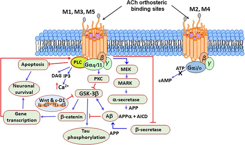
Iarkov et al.                                                                                                Cholinergic Strategy for Parkinson’s Disease

INTRODUCTION                                                                 (Alexander et al., 1986; Bjorklund and Dunnett, 2007; Rodriguez-
                                                                             Sabate et al., 2021). In other words, an enhanced excitatory output
Parkinson’s disease (PD) is a severe neurodegenerative condition             from the subthalamic nucleus (STN) increases the activity of the
characterized by the death of dopaminergic motor and non-                    GP that induces an anomalous inhibitory outflow to the thalamus
motor symptoms leading to locomotor impairment, loss                         and brain stem areas (Braak and Braak, 2000; Cerasa et al., 2016;
of cognitive function, dementia, psychiatric disorders, and                  Iarkov et al., 2020; van Nuland et al., 2020). The inhibition
premature death (Gelb et al., 1999; Leverenz et al., 2009;                   of the thalamus affects the thalamocortical communication
Subramaniam and Chesselet, 2013; Grover et al., 2015; Aarsland               triggering movement abnormalities characteristic of PD, such as
et al., 2017; Martin-Jimenez et al., 2017; Poewe et al., 2017; Walker        bradykinesia (DeLong, 2000; Poewe et al., 2017; Wichmann, 2018;
et al., 2019; Wichmann, 2019; Hussein et al., 2021).                         Reich and Savitt, 2019).
    More than a century of studies in PD achieved breakthrough                   Though PD is a progressive neurodegenerative disease that
discoveries in the etiology of this disease and the role of                  is mainly considered a clinically dominant movement disorder,
cholinergic neurons (Quik et al., 2015b; Jurado-Coronel et al.,              it also has noticeable non-motor symptoms such as psychiatric
2016; Rizzi and Tan, 2017; Tanimura et al., 2018; Ztaou and                  signs of depression, anxiety, and cognitive impairment, some of
Amalric, 2019; Iarkov et al., 2020; Liu, 2020). These studies                which may appear even before the motor ones (Hemmerle et al.,
indicated that the etiology of PD is not entirely clear and has              2012; Lindqvist et al., 2012; Lohle et al., 2019; Mendonca et al.,
a complex and multifactorial nature (Olanow and Tatton, 1999;                2020; Carey et al., 2021; Hussein et al., 2021; Kwon et al., 2021).
Takahashi and Yamada, 1999; Thomas and Beal, 2007; Guttuso                       Many research groups have investigated the prevalence,
et al., 2019b; O’Callaghan and Miller, 2019; Hasan et al., 2020).            sex differences, morphological and functional changes, and
Etiological risk factors are considered a combination of age,                biomarkers to predict the progression from cognitive impairment
gender, genetic background, and environmental factors (Carvey                to dementia in PD (Braak et al., 2005; Anang et al., 2017;
et al., 2003; Thomas and Beal, 2007; Pavlou and Outeiro, 2017;               Hoogland et al., 2017; Ye et al., 2017; Cholerton et al., 2018;
Videira and Castro-Caldas, 2018; Guo et al., 2019; Mehra et al.,             Friedman, 2018; Hussain and Camicioli, 2018; Lanskey et al.,
2019; Delic et al., 2020; Kline et al., 2020). Nevertheless, less            2018; Renouf et al., 2018; Zou et al., 2018; Agelink van Rentergem
than 15% of PD cases have a family history, and most of them                 et al., 2019; Berman and Miller-Patterson, 2019; Chondrogiorgi
are sporadic and seemingly caused by deleterious environmental               et al., 2019; Chung et al., 2019; Palermo et al., 2019a,b; Yoo et al.,
factors acting synergically with susceptibility genes to affect the          2019; Byeon, 2020).
striatum activity (Foltynie et al., 2002a,b; Ferrer et al., 2011;                Almost two decades ago, Braak et al. (2005) found an
Wirdefeldt et al., 2011; Deng et al., 2018; Guo et al., 2019).               association between cognitive status and the neuropathologic
However, much remains unclear, and effective treatments have                 stages of PD in patients with the sporadic form of the disease.
yet to be developed based on innovative new strategies (Liu                  The authors assessed Lewy bodies (LBs) immunoreactive for
et al., 2010; Campos et al., 2011; Schapira, 2011; Irwin et al.,             α-SYN and neuropathological markers for comorbidities such as
2016; Wang et al., 2017; Shimohama and Kawamata, 2018;                       Alzheimer’s disease (AD) that could be contributing to cognitive
Iarkov et al., 2020). A decline of the flow of information from              decline. The authors divided the patients into groups from
midbrain dopaminergic neurons to the striatum, limbic, and                   marginally impaired cognition to severe dementia according to
cortical regions and a deficiency of dopamine (DA) in these                  the Mini-Mental State Examination (MMSE) scores. The results
structures are central events triggering PD (Alexander, 2004;                showed that MMSE scores positively and linearly correlated
Cerasa et al., 2016; Anderkova et al., 2017; Galantucci et al., 2017).       with ascending neuropathologic stages (Braak et al., 2005).
The decrease of incoming dopaminergic input disrupts complex                 Cognitively impaired patients showed higher levels of AD-
regulatory mechanisms in the overlying structures (Gale et al.,              like neuropathology, including beta-amyloid (Aβ) deposition
2008; Schapira and Jenner, 2011; Singh et al., 2017). Deficiency of          than cognitively intact patients. MMSE scores did not correlate
DA can arise due to the neuronal death or synaptic dysfunction               significantly with disease duration, age at disease onset, or death.
of dopaminergic neurons in the midbrain (Schulz-Schaeffer,                   The authors concluded that a decrease in MMSE scores between
2010, 2015; George et al., 2013). The striatum contains mainly               the disease stages 3 to 6 raises the risk of developing dementia
GABAergic medium spiny neurons (MSN) and large aspiny                        during PD progression (Ross et al., 1996; Braak et al., 2005).
choline interneurons ChIs (Alexander et al., 1986; Conti et al.,             However, in some patients, cognitive decline develops in the
2018; Tanimura et al., 2018; Martel and Apicella, 2021). At the              absence of substantial PD-related cortical pathology and, on the
cellular level, DA deficiency induces an imbalance of the activity           contrary, in other patients, extensive cortical neuropathology
of different MSN populations resulting in motor and behavioral               does not unavoidably lead to cognitive decline and dementia
disturbances (Alexander, 2004; Taverna et al., 2008; Tozzi et al.,           (Green et al., 2002; Braak et al., 2005; Leverenz et al., 2009).
2011; Gallo, 2019; Iarkov et al., 2020). MSNs expressing the                 Further studies have given more insight into the mechanisms and
DA receptor 2 (D2R) will decrease their activity, while neurons              morphological correlations of cognitive impairment progression
expressing the DA receptor 1 (D1R) will increase it (Taverna et al.,         to dementia (Aybek et al., 2009; Foster et al., 2013; Aarsland et al.,
2008; Tozzi et al., 2011; Gallo, 2019). Alteration of the indirect           2017; Lanskey et al., 2018).
and direct pathways to globus pallidus internal (GPi)/Substantia                 On the other hand, other non-motor symptoms, including
nigra pars reticulata (SNpr) impairs the communication of the                anxiety and depression, and impulse control disorder, and
thalamus with the motor cortex resulting in motor dysfunction                psychosis, affect many patients, with a prevalence of 50–80%,

Frontiers in Neuroscience | www.frontiersin.org                          2                                 September 2021 | Volume 15 | Article 665820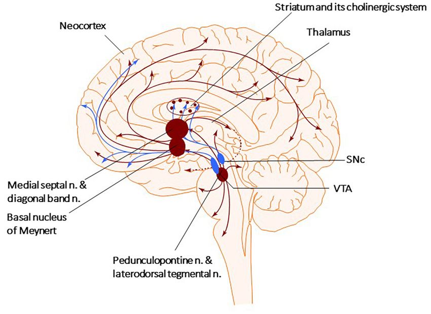
Iarkov et al.                                                                                             Cholinergic Strategy for Parkinson’s Disease

often appearing at the early stages of the disease is only partially       inside neurons and glia (Braak et al., 1998; Martin et al., 2012).
treated by conventional treatments such as L-DOPA and new                  Although the role of Lewy bodies in the development of PD is
treatments have been tested (Bonito-Oliva et al., 2014; Titova             still unknown, the neuropathological diagnosis of PD was base
and Chaudhuri, 2018; Eisinger et al., 2019; Hussein et al.,                on its detection and quantification (Beach et al., 2008, 2009).
2021). Experimentally, non-motor symptoms can be induced                   Intriguingly, not always neurodegeneration of dopaminergic
in mice by bilateral injection of the toxin 6-hydroxydopamine              neurons is accompanied by Lewy bodies (Tompkins et al.,
(6-OHDA) in the dorsal striatum. This mouse model of PD-                   1997; Burke and O’Malley, 2013). Patients with mutations in
like pathology shows only slight gait modifications, with no               α-SYN present [Parkinson disease (PARK)1, PARK3/4/5] or
horizontal motor activity changes as tested in the open-field              not present (PARK2 and PARK8) Lewy bodies associated with
test. However, The treated mice showed depressive-like behavior            nigral degeneration (Foltynie et al., 2002b; Duce et al., 2017).
such as increased immobility in the forced swim and tail                   Mutations such as PARK1 lead to amino acid changes such
suspension tests.                                                          as A53T that increase α-SYN aggregation to form oligomers
   Additionally, mice showed anxiety, expressed as a reduced               and fibrils (Duce et al., 2017). DA inhibits the transition of
time spent in the open arms in the classic anxiety test elevated           the protein oligomers neurotoxic to filaments, a property that
plus maze test and increased thigmotaxis in the open-field test.           may clarify the higher vulnerability of dopaminergic cells to
L-DOPA did not decrease depressive- and anxiety-like behaviors.            neurodegeneration in PD (Foltynie et al., 2002b). Moreover,
Reboxetine, a noradrenaline reuptake inhibitor, reverted the               neurons in the SN, regardless of whether they contain Lewy
depressive and anxiogenic effects. However, desipramine used               bodies or not, present morphological dendritic abnormalities or
to preserve noradrenaline neurons, when administered before                biochemical changes, indicating that all neurons are involved
injection of 6-OHDA, did not modify the resultant depressive-              in the disease process (Patt et al., 1991; Bergeron et al., 1996;
and anxiety behaviors. The authors concluded that mood-related             Hill, 1996; Devi et al., 2008). Due to its structure, α-SYN
disorders were not due to a decrease in noradrenaline (Bonito-             can interact with anionic lipids, which leads to conformational
Oliva et al., 2014). Last decade studies have indicated the                changes that facilitate its aggregation into toxic species (Schapira
involvement of alteration of the serotoninergic system and its             and Jenner, 2011; Bose and Beal, 2016; Shamoto-Nagai et al.,
components, such as the serotonin receptors, with the appearance           2018; Zeng et al., 2018; Gilmozzi et al., 2020). For instance,
of depression in PD (Ballanger et al., 2012; Bonito-Oliva et al.,          the accumulation of mutant forms of α-SYN in the inner
2014; Maillet et al., 2016). One of these studies used positron            mitochondrial membrane disrupts complex I, increasing the
emission tomography (PET) and (18)F (Roselli et al., 2010)                 production of reactive oxygen species (ROS) and contributing
MPPF, a selective serotonin 1A receptor antagonist, to investigate         to neuronal apoptosis (Devi et al., 2008). ROS influence cellular
whether changes in this receptor activity at the postsynaptic site         self-defenses by promoting the cytoprotective effects of DJ-1 and
were involved in the pathophysiology of depression. Compared               PTEN-induced putative kinase 1 (PINK1) while inducing Akt
with non-depressed parkinsonian patients, depressed patients               dysregulation (Zhao et al., 2017).
showed a lower tracer uptake in the left hippocampus, the right
insula, the left superior temporal cortex, and the orbitofrontal
cortex. Compared with controls, non-depressed parkinsonian                 WHY ARE DOPAMINERGIC NEURONS IN
patients presented a reduced F-18 MPPF uptake bilaterally in               THE MIDBRAIN SO VULNERABLE?
the frontal cortex and the right ventral striatum and insula.
Compared with controls, F-18 MPPF uptake was decreased                     Dopaminergic neurons in the midbrain have unique
in depressed parkinsonian patients in the left dorsal anterior             morphological characteristics that may contribute to their
cingulate and orbitofrontal cortices, in the right hippocampal             enhanced vulnerability (Carlsson and Fornstedt, 1991; Chung
region, and the temporal cortex. The imaging data suggest that             et al., 2005; Alavian et al., 2008; Hegarty et al., 2013). For
serotonin 1A receptor dysfunction in the limbic system may                 example, DA neurons have long unmyelinated axons and
underly depression in patients with PD (Ballanger et al., 2012;            massive dendrites that branch out into SNpr, with their somas
Bonito-Oliva et al., 2014; Maillet et al., 2016). The mechanism            being less than 1% of the total volume of these cells (Iarkov
of action of various neuroprotective strategies to prevent PD              et al., 2020). Due to this morphology, a relatively small number
is under investigation; however, efficacious new therapeutic               of neurons provide massive dopaminergic innervation of the
approaches still need to be discovered (Guo et al., 2019; Jurado-          striatum (Sulzer, 2007). It has been calculated that each neuron
Coronel et al., 2019; Iarkov et al., 2020).                                in the SN may have up to 150,000 presynaptic terminals in
                                                                           the striatum (Oorschot, 1996; Sulzer and Schmitz, 2007). The
                                                                           normal functioning of such neurons requires highly active
THE ROLE OF LEWY BODIES IN PD                                              axonal transport through microtubules to support metabolic and
                                                                           reparative processes, synaptogenesis, removal of cellular waste,
The progressive appearance of protein deposits called Lewy                 and communication with other brain cells (Prots et al., 2013,
bodies often accompanies the loss of dopaminergic neurons                  2018; Lu et al., 2014). These cellular process demands high levels
in various brain regions (Schulz-Schaeffer, 2010; Mehra et al.,            of ATP, turning DA neurons in the SN exceptionally susceptible
2019). These deposits contain elevated misfolded α-synuclein (α-           to mitochondrial dysfunction during the development
SYN) oligomers and aggregates, neurofilaments, and ubiquitin               of PD (Horowitz et al., 2011; Venkateshappa et al., 2012;

Frontiers in Neuroscience | www.frontiersin.org                        3                                 September 2021 | Volume 15 | Article 665820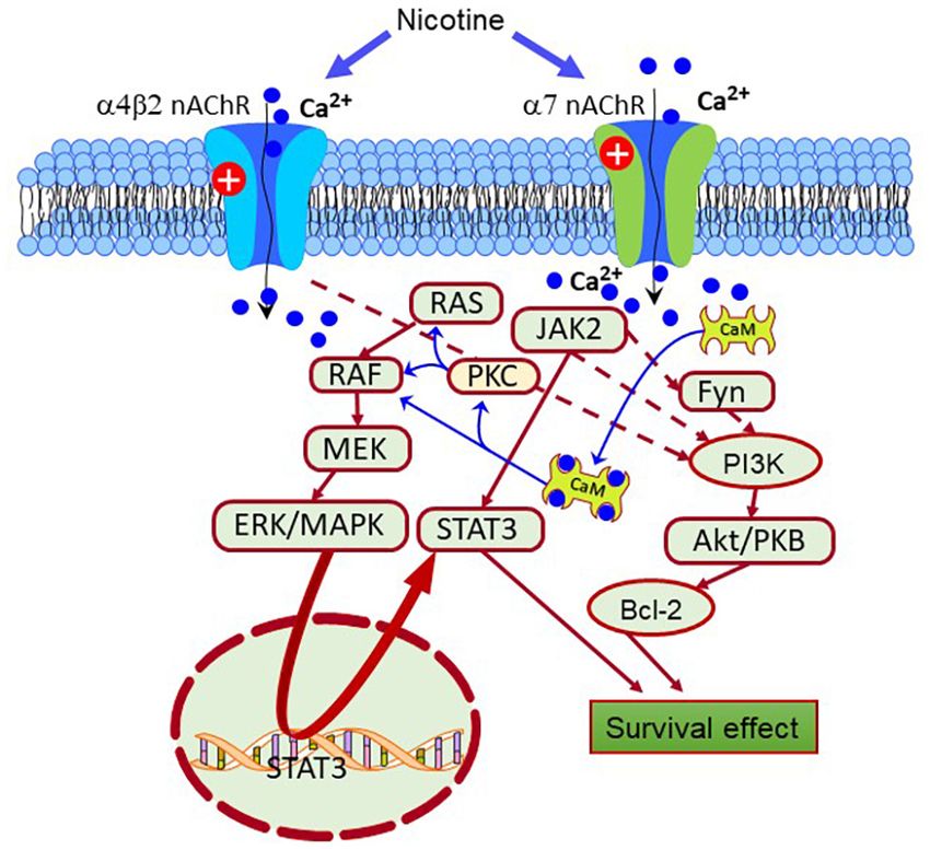
Iarkov et al.                                                                                               Cholinergic Strategy for Parkinson’s Disease

Vanhauwaert and Verstreken, 2015; Course and Wang, 2016;                    effects of alkaloids such as nicotine and other nicotinoids from
Burbulla et al., 2017).                                                     tobacco plants with positive results (Maggio et al., 1998; Linert
                                                                            et al., 1999; Court et al., 2000; Mihailescu and Drucker-Colin,
                                                                            2000; Quik and Kulak, 2002; Soto-Otero et al., 2002; Quik et al.,
MOLECULAR MECHANISMS                                                        2006; Park et al., 2007; Bordia et al., 2008; Huang et al., 2009). This
ASSOCIATED WITH PD                                                          effect has been attributed mainly to nicotine or its metabolites
                                                                            acting on the AChRs (Bordia et al., 2008; Huang et al., 2009).
It is reasonable to postulate that an accumulation of risk factors          On the other hand, nAChRs are expressed on every cell of the
above the repair capacity of DA neurons triggers mitochondrial              dopaminergic system and exert many neuroprotective effects. For
dysfunction, abnormal accumulation of misfolded proteins,                   this reason, modulators of the nAChRs may act as preventative
oxidative stress, and tau hyperphosphorylation in the PD brain              drugs against PD deserve more in-depth consideration (Parain
(Alexander, 2004; Perier and Vila, 2012; Franco-Iborra et al.,              et al., 2001, 2003; Soto-Otero et al., 2002; Bordia et al., 2008;
2016; Jiang and Dickson, 2018). Tau dysfunction disrupts the                O’Leary et al., 2008; Riveles et al., 2008; Hong et al., 2009; Huang
potential of the mitochondrial membrane, impairs the activity               et al., 2009, 2011b; Bordia et al., 2010; Quik et al., 2012, 2013a;
of respiratory enzymes, resulting in a decreased ATP production             Barreto et al., 2014; Iarkov et al., 2020).
and energy supply as well as increased reactive oxygen species                  On the other hand, other authors have attributed these
(ROS) production (O2− and H2 O2 ) (Bose et al., 2011; Keane                 potential positive effects of tobacco consumption in decreasing
et al., 2011; Sutachan et al., 2012). Oxidative stress damages              the risk for PD to the content of lithium in the cigarettes
cellular organelles and the DNA, an event that is particularly              (Guttuso, 2019; Guttuso et al., 2019a,b). These effects have been
dangerous for mitochondrial DNA that does not have protective               linked to changes in the activity of beta-Catenin, a transcriptional
histones and therefore is more vulnerable to ROS damage than                cofactor that upregulates the expression of canonical Wnt target
nuclear DNA (Dexter and Jenner, 2013). Once started, the                    genes, that it has been found reduced in sporadic PD and
disease develops on the principle of positive feedback; oxidative           cell carrying Leucine-rich repeat serine/threonine-protein kinase
stress can potentiate different risk factors, such as age and               (LRPK)2 and beta-glucosidase PD-linked mutations (Marchetti,
unfavorable environmental conditions to induce mutations in                 2018). Also, smokers’ brains have significantly lower alpha-
both cellular and mitochondrial DNA (Bandy and Davison,                     synuclein levels. Tobacco contains very high lithium levels
1990). Although mitochondria contain the genetic information                compared to other plants. Lithium has a broad array of
to produce proteins, most mitochondrial proteins, including                 neuroprotective actions, including enhancing autophagy and
those involved in DNA transcription, translation, and repair, are           reducing intracellular alpha-synuclein levels, and is effective
encoded by nuclear DNA and transported to mitochondria from                 in neurotoxin and transgenic preclinical PD models (Guttuso,
the cytosol (Lenka et al., 1998; Lee et al., 2005). DNA mutations           2019; Guttuso et al., 2019a,b; Vallee et al., 2021). One of the
affecting genes involved in mitochondrial electron transport,               lithium’s neuroprotective actions is the enhancement of beta-
glucose utilization, and glucose sensing may correlate with PD              catenin-mediated activity, leading to increased Nurr1 expression
occurrence (Blanch et al., 2016; Requejo-Aguilar and Bolanos,               through its ability to inhibit glycogen synthase kinase-3 beta
2016; Grunewald et al., 2019). It has been found that 28 sets of            (GSK3β) (Zhu et al., 2014; Guttuso, 2019; Guttuso et al., 2019b;
genes are linked to PD, likely playing a pathogenic role at the early       Vallee et al., 2021). The authors hypothesized that inhaled
stages of the disease (Anderson and Becker, 1981; Zheng et al.,             lithium from smoking might account for the associated reduced
2010; Keane et al., 2011). Currently, a more extensive list of genes        rates of PD, a beneficial effect mediated by the inhibition of
is associated with the onset of PD, supporting the multifactorial           GSK3β and activation of beta-catenin, two factors that could
etiology of both familial and sporadic cases of PD (Scott et al.,           be effective therapeutic targets against PD, for neuroprotective
2017; Lu et al., 2018; Zeng et al., 2018; Kline et al., 2020; Wang          drugs, including the ones modulating the α7nAChRs (L’Episcopo
et al., 2020b; Allende et al., 2021; Martinez-Banaclocha, 2021).            et al., 2014; Liu et al., 2017; Guttuso, 2019; Guttuso et al., 2019b;
    An early study investigating changes in the binding of the              Vallee et al., 2021).
α4β2 nAChR tracer 5- (125)I-A-85380 in PD found a loss of
striatal 5-(125)I-A-85380 binding that correlated with the loss
of nigrostriatal dopaminergic markers (Pimlott et al., 2004).               NEUROTRANSMITTER SYSTEMS IN THE
Similar changes were observed in subjects with dementia with                STRIATUM ALTERED BY PD
Lewy bodies (DLB) that showed a reduced striatal 5-(125)I-A-
85380 binding density, which the authors interpreted as an early            The striatum receives many synaptic inputs from all cortical
degeneration in nigrostriatal inputs. These results suggest the             regions and the thalamus providing excitatory glutamatergic
involvement of the nAChRs on PD etiology (Pimlott et al., 2004).            afferents (Aosaki et al., 2010; Ferre et al., 2010; Huang et al.,
In agreement with this idea, multiple epidemiological studies               2011a,b). At the same time, the nigrostriatal pathway delivers
have shown that active smokers have a lower risk of developing              modulatory neurotransmitters such as DA, ACh, GABA, nitric
PD (Fratiglioni and Wang, 2000; Quik, 2004; Chapman, 2009;                  oxide, and adenosine (Calabresi et al., 2000a; Morelli et al., 2010;
Chen et al., 2010; Greenbaum et al., 2013; Gallo et al., 2019; Cheng        Parent et al., 2011; Tripathy et al., 2015; Sanjari Moghaddam
and Wang, 2020; Kim et al., 2020; Mappin-Kasirer et al., 2020)              et al., 2017; Lopes et al., 2019). All these neurotransmitter systems
encouraging the investigation of the potential neuroprotective              modulate the efficacy of the synaptic transmission in the striatum,

Frontiers in Neuroscience | www.frontiersin.org                         4                                  September 2021 | Volume 15 | Article 665820
Iarkov et al. Cholinergic Strategy for Parkinson’s Disease
which processes excitatory glutamatergic signals from cortical and pars dissipatus (PPNd), and it is involved in starting and
and thalamic afferents and modulates signals from dopaminergic modulating stereotyped movements, including gait (Garcia-Rill,
neurons of the midbrain, aspiny GABAergic, and cholinergic 1986; Snijders et al., 2016; Dos Santos et al., 2021). Glutamatergic
interneurons (Bolam et al., 2000; Kreitzer and Malenka, 2008; neurons of the PPNd (pars dissipatus) regulates the basal ganglia
Gerfen and Surmeier, 2011). These signals are received and and spinal cord. In contrast, the cholinergic pars compacta
processed by the dorsal striatum MSN, which make up 90– (PPNc) is part of the loop connecting the spinal cord and limbic
95% of the striatum neuron population (Tepper et al., 2007). areas with the basal ganglia and thalamus (Bohnen et al., 2011;
The remaining 5–10% of striatum neurons are interneurons, French and Muthusamy, 2018; Bertino et al., 2020). Non-bursting
including the GABA and ACh interneuron (ChI) populations, cholinergic PPNc neurons are considered key to sustaining
which are significant regulators of both MSN and striatal afferents steady-state locomotion (Brimblecombe et al., 2018; Sharma
(Durieux et al., 2011; Munoz-Manchado et al., 2018). Among et al., 2020; Huerta-Ocampo et al., 2021). Additionally, small
them, the most important are ChIs, which closely interact with cholinergic neurons are present in the reticular formation, the
DA afferents of the midbrain (Kim et al., 2019; Dautan et al., medial habenula, and the cortex (Mesulam et al., 1983, 1992;
2020). The glutamatergic, serotonergic, cholinergic, GABAergic, Terenzi et al., 1992; Ballinger et al., 2016).
noradrenergic systems are involved in modulating the striatum’s The cholinergic neurons in the striatum play one of the
output signals (Calabresi et al., 2000a; Do et al., 2012; Zhai most critical roles in developing symptoms in PD, and their
et al., 2019). In addition, opioids, neuropeptides, steroids, and stimulation decrease PD symptomatology (Bohnen et al., 2009;
adenosine receptors families are present in the dorsal striatum Dautan et al., 2014; Kucinski and Sarter, 2015; Osada and
(Aosaki et al., 2010; Ferre et al., 2010; Huang et al., 2011b; Iwasaki, 2017; Chambers et al., 2019; Lieberman et al., 2019).
Moreno et al., 2011; Quik et al., 2012, 2013a; Almey et al., 2015; As mentioned above, the striatum contains giant aspiny ChIs,
Iarkov et al., 2020). Due to the presence of such a variety of connecting to medium spiny neurons. Although giants ChIs
modulators, DA deficiency could be surmounted by modulating account for only 1–3% of striatal neurons, they have highly dense
these receptors (Quik and McIntosh, 2006; Quik et al., 2007; axonal arbors that overlap with those of dopaminergic neurons
Livingstone and Wonnacott, 2009; Avena and Rada, 2012; projecting from the SNpc (Dautan et al., 2014, 2020; Mallet
Goldberg et al., 2012; Mathur and Lovinger, 2012; Myslivecek et al., 2019; Martel and Apicella, 2021). Thus, the high density of
et al., 2017; Ferre and Ciruela, 2019; Ztaou and Amalric, 2019; striatal cholinergic markers reveals the vital role of the cholinergic
Liu, 2020). neurotransmission in modulating striatal function (Phelps et al.,
1985; Calabresi et al., 2000a,b; Mallet et al., 2019; Martel and
Apicella, 2021).
INTERACTION OF THE CHOLINERGIC Research over the past decade shows that striatal ChIs
AND DOPAMINERGIC SYSTEMS IN THE maintain synaptic plasticity and are involved in memory and
STRIATUM other cognitive functions mediated by the posterior striatum,
such as attention and motivation (Bennett et al., 2000; Bohnen
Both the dopaminergic and cholinergic systems belong to the et al., 2011; Havekes et al., 2011; Deffains and Bergman, 2015).
regulatory systems of the brain, the neurons of which are actively ChIs display a constant spiking activity in the absence of synaptic
involved in maintaining the body’s homeostasis (Picciotto et al., inputs (Bennett et al., 2000; Goldberg and Reynolds, 2011).
2012; Rizzi and Tan, 2017). They have a similar anatomical Changes in ChI activity occur during associative conditioned
structure with the neuronal bodies of both systems located in the learning (Robinson et al., 2011; Jiang et al., 2016). For example,
brain stem, midbrain, and subcortical structures of the forebrain, in classical conditioning studies, the temporal pattern of ChIs
and they send their axons throughout the forebrain toward the activity has been investigated. In these experiments, animals
cortex hippocampus, and limbic structures (see Figure 1). Both learned to associate a conditioned stimulus (CS) (a tone)
express several different types of receptors that can generate a with an unconditioned stimulus (US) (a food reward) (Wilson
wide range of cellular responses (Rizzi and Tan, 2017). It is and Fadel, 2017; Kellis et al., 2020). The results showed that
essential to keep in mind that during the development of PD, the during conditioning, shortly after the presentation of the CS,
brain loses not only dopaminergic neurons but also cholinergic ChIs typically responded with a pause in firing that lasted
and serotonergic neurons (Reader and Dewar, 1999; Roselli et al., approximately 200 ms. This pause was preceded or followed by
2010; Ferrer et al., 2012; Myslivecek, 2021). an excitatory burst response (Mallet et al., 2019). This evidence
There are four primary sources of cholinergic projections supports the view that ChIs are involved in acquiring contextual
in the mammalian brain: the pedunculopontine (PPN) and information during conditioning learning (Aosaki et al., 1994a,b;
laterodorsal tegmental nuclei, the thalamic nuclei, the striatum, Apicella, 2017). ChIs modulation with a cholinergic agonist
where a small number of giant cholinergic neurons play the mimicked the electrical oscillations in the striatum of PD brains
role of local interneurons, and the basal forebrain nuclei, that (McCarthy et al., 2011). Furthermore, Opto-excitation of ChIs in
collectively serve as crucial sources of cholinergic neurons healthy animals resulted in PD-like motor deficits (Kondabolu
projecting toward the neocortex, hippocampus, and amygdala et al., 2016), while ChIs Opto-inhibition decreased them in PD
(Figure 1; Garcia-Rill, 1986; Charara and Parent, 1994; Bohnen mice (Maurice et al., 2015). Other scientists have investigated
et al., 2011; Stein and Aziz, 2012; Dautan et al., 2014). The PPN how the temporality of ChIs activity shapes striatal function
can be divided into two subnuclei, the pars compacta (PPNc) using optogenetics coupled to the direct infusion of cholinergic
Frontiers in Neuroscience | www.frontiersin.org 5 September 2021 | Volume 15 | Article 665820Iarkov et al. Cholinergic Strategy for Parkinson’s Disease
FIGURE 1 | Cholinergic and Dopaminergic systems. The diagram describes the Cholinergic (brown) and Dopaminergic (blue) systems. There are four primary
sources of cholinergic projections in the mammalian brain. These include pedunculopontine nucleus and laterodorsal tegmental nuclei; a set of thalamic nuclei;
striatum, where few cholinergic neurons are local interneurons; and the basal forebrain nuclei, which collectively serve as the primary sources of cholinergic
projection neurons in the neocortex, hippocampus, and amygdala. SNc-Substantia nigra pars compacta, VTA, ventral tegmental area.
modulators in the striatum (Mallet et al., 2019). Overall, this disorder induced by a dysbalance between Dopaminergic and
evidence suggests that both ChI and dopaminergic neurons work cholinergic systems (Bohnen and Albin, 2011; Tata et al., 2014;
together to regulate some motor and cognitive functions and McKinley et al., 2019).
represent promising targets for alleviating the symptoms in PD Parkinson’s disease develops as an imbalance between
(Iarkov et al., 2020). dopaminergic inputs and cholinergic interneurons as well as
between the serotoninergic and histaminergic systems, increasing
the histaminergic tone and decreasing the serotoninergic and
dopaminergic activities (Fahn, 1989; Przuntek and Muller,
THE DEVELOPMENT OF AN IMBALANCE 1999; Aquino-Miranda et al., 2012; Johnston et al., 2019).
BETWEEN DIFFERENT SYSTEMS IN THE This hypothesis is coherent because PD symptoms can be
STRIATUM AS THE MAIN successfully relieved by anticholinergics and anti-histamine drugs
CONTRIBUTING FACTOR IN PD such as Benadryl (Barbeau, 1962). However, the prescription
of anticholinergic drugs stopped due to their undesired side
Dopamine deficiency in the striatum causes an imbalance of effects, including the impairment of cognitive abilities (Cooper
activity between two MSN populations, each expressing only et al., 1992; Herzallah et al., 2010; Crispo et al., 2016). These
one type of receptor (D1R or D2R). Each of both MSN groups results suggest that a deterioration of the ascending cholinergic
has a unique path to the GPi/SNpr neurons. MSNs expressing neurons observed post-mortem in PD brains might underly the
D1R form a direct pathway, while those expressing D2R form behavioral deficits in tasks depending on the subcortical frontal
an indirect pathway via the GPe and the subthalamic nucleus cortex. After the work of Alexander and DeLong (Alexander
(STN) (Alexander, 2004; Tozzi et al., 2011; Lu et al., 2021). DA et al., 1986; DeLong, 2000), the concept changed, but now many
deficiency causes a decrease in the activity of MSN expressing researchers are again paying attention to it, and, considering that
D1R and increases the activity of neurons expressing D2R, an imbalance between the ChIs activity and DA input signals
thereby causing motor and cognitive dysfunctions (Alexander, contributes to the development of PD (Threlfell et al., 2012; Ztaou
2004; Tozzi et al., 2011; Wang et al., 2019). The balance and Amalric, 2019). Fortunately, over the last decade, results
between the dopaminergic and cholinergic systems is vital for obtained with new research methods have clarified the main
the correct functioning of the striatum (Aosaki et al., 2010; aspects of the complex relationship between these two systems,
Lester et al., 2010; Crans and Ciruela, 2021). PD symptoms clarifying that ChIs is modulated mainly by dopaminergic
such as tremor and rigidity are ameliorated by L-DOPA and neurons located in the SN and the VTA (Threlfell and Cragg,
anticholinergic drugs, suggesting that PD is a hypercholinergic 2011; Gonzales and Smith, 2015).
Frontiers in Neuroscience | www.frontiersin.org 6 September 2021 | Volume 15 | Article 665820Iarkov et al. Cholinergic Strategy for Parkinson’s Disease
Direct pathway MSNs are activated by dopaminergic signals showed that activation of nAChRs, stimulated the GABAergic
via D1R and inhibited by ChIs signals via M4 mAChRs in these interneurons (English et al., 2011; Luo et al., 2013), indirectly
cells expressing D1R (Bonsi et al., 2011; Gerfen and Surmeier, affecting the striatal dopaminergic activity (Adermark et al., 2011;
2011; Oldenburg and Ding, 2011). The MSNs forming the Clarke and Adermark, 2015).
indirect pathway are inhibited by inputs from dopaminergic
neurons through D2R but activated by inputs coming from
ChIs stimulating the M1 mAChRs (expressed in both MSNs
expressing D1R and D2R) (Bonsi et al., 2011; Gerfen and
WHAT ARE THE MECHANISMS
Surmeier, 2011; Oldenburg and Ding, 2011; Goldberg et al., INVOLVED IN THE PUTATIVE
2012; Rizzi and Tan, 2017). Thus, the dopaminergic system of NEUROPROTECTIVE EFFECTS OF
the midbrain and striatal ChIs modulate each other to maintain TOBACCO CONSUMPTION?
the functional balance between the direct and indirect pathways,
precisely controlling the movement (Liu, 2020). At the same time, The nAChRs have attracted particular interest among researchers
dopaminergic control of ACh release depends on dopaminergic after numerous epidemiological studies have confirmed the low
neurons on ChIs expressing D2Rs that decrease ACh release incidence of PD in active smokers (Chen et al., 2010; Gallo et al.,
(Stoof et al., 1992; Consolo et al., 1993; Yan et al., 1997; Pisani 2019). The activation of the cholinergic system is the best target
et al., 2000). Only a tiny fraction of ChI expresses D1Rs, which to induce neuroprotection by nicotine-derived compounds in PD
increases ACh release (Damsma et al., 1991; Di Chiara et al., 1994; (Quik et al., 2011, 2015a,b; Zhang et al., 2013; Bordia et al., 2015;
Steinberg et al., 1998; Acquas and Di Chiara, 1999; Lim et al., Jurado-Coronel et al., 2019; Iarkov et al., 2020). ACh and other
2014; Gonzales and Smith, 2015). On the other hand, the control ligands acting on the AChRs stimulate the release of DA in the
by the cholinergic system of DA release depends on the activation striatum, reduce neuroinflammation and gliosis, and promote
of presynaptic nAChRs and the modulation by mAChRs (Acquas neuronal survival and synaptic plasticity in the brain (Zhou et al.,
and Di Chiara, 1999; Aosaki et al., 2010; Goldberg et al., 2012). 2003; Quik et al., 2007; Goldberg et al., 2012; Jurado-Coronel
Thus, the initial view of an antagonistic relationship between et al., 2016; Abudukeyoumu et al., 2019; Bordia and Perez, 2019;
these two systems has evolved. New studies have shown an even Liu, 2020). Distinctively, mAChRs are expressed exclusively in
higher complexity in their mutual influence that depends on an the MSNs (Zhou et al., 2003; Goldberg et al., 2012).
organism’s physiological state (Ztaou and Amalric, 2019; Liu, The nAChRs are present in dopaminergic neurons,
2020). The interaction of these two systems in the striatum is glutamatergic neurons, cholinergic interneurons (ChIs),
perceived instead not as enmity but as a dynamic interplay in a and GABAergic interneurons in the striatum (Tanaka et al., 2010;
virtual “neurotransmitter dance” (Surmeier and Graybiel, 2012). Searles Nielsen et al., 2012; Mappin-Kasirer et al., 2020).
Since tobacco smoke contains nicotine, an agonist of the
nAChRs, its protective effect has been related to the activity of
THE NICOTINIC RECEPTORS IN THE nicotine on these receptors (Quik and McIntosh, 2006; Quik
STRIATUM et al., 2007, 2009, 2012, 2013a,b; O’Leary et al., 2008; Riveles
et al., 2008; Hong et al., 2009; Huang et al., 2009, 2011b; Kyaw
In vertebrate species, 17 different subunits of the nAChRs et al., 2013; Barreto et al., 2014; Tiwari et al., 2015). In the
have been identified (α1–10, β1–4, δ, ε, γ) (Millar, 2003; last decades, new evidence highlights cotinine’s neuroprotective
Dineley et al., 2015; Papke and Lindstrom, 2020). The subunits actions, a derivative of nicotine that is a positive modulator of
form homo-and heteropentameric receptors, and the different the α7nAChRs (Soto-Otero et al., 2002; Buccafusco and Terry,
combinations change their specific pharmacological properties 2003; Terry et al., 2005; Echeverria and Zeitlin, 2012; Barreto
(Wu and Lukas, 2011; Dani, 2015). The nAChRs are composed et al., 2014; Gao et al., 2014; Boiangiu et al., 2020; Iarkov
of α4, α6, α7, β2, and β3 subunits, with preferential expression et al., 2020). Cotinine, acting on the α7nAChRs, stimulates
of the α4β2 and α6β2 receptors (Marchi et al., 2002; Millar mechanisms of neuroprotection acting on glial cells (Morioka
and Gotti, 2009; Quik and Wonnacott, 2011; Siciliano et al., et al., 2018; Oliveros-Matus et al., 2020). The actual evidence
2017). Under basal conditions, binding of ligands to the suggests that activated microglia (M1 microglia) contributes
α7nAChR induces a conformational change of the receptor to PD development (Bayarsaikhan et al., 2015; Saitgareeva
that opens the central channel permitting the influx of et al., 2020). Thus, decreasing microglial activation could be an
sodium and calcium ions and the efflux of potassium ions excellent therapeutic strategy for preventing or treating PD.
(Albuquerque et al., 2009). The α7nAChRs are detectable in α7nAChRs are unique targets to diminish the synthesis of
cortical glutamatergic terminals, potentially directly modulating proinflammatory molecules and neuroinflammation due to their
corticostriatal transmission (Howe et al., 2016). In the striatum, ability to inhibit microglial activation (Kim et al., 2015; Lee et al.,
the nAChRs are not expressed in the MSN; however, they 2015). Also, in microglia, the glutamate transporter (GLAST) is
are extensively distributed on GABAergic interneurons and upregulated by α7nAChRs stimulation through the activation of
dopaminergic and glutamatergic terminals, thereby having the both inositol triphosphate-Ca2+/ calmodulin-dependent protein
physiological mechanism for a fine-tuned modulation of the kinase II (CaMKII) and fibroblast growth factor-2 (FGF2)
striatum (Marshall et al., 1997; Kaiser and Wonnacott, 2000; pathways (Morioka et al., 2014). The activation of microglial
Howe et al., 2016; Siciliano et al., 2017). Additional studies α7nAChRs is neuroprotective by inhibiting the expression of
Frontiers in Neuroscience | www.frontiersin.org 7 September 2021 | Volume 15 | Article 665820Iarkov et al. Cholinergic Strategy for Parkinson’s Disease
proinflammatory molecules and preventing excitotoxicity by The two main subtypes of nAChRs are the heteropentameric
promoting glutamate clearance (Morioka et al., 2018). α4β2, α3β2, α7β2 receptors, and homopentameric α7 receptor,
all of which are channels with high permeability to Ca2+
(Akaike and Izumi, 2018). An increase in the intracellular
THE NEUROPROTECTIVE ROLE OF THE Ca2+ activates critical signaling pathways, linking input signals
nAChRs from the extracellular environment to a cellular response to
maintain homeostasis (Gotti et al., 2009; Wang et al., 2012). Some
The cholinergic system plays a vital role in controlling the release studies have shown that activation of the nicotinic receptors can
of neurotransmitters, decreasing neuroinflammation, promoting be neuroprotective by activating signaling pathways stimulated
synaptic plasticity, and neuronal survival in the brain (Van by Ca2+ (Kihara et al., 2001; Dajas-Bailador and Wonnacott,
Beek and Claassen, 2011; Hampel et al., 2018). In addition, the 2004; Rehani et al., 2008; Shimohama and Kawamata, 2018;
cholinergic system modulates both innate and adaptive immune Takahashi, 2020). For example, nicotine induces neuroprotection
responses (Bosmans et al., 2017; Halder and Lal, 2021; Pohanka, throughout the α7- and α4β2 receptors that, when activated,
2021). The cholinergic system affects immune cell proliferation, stimulate the expression of pro-survival genes that inhibit
T-helper differentiation, antigen presentation, and cytokine apoptosis and support synaptic function (Mudo et al., 2007;
production (Fujii et al., 2017; Halder and Lal, 2021; Lu and Buckingham et al., 2009; Shimohama and Kawamata, 2018;
Wu, 2021). In agreement with these functions, the α7nAChRs Papke and Lindstrom, 2020). For example, the increase in
are expressed in basophils, dendritic cells, macrophages, mast Ca2+ ion levels induced by ligands of the α7nAChRs activates
cells, and T and B lymphocytes (Sato et al., 1999; Skok et al., the phosphatidylinositol-3-kinase (PI3K) signaling pathway
2003; Sudheer et al., 2006; de Jonge and Ulloa, 2007; Mashimo (Kihara et al., 2001; Dajas-Bailador and Wonnacott, 2004;
et al., 2020). In addition, in neurons, presynaptic nAChRs Rehani et al., 2008; Shimohama and Kawamata, 2018). For
modulate neurotransmitter release, while postsynaptic nAChRs instance, downstream of the α7nAChR the protein kinase
increase neuronal firing rate, promoting long-term potentiation, Fyn, a member of the Src family, stimulates the PI3K
considered a cellular mechanism of memory formation (Wayner (Gergalova et al., 2014). PI3K phosphorylates Akt, which
et al., 1996; Fujii et al., 1999; Chen et al., 2006; Huang et al., 2008; activates by phosphorylation the transcription factor cAMP
Albuquerque et al., 2009; Kroker et al., 2011; Srivareerat et al., response element-binding protein (CREB) (Leinninger et al.,
2011; Nees, 2015; Echeverria et al., 2016b). 2004; Wang et al., 2020a; Liu et al., 2021). This transcription,
Epidemiological studies have shown a lower rate of in turn, increases the expression of the cell survival factor
development of PD in people who use tobacco products, Bcl-2 (Rehani et al., 2008). Also, Akt inhibits the pro-
suggesting that one or more tobacco-derived compounds may apoptotic factor GSK3β and, therefore, the phosphorylation
have neuroprotective effects (Fratiglioni and Wang, 2000; Parain of the microtubule-associated protein Tau (Rajmohan and
et al., 2003; Hong et al., 2009). Various studies have shown Reddy, 2017; Sayas and Avila, 2021). These actions are
that nicotine reduces the damage of cultured dopaminergic relevant to preventing dementia because hyperphosphorylated
neurons (Riveles et al., 2008; Toulorge et al., 2011; Getachew Tau inhibits axonal transport and induces energy deficits
et al., 2019). Other studies have shown that nicotine and its in the brain leading to oxidative stress, synaptic deficits,
major metabolite, cotinine, have neuroprotective effects against and neuronal cell death (Perez et al., 2018; Compta and
6-hydroxydopamine (6-OHDA) toxicity in cultured SH-SY5Y Revesz, 2021; Koziorowski et al., 2021). Coherent with this
neuroblastoma cells expressing nAChRs (Pogocki et al., 2007; idea, the positive modulation of the α7nAChR by cotinine
Riveles et al., 2008). Nicotine also showed neuroprotective effects is neuroprotective against amyloid-β peptide (Aβ) toxicity
in animal models of PD when administered before nigrostriatal in vivo, inhibiting GSK3β-mediated Tau phosphorylation,
damage occurs (Linert et al., 1999; Salminen et al., 1999; Huang and activating the transcription factor CREB (required for
et al., 2009; Quik et al., 2009, 2015a). The neuroprotective long-term memory storage), improved memory abilities in
effects of other nAChR modulators have also been investigated mouse models of AD (Echeverria et al., 2011; Echeverria
with promising results (Pogocki et al., 2007; Tiwari et al., 2013; and Zeitlin, 2012; Patel et al., 2014; Grizzell and Echeverria,
Jurado-Coronel et al., 2019). 2015; Grizzell et al., 2017). Other mechanisms of nAChR-
mediated neuroprotection include activating the extracellular
signal-regulated protein kinase/mitogen-activated protein kinase
THE NICOTINIC RECEPTORS AND (ERK/MAPK) pathway (Grizzell and Echeverria, 2015). The
NEURONAL SURVIVAL IN THE PD BRAIN α7nAChRs activate neuroprotective factors stimulated by Ca2+ ,
including the protein kinase C (PKC) and the Ca2+ /calmodulin-
Numerous studies in this area have shown that these pathways dependent protein kinase (CaMK), both of which activate CREB
promote neuronal survival, proliferation, and neurite growth as (Kawamata and Shimohama, 2011; Kawamata et al., 2011, 2012;
well as neurotransmitter release in the brain and other tissues Sutachan et al., 2012; Albert-Gasco et al., 2020).
(Eneroth et al., 1977; Fuxe et al., 1979; Aizenman et al., 1991; Evidence obtained using agents inducing PD-like pathologies
Sastry, 1995; Parain et al., 2001; Buccafusco and Terry, 2003; Koh such as Rotenone, 6-OHDA, and 1-methyl-4-phenyl-1,2,3,6-
et al., 2003; de Aguiar et al., 2013; Gao et al., 2014; Tiwari et al., tetrahydropyridine (MPTP), enlightened potential mechanisms
2015; Majdi et al., 2018). of nAChRs-mediated neuroprotection (Kawamata et al., 2011).
Frontiers in Neuroscience | www.frontiersin.org 8 September 2021 | Volume 15 | Article 665820Iarkov et al. Cholinergic Strategy for Parkinson’s Disease
These mechanisms involve activating the signal transducer Echeverria and Zeitlin, 2012; Kawamata et al., 2012; Pillai and
and activator of transcription (STAT)1/3/5, and the Chellappan, 2012; Echeverria et al., 2016a,b).
Fyn/PI3K/Akt/Bcl2, Janus kinase 2 (JAK2)/PI3K/Akt (Bharadwaj
et al., 2020). Besides, the stimulation of the α4β2 and α7nAChRs
triggers other neuroprotective signaling cascades without the THE MUSCARINIC RECEPTORS IN THE
direct involvement of the PI3K system, such as the ERK/MAPK STRIATUM
and JAK2/STAT3 pathways, PKC/Raf/MEK/ERK/STAT3, and
Ras-Raf-ERK signaling pathways (Figure 2; Buckingham et al., In the striatum, every type of neuron expresses different
2009; Zdanowski et al., 2015). subtypes of both mAChR and nAChRs (Threlfell et al., 2010;
The α7nAChRs also stimulate the expression and activity Lim et al., 2014). The mAChRs are metabotropic receptors
of various growth factors and their receptors, such as the that indirectly control the activity of membrane ion channels
vascular endothelial factor (VEGF) and the VEGF receptor 2 through heterotrimeric G-proteins (Kruse et al., 2014; Roth,
(VEGFR2), the platelet-derived growth factor (PDGF) and the 2019). These G-proteins are composed of Gα and Gβg subunits
PDGF receptor (PDGFR), and the epidermal growth factor classified according to the type ofα subunit, which determines
receptor (EGFR) (Pillai and Chellappan, 2012). EGFR activates their association to specific G-protein coupled receptors (GPCR)
the Akt pathway and its downstream effectors, X-linked inhibitor (Huang and Thathiah, 2015; Roth, 2019).
of apoptosis protein-survivin and the nuclear factor kappa In the CNS, the mAChRs are categorized into five subtypes
B (NFκB) (Zdanowski et al., 2015). In general, the nAChRs groups (M1 to M5) (Lim et al., 2014; Mallet et al., 2019).
can activate neuroprotective signaling cascades in neurons, These receptors show significant differences in expression
astroglia, and microglia to promote cell survival, synaptic M1 > M2 > M4 > M3 and M5 (Zhou et al., 2003;
plasticity and maintain brain homeostasis (Picciotto et al., Graef et al., 2011).
2000; Echeverria and Zeitlin, 2012; Kawamata et al., 2012). For A study using atropine to inhibit M2 and M3 mAChRs present
example α7nAChRs upregulate the transcription factors hypoxia- on the glutamatergic terminals revealed a small but significant
inducible factor-1 (HIF-1), GATA-3, NFκB, and signal transducer increase in corticostriatal transmission, suggesting the existence
and activator of transcription (STAT) 1 (Picciotto et al., 2000; of tonic cholinergic presynaptic inhibition of this excitatory
afferents inputs (Pakhotin and Bracci, 2007; Mallet et al., 2019).
At different, the M1 mAChR blocker pirenzepine decreased
corticostriatal transmission (Tozzi et al., 2011).
In PD, anti-muscarinic receptor drugs were the first
symptomatic PD treatment before discovering L-DOPA (Fahn,
1989). mAChR antagonists were used as early treatments
and are still under use in PD (Langmead et al., 2008a,b;
Thomas et al., 2009). The muscarinic antagonists decrease
the hyperactivity of ChIs and corticostriatal glutamatergic
neurotransmission after nigrostriatal denervation (Lim et al.,
2014). However, while they provide some benefits, these drugs
are not without side effects, including cognitive impairment
(Drachman and Leavitt, 1974; Yamamoto et al., 2011). Therefore,
there is a need for more selective cholinergic modulators with
improved therapeutic properties. In addition, therapies with
more selective modulators of the cholinergic receptors may
permit more target specificity and improved pharmacokinetics
compared to ACh.
THE MUSCARINIC AChRs SIGNALING
AND THEIR ROLE IN MAINTAINING
BRAIN HOMEOSTASIS
FIGURE 2 | Scheme depicting different pro-survival signaling pathways
activated by the nAChRs. Nicotine-induced neuroprotection is mediated by The M1, M3, and M5 mAChRs are associated with G-proteins’
receptors, primarily through the α7 and α4β2 receptors. Also, the nAChRs can
activate intracellular pathways that enhance the expression of pro-survival
Gq/11 subfamily (Sil’kis, 2003; Tobin and Budd, 2003; Santiago
proteins that inhibit apoptosis. From them, Janus kinase 2 (JAK2), Fyn, protein and Abrol, 2019). When activated by these receptors, Gq
kinase C (PKC), and calcium-calmodulin kinase (CaMK) are crucial protein interchanges GTP for GDP and dissociates in their constituent
factors triggering the activation of the extracellular signal-regulated protein subunits that become free to activate downstream effectors such
kinase (ERK), JAK2/signal transducer, and activator of transcription 3 (STAT3),
as the phospholipase C (PLC) (Falkenburger et al., 2010). PLC
and phosphatidylinositol 3-kinase (PI3K)-Akt pathways. In turn, these
pathways enhance the expression of antiapoptotic factors such as Bcl-2.
activity results in the release of inositol-triphosphate 3 (IP3)
and diacylglycerol (DG) (Berridge, 2016). IP3 binds to the IP3R
Frontiers in Neuroscience | www.frontiersin.org 9 September 2021 | Volume 15 | Article 665820Iarkov et al. Cholinergic Strategy for Parkinson’s Disease
in the endoplasmic reticulum (ER), triggering the release of consequently promoting mitochondrial apoptosis (Gergalova
Ca2+ from intracellular stores, and DG stimulates the PKC et al., 2014; Lykhmus et al., 2014; Muramatsu et al., 2018). New
(Rehman and Dimri, 2020). studies indicate that the mAChRs, the cannabinoid receptor,
The activation of M2 and M4 receptors activates the Gi/o and the metabotropic glutamate receptor 5 (mGluR5) also
subfamily of G proteins, increasing the opening time of localize intracellularly in the membranes of various organelles
potassium channels and decreasing cAMP production (Santiago (Jong et al., 2018). In these intracellular locations, they can
and Abrol, 2019). Since this second messenger activates the pro- transmit signals from structures such as endosomes, Golgi
survival PKA/CREB pathway, this decrease inhibits cell survival apparatus, endoplasmic reticulum, mitochondria, and nucleus
and synaptic plasticity in the brain (Resende and Adhikari, 2009). (Boivin et al., 2008; Jong et al., 2009; Benard et al., 2012;
The M2 mAChRs also weakly bind to Gs and Gq, acting as a Gergalova et al., 2014; Lykhmus et al., 2014). Recent studies
negative autoreceptor leading to decreased ACh release (Quirion have shown that approximately half of the M1 mAChRs are
et al., 1990; Wess et al., 2007; Santiago and Abrol, 2019). in the intracellular part of the membrane in neuronal cells
On the other hand, the activation of the M1 mAChRs (Anisuzzaman et al., 2013; Uwada et al., 2014). Expression
increases the non-amyloidogenic processing of the amyloid studies using immunohistochemistry methods indicated that the
precursor protein (APP) to generate sAPPα, a proteolytic product mAChRs could also localize in the Golgi apparatus (Muramatsu
that promotes neuroprotection by stimulating neuritogenesis, et al., 2018). Interestingly, the intracellular localization of the
neurogenesis, synaptic plasticity, and memory formation while mAChRs requires a C-terminal tryptophan motif that is only
reducing Aβ and Tau pathology in the brain (Deng et al., 2015; present in the M1 subtype. The M1 mAChRs are also present
Habib et al., 2017). in postsynaptic neurons (Anisuzzaman et al., 2013; Morishima
Some drugs, including non-steroidal anti-inflammatory et al., 2013; Uwada et al., 2014; Muramatsu et al., 2018). Figure 4
compounds, have neuroprotective effects by shaping APP shows a simplified schematic view of the plasma membrane and
processing (Avramovich et al., 2002; Yogev-Falach et al., 2003; intracellular M1 mAChRs in the pre-and postsynaptic neurons,
Kalaitzakis et al., 2008; Miklossy et al., 2008; Shimohama their predicted signal transduction pathways, and the elicited
and Kawamata, 2018). Also, other kinases such as PKC and physiological responses. Studies with the GABAA receptor’s
ERK1/2 stimulate the non-amyloidogenic processing of APP by competitive antagonist bicuculline suggested that M3 and M4
α-secretase (Wang et al., 2016; Zhang et al., 2019). receptors modulate DA release via facilitation or inhibition of
Also, M1 mAChR activation upregulates the expression striatal GABA release.
of β-secretase 1 (BACE1) through a mechanism involving
the activation of MEK/ERK, by a mechanism prevented by
M2 mAChRs activation (Zuchner et al., 2004). Besides, the
stimulation of M1 mAChRs counteracted the Aβ-induced
EFFECT OF COTININE PREVENTING
inhibition of Wnt signaling by GSK3β, resulting in the AMYLOID-β PEPTIDES ACCUMULATION
stabilization of β-catenin and increased expression of survival AND PROMOTING SYNAPTIC
genes (Farias et al., 2004; Jiang et al., 2014b; Wysocka et al., PLASTICITY IN THE BRAIN
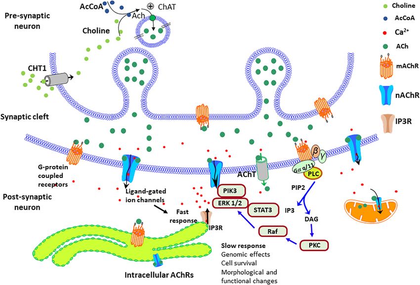
2020). Additionally, the M1 mAChRs, by inhibiting the tau
kinase GSK3β also prevented tau hyperphosphorylation and Despite their structural similarities, nicotine, and cotinine, differ
toxicity (Jiang et al., 2014b). Figure 3 represents a possible in their mechanism of action, properties, behavioral effects, and
mAChR signaling mechanism that protects brain cells from toxicity profile (nicotine is one hundred times more toxic than
mitochondrial dysfunction, caspase activation, oxidative stress, cotinine) (Buccafusco and Terry, 2003; Grizzell and Echeverria,
and DNA damage in PD. 2015; Majdi et al., 2018). The last decade of research has shown
In agreement with this effect, cell studies indicate that that cotinine has unique pharmacokinetic and pharmacodynamic
Wnt/β-catenin pathway inhibition mediates manganese-induced properties, acting as a very weak nAChR agonist but a positive
neurotoxicity (Jiang et al., 2014a). In AD, increased levels of modulator of the α7nAChRs (Moran, 2012; Grizzell et al., 2014;
aggregated forms of Aβ seem to interfere with the function of Sadigh-Eteghad et al., 2020). Different from nicotine, cotinine
M1 mAChRs by uncoupling the receptor-G protein complex is safe and does not elicit addictive behaviors in mammals,
(Janickova et al., 2013). including humans (Yim and Hee, 1995; Hatsukami et al.,
On the other hand, the cellular localization of these cholinergic 1997; Vainio et al., 1998; Zevin et al., 2000; Echeverria and
receptors also may play a key role in their function and cellular Zeitlin, 2012; Thomopoulos et al., 2013). Cotinine has shown
effects (Anisuzzaman et al., 2013; Uwada et al., 2014; Uspenska to protect astrocytes from the toxic effects of chronic and
et al., 2017; Jong et al., 2018; Muramatsu et al., 2018). Although acute stress in vivo (Alvarez-Ricartes et al., 2018; Mendoza
most cholinergic receptors are located in the plasma membrane et al., 2018; Oliveros-Matus et al., 2020) and to prevent the
to convert extracellular signals into intracellular ones, several loss of presynaptic proteins such as synaptophysin in the
studies have reported nAChRs in other neuronal organelles PFC and hippocampus of mice subjected to chronic stress
like mitochondria (Skok and Lykhmus, 2016). The current (Grizzell et al., 2014; Grizzell and Echeverria, 2015). In addition,
evidence suggests that intracellular α7β2 receptors mainly cotinine has shown to reduce the activation of macrophages
stimulate the PI3K/Akt pathway, while α3β2 and α4β2 receptors (Rehani et al., 2008) and be neuroprotective, reducing plaque
inhibit Akt signaling and Ca2+ /calmodulin-dependent pathways, deposition, tau hyperphosphorylation, and cognitive impairment
Frontiers in Neuroscience | www.frontiersin.org 10 September 2021 | Volume 15 | Article 665820You can also read