Membrane Trafficking of Integral Cell Junction Proteins and its Functional Consequences - arXiv
←
→
Page content transcription
If your browser does not render page correctly, please read the page content below
Membrane Trafficking of Integral Cell Junction Proteins and its
                                                                        Functional Consequences∗
                                                                                              Arie Horowitz†
                                                                            Faculty of Medicine, Normandy University, France
                                                    Though membrane trafficking of cell junction proteins has been studied extensively for more than two decades,
                                                 the accumulated knowledge remains fragmentary. The goal of this review is to synthesize published studies on
                                                 the membrane trafficking of the five major junction transmembrane proteins: claudins, occludin, and junction ad-
                                                 hesion molecules (JAMs) in tight junctions; cadherins and nectins in adherens junctions; to identify underlying
                                                 common mechanisms; to highlight their functional consequences on barrier function; and to identify knowledge gaps.
                                                 Clathrin-mediated endocytosis appears to be the main, but not exclusive, mode of internalization. Caveolin-mediated
                                                 endocytosis and macropinocytosis are employed less frequently. PDZ-domain binding is the predominant mode of
arXiv:2101.05221v6 [q-bio.SC] 12 Apr 2021

                                                 interaction between junction protein cytoplasmic tails and scaffold proteins. It is shared by claudins, the largest
                                                 family of junction integral proteins, by junction adhesion molecules A, B, and C, and by the three nectins. All
                                                 eight proteins are destined to either recycling via Rab4/Rab11 GTPases or to degradation. The sorting mechanisms
                                                 that underlie the specificity of their endocytic pathways and determine their fates are not fully known. New data is
                                                 presented to introduce an emerging role of junction-associated scaffold proteins in claudin membrane trafficking.

                                                              1. INTRODUCTION1                                       ous signaling pathways and which interface with the cy-
                                                                                                                     toskeleton. A further layer of complexity is conferred by
                                               Despite being no more than a single-cell thick, the en-               the diversity of membrane trafficking routes and endo-
                                            dothelial and epithelial cell layers that form the lumi-                 somes encountered by different junction transmembrane
                                            nal surface of blood and lymph vessels and of numerous                   proteins.
                                            tubular organs (e.g., the tracheal, digestive, and ureteric                 Much of the data on cell junction dynamics reported
                                            systems), respectively, are the only barriers that pre-                  to date was derived from epithelial cells and the organs
                                            vent breaching of the walls of each system and, con-                     they populate. Though it cannot be assumed by default
                                            comitantly, its dysfunction. The cytoplasmic faces of                    that all the molecular mechanisms that control cell junc-
                                            adherens [291] and tight [300] junctions that maintain                   tion dynamics in epithelial cells are identical to the anal-
                                            monolayer integrity harbor protein complexes which pro-                  ogous mechanisms in endothelial cells (ECs), their ob-
                                            vide structural support and continuity from the junction                 served similarities suggest that they are shared to a large
                                            transmembrane proteins to the cytoskeleton. These intri-                 extent. In a few cases, the review considers data obtained
                                            cate molecular assemblies recycle constantly even in qui-                from fibroblasts used to express epithelial or EC junction
                                            escent cells [57, 64, 236] and undergo extensive remod-                  proteins free of the confounding effects of the endogenous
                                            eling in response to agonists such as vascular endothe-                  proteins. The review does not address desmosomes, which
                                            lial growth factor (VEGF), transforming growth factor                    form in epithelial cells but not in ECs, or gap junctions,
                                            (TGF)-β, sphingosine-1-phosphate (S1P), and platelet-                    which have been studied primarily as electrophysiological
                                            derived growth factor (PDGF) [74, 132, 179, 195]. The                    interfaces.
                                            molecular mechanisms that determine their membrane                          Intercellular junction proteins redistribute from the
                                            trafficking from and to the cell junctions are not fully                 plasma membrane (PM) to the cytoplasm constitutively
                                            known.                                                                   or in response to agonists such as VEGF in ECs [76],
                                               Survey of studies reported mostly during the last two                 or epithelial growth factor (EGF) in epithelial cells [25].
                                            decades reveal that despite the attention given to the                   Once internalized, they have one of two possible fates:
                                            subject of intercellular junction remodeling, the grasp                  recycling back to the PM [171, 200], or proteolysis in
                                            of this process is fragmentary. Conceptual progress on                   lysosomes or proteosomes [45, 282]. Membrane trafficking
                                            cell junction dynamics is hampered by their structural                   of the integral cell junction proteins confers the plastic-
                                            and functional complexities. While there are only two                    ity that cell junctions require to remodel in response to
                                            or three species of junction transmembrane proteins in                   physiological and pathological stimuli.
                                            adherens [164] and in tight junctions [300], respectively,                  For reasons that are related in part to the multiple
                                            some of them consist of protein families of multiple mem-                functions of the catenins that bind their cytoplasmic do-
                                            bers. Moreover, they bind cytoplasmic protein complexes                  mains [161], the membrane trafficking of cadherins is bet-
                                            of varying compositions and sizes that mediate numer-                    ter known than that of all other intercellular junction
                                              ∗ Has  not been submitted for publication                              transmembrane proteins, whereas that of nectins, the sec-
                                               † Correspondence: Faculty of Medicine, 22 Boulevard Gambetta,         ond adherens junction transmembrane protein species, is
                                            76183 Rouen Cedex, Normandy, France; arie.horowitz@inserm.fr             known the least.
                                                1 Nomenclature: names of the proteins mentioned in the review

                                            follow current nomenclature rather than names used in the reviewed
                                                                                                                        The objective of this review is not to cover exhaustively
                                            studies to increase clarity and uniformity (e.g., catenin p120 is        all studies that have a bearing on membrane trafficking
                                            named δ-catenin).                                                        of junction proteins, but, to the extent possible, extract

                                                                                                                 1
patterns and derive organizational principles. Because of            clathrin adaptor protein α-adaptin (AP2A1) [22, 112].
the volume of the data and to maintain focus, the review             Their removal from the cell junctions was blocked by
addresses only the five major integral proteins of tight             cytosolic acidification, sucrose-induced hypertonic stress,
and adherens cell junctions, i.e., claudins, occludin, and           and phenylarsine oxide, all of which block clathrin-
JAMs, and cadherins and nectins, respectively. The cell              mediated endocytosis [95, 174, 223]. The endocytosed
systems, agonists, and experimental methods are speci-               claudins were recruited to early endosomes [148]. Because
fied in order to facilitate evaluation of the relevance and          claudins 1 and 4 collocated in that compartment with
validity of the conclusions of the reviewed studies.                 syntaxin-4, a t-SNARE that mediates docking of trans-
   The accompanying schemes in figures 1-5 represent syn-            port vesicles to the PM [16], it appears they were readied
theses of the reviewed studies on each of the five tight and         for reincorporation in the PM once intercellular junctions
adherens junction transmembrane proteins. They are in-               were restored [112]. In contrast to the effect of hypertonic
tended to provide an ‘at a glance’ overall view of these             stress, the induction of hypotonic stress by halving the os-
pathways while maintaining the connection with the text              molarity of the medium induced endocytosis of claudin-1
by associating each component with one or more rele-                 and -2 in Madin-Darby canine kidney (MDCK) cells. Its
vant studies. Figure 6 presents new data which indicates             blockage by pharmacological inhibition of dynamin, a GT-
that the large junction-associated scaffold protein multi-           Pase required for the scission of clathrin-coated vesicles
ple PDZ domain protein (MPDZ) is involved in claudin                 [42], or of clathrin polymerization, indicated that claudin-
membrane trafficking.                                                1 and -2 underwent clathrin-mediated endocytosis [67].
                                                                        The homologous carboxy-terminus PDZ-binding mo-
 2. MEMBRANE TRAFFICKING OF TIGHT
                                                                     tif shared by 20 members of the claudin family under-
     JUNCTION INTEGRAL PROTEINS
                                                                     lies the similarity of their membrane trafficking path-
                                                                     ways. A missense mutation that replaced threonine in
                    2.1. CLAUDINS
                                                                     the -2 position of the motif to an arginine (T233R) was
   Encoded by 27 known human genes, claudins are the                 linked to familial hypomagnesaemia with hypercalciuria
second largest family of intercellular junction transmem-            and nephrocalcinosis (FHHNC) [176], an inherited kid-
brane proteins [86, 143] after the cadherins. Claudins are           ney disorder. Unlike native claudin-16, the mutant did
a major tight junction structural component. Their vari-             not bind zona occludens (ZO)-1 when expressed in MDCK
ety may reflect tissue-specific expression of their genes,           cells. When triggered to undergo constitutive endocyto-
and/or differences in the extent of sealing they confer              sis by temperature elevation from 4◦C to 37◦C, claudin-16
on tight junctions. Claudins are 210-305 amino-acid-long             appeared in lysosomes instead of intercellular junctions.
tetraspan proteins with cytoplasmic amino- and carboy-               Though junction recruitment appeared to require the
termini (Fig. 1). The majority of claudins harbor a post-            PDZ-binding motif, endocytosis evidently occurred in its
synaptic density-95/Discs large/Zonula occludens (PDZ)-              absence because a claudin-16 with a missense L203X mu-
binding motif at their carboxy termini [86], implying that           tation, which truncates most of its intercellular carboxy-
their recruitment to tight junctions or to trafficking path-         terminus domain including the PDZ-binding motif, was
ways is mediated by proteins that harbor PDZ domains.                retained at the cell junctions when constitutive clathrin-
Whereas most members of the claudin family genes are ex-             mediated endocytosis was inhibited [122, 176]. However,
pressed in epithelial cells, their subsets vary among host           the abundance of the L203X mutant at the cell junctions
organs. A smaller number of claudin genes are expressed              was substantially lower than the wild-type (WT) variant
in ECs. In mouse brain capillaries, the expression levels of         and, unlike the latter, was present throughout the cyto-
Cldn5 and Cldn11 are predominant, but multiple sources               plasm (ibid.).
indicate that Cldn1, 3, 10, 12 and 20 are also expressed                Clathrin-mediated endocytosis of claudins was induced
at significant levels in these cells [192]. Claudins seal the        by several physiological and pharmacological agonists.
tight junctions by forming elongated peripheral strands              EGF induced claudin-2 binding to the clathrin heavy
on the cell’s surface [72, 172]. In endothelial [243, 284], or       chain and α-adaptin in MDCK epithelial cells [107]. The
epithelial [149, 235] cells, claudin removal by endocytosis          endocytosed claudin-2 traversed early endosomes and
from the tight junctions invariably increased monolayer              collocated with lysosome-associated membrane protein
permeability. Claudin strands on abutting cells can form             (LAMP)-1 [33], but not with the Golgi apparatus, sug-
heterophilic interactions, though only a small number of             gesting it was destined to lysosomal degradation rather
combinations had been reported [73]. Claudin-1, a pro-               than recycling. Cevimeline, a specific agonist of the M1
totypical member of the family, underwent constitutive               and M3 muscarinic acetylcholine (Ach) receptors [9], as
recycling in several epithelial cell types [57] . Further            well as carbachol, a non-selective Ach receptor agonist
observations recounted bellow suggested this behavior is             analog, induced claudin-4 binding to clathrin and to the
shared by numerous claudins in epithelial and endothelial            endocytic sorting protein arrestin-β2 in immortalized rat
cells.                                                               salivary gland epithelial cells [40]. This effect was spe-
   2.1.1. Endocytosis                                                cific to claudins, as the interaction of occludin and ep-
   The predominant endocytic pathway of the claudins is              ithelial (E)-cadherin with arrestin-β2 was not altered by
clathrin-mediated. Calcium depletion of human colon car-             carbachol. The latter finding is in agreement with the
cinoma epithelial cells resulted in claudin-1 and claudin-4          M3 receptor specificity to arrestin-β2 [117]. The clathrin
collocation with the clathrin heavy chain and with the               dependence of claudin-4 endocytosis was established by

                                                                 2
1 7 19 21
ACh EGF TGFβ1 CCL2
transcytosis
CLDN3,4,5
5 15
19
CLDN1,2,4,5,16
CLDN5
RAB4 16 4 5 6 1 9 7
9 16 20 arrestin-β2 α-adaptin
CLDN3 CLDN16
EE CLDN1,4
RAB11 7
RAB11
21
CLDN16
9
CLDN1,4 syntaxin-4
12
RE
3 junctions
PIKfyve 5
T191 p38 CLDN1
CHMP3 RAB7 CLDN1,5 1
S195 MAPK1/3 CLDN4 arrestin-β2
2 8 15
MICALL2 K199 Ub-CLDN5
CLDN1,2,3,4,5 18 13 endocytosis
23 junctions 22
cPLA2α RAB13 LNX1 CLDN2
S208
TGN 17 26 27
CLDN2 ? 24 S217 LNX2 CLDN5
LE
early endosome PDZRN3 CLDN16
4 24 25 Golgi 18
CLDN16 PKA
LAMP1 LAMP2 6 endocytosis
LIMP2 10 syntaxin-8
6 10 11 27 Lys
CLDN1,4,5,11,16,19 SEC24C
Lys
Claudins Clathrin
Interacting protein Caveolin
ER Kinase
Agonist 1 Referene number
Nucleus
PDZ-binding motif
Figure 1: Claudin membrane trafficking pathways. Relevant studies are amalgamated rand coupled to the illus-
trated events by the circled numbers. Claudins were internalized via either clathrin caveolin or mediated endocytosis, or via
macropinocytosis. It was partially retrieved to the PM via Rab4, Rab11 and Rab13-mediated recycling or diverted to lysosomal
degradation. The claudin isoforms addressed in the reviewed studies are indicated. The inset presents phosphorylation sites
that regulate claudin membrane trafficking, its carboxy-terminus PDZ-binding motif, and the ubiquitin ligases that bind it.
EGF and CCL2-induced claudin endocytosis increased tight junction permeability (red arrows). The two adjoining PMs in
this figure and in figures 2-5 represent two abutting cells. Dashed lines indicate speculative features. EE, early endosome; LE,
late endosome; Lys, lysosome; RE, recycling endosome. Numbers correspond to the following references: (1) [40], (2) ([57], (3)
[58], (4) [67], (5) [77], (6) [106], (7) [107], (8) [108], (9) [112], (10) [144], (11) [145], (12) [149], (13) [155], (14) [157], (15) [159],
(16) [177], (17) [188], (18) [214], (19) [227], (20) [243], (21) [245], (22) [253], (23) [257], (24) [268], (25) [269], (26) [286], (27)
[292].
showing that it was blocked by pharmacological inhibition entry mechanism of claudin-3 were not specified. Putative
of dynamin [152], by siRNA-mediated knockdown of the nuclear recognition signals had been identified in a major-
clathrin heavy chain, and by hypertonic sucrose medium. ity of the claudins, and some of them had been observed
Endocytosed claudin-4 was ubiquintinated and underwent in nuclei, but only in the context of cancer [87].
proteasomal degradation. Dissociation of trans-binding Aside from endocytosis into the parental cell, claudins
claudins located on adjoining Caco-2 and human embry- underwent transcytosis into adjoining cells. During
onic kidney (HEK)-293 epithelial cells by peptidomimet- wound closure of monolayers of Eph4 or MDCK cells
ics of the first extracellular loop of claudin-1 induced expressing GFP or FLAG (DYKDDDDK), respectively,
clathrin-mediated endocytosis of endogenous claudin-1 fused to the amino-terminus of claudin-3 [77], cells of ei-
and -5 [243]. Because the endocytosis was a direct re- ther type harbored cytoplasmic vesicles containing both
sult of the release of the mechanical coupling between endogenous claudin-3 as well as GFP or FLAG-fused
claudins rather than the downstream effect of specific ag- claudin-3. This indicated that there was no preference be-
onists or cell culture conditions, this result is arguably the tween parental and transfected cells or between WT and
most generic indication that the default internalization recombinant claudins in the direction of transcytosis. The
mechanism of claudins is clathrin-mediated endocytosis. frequency of transcytosis was higher among dispersing
Uniquely among all the junction transmembrane proteins subconfluent cells, suggesting that transcytosis resulted
discussed in this review, claudin-3 redistributed from the from mechanical tension along the intercellular junctions
tight junctions of primary human bronchial epithelial cells produced by cells pulling apart from each other. While
to their nuclei in response to TGFβ1 downstream of TGF the claudins were pinched off from the PM into tubular
receptor 1 [227]. The endocytic pathway and the nuclear structures together with JAMA, occludin, and ZO1, they
3were sorted subsequently into claudin-only vesicles. Binding of claudin-4 to arrestin-β2 in carbachol-treated
Constitutive transcytosis of claudin-4 or -5 overex- immortalized rat salivary gland epithelial cells was linked
pressed in MDCK cells, each fused at its amino-terminus to the phosphorylation of S195 in the carboxy-terminus
to a different species of fluorescent protein, internalized cytoplasmic domain of claudin-4 [40]. The phosphoryla-
simultaneously all the transmembrane proteins present tion was attributed to MAPK1/3 because it was inhib-
on the transcytosed PM region . The endocytic vesicles ited by PD98059, its pharmacological inhibitor [4], and
contained both species of claudins as well as endogenous by U0126, a pharmacological inhibitor of the phosphory-
claudin-1, -2, or -7, or occludin [77]. Single amino-acid lation and activation of MAPK1/3 by MAPKK1/2 [55].
substitutions F147A and Q156E in the second extracel- Claudin-4 phosphorylation on S195 was required for its
lular loop of claudin-5, which impair homophilic trans- binding to arrestin-β2, which, in turn, recruited clathrin.
binding [205], reduced transcytosis frequency. Remark- Hence, the phosphorylation was required for the clathrin-
ably, transcytosis was inhibited by either chlorpromazine, mediated endocytosis of claudin-4. Hypotonic stress-
an inhibitor of adaptor protein (AP)-2 complex bind- induced phosphorylation and endocytosis of claudin-1 and
ing to clathrin [274], or by filipin, which interferes with -2 in MDCK cells [67] was attributed to MAPK p38
caveolae assembly by removal of cholesterol from the PM (MAPK14) because it was blocked by SB202190, its spe-
[229]. Paradoxically, the dynamin-specific pharmacologi- cific pharmacological inhibitor [62]. Based on previous
cal inhibitor dynasore [152] appeared to have no effect on studies [2, 239, 268], the phosphorylated residues were
transcytosis, in conflict with the effective chlorpromazine- assumed to be T191 in claudin-1 and S208 in claudin-2.
induced inhibition. Collocation with the microtubule- This premise was supported by the resistance of the phos-
associated protein 1A/1B-light chain 3 (MAP1LC3B) and phomimetic claudin-1 and -2 mutants T191E and S208E,
with autophagy related protein 16L (ATG16L) indicated respectively, to their removal from the cell junctions by
the transcytosed claudins were targeted to autophago- hypotonic stress. Conversely, the dephosphomimetic mu-
somes. Accumulation in the cytoplasm upon administra- tants T191A and S208A were segregated to the cytosol
tion of the lysosome inhibitor chloroquine [277] suggested and collocated with the late endosome marker Rab7 and
that they were destined to lysosomal degradation, similar the lysosomal marker LAMP1.
to the observation of Matsuda et al. [159]. Claudin-5 underwent caveolin/lipid raft-mediated en-
While there is abundant evidence for the dependence docytosis in mouse brain primary ECs in response to
of monolayer barrier function on claudin phosphorylation the monocyte chemoattractant [3] chemokine C-C ligand
[8, 51, 52, 66, 285], the specific effect of phosphorylation (CCL)-2 [245] based on the collocation of claudin-5 with
on membrane trafficking is less documented. In quiescent cholera toxin, a marker of caveolin-mediated endocytosis
MDCK cells, claudin-16 was phosphorylated constitu- and on the inhibition of claudin-5 endocytosis by choles-
tively [106] by cyclic adenosine monophosphate (cAMP)- terol depletion. Collocation with either caveolin or a lipid
dependent protein kinase (PK)-A, as the phosphoryla- raft marker was not shown.
tion was blocked by several PKA and adenylyl cyclase 2.1.2. Recycling
inhibitors and restored by cAMP. Single site mutations The recruitment of claudins from the Golgi apparatus
of potentially phosphorylatable serines pinpointed the to intercellular junctions required the activity of cytosolic
PKA-phosphorylation to S217 in the claudin-16 carboxy- phospholipase A2 (cPLA2)-α, an arachidonic acid gen-
terminal cytoplasmic domain. Dephosphorylated claudin- erating enzyme involved in the formation of cytoplasmic
16 did not associate with ZO1 and was located predom- tubular membranes [228]. In subconfluent human um-
inantly in the cytoplasm or in lysosomes. Claudin-16 bilical cord ECs (HUVECs) cPLA2α and claudin-5 were
phosphorylated on S217 was recruited to the cell junc- located in separate cytoplasmic punctae [214]. There was
tions and bound the soluble N-ethylmaleimide-sensitive a reciprocal relationship between the cellular locations of
factor attachment protein receptor (SNARE) syntaxin-8 cPLA2α and claudin-5: when the cells reached conflu-
[207], which mediates endocytosis of cell membrane pro- ence, inactive cPLA2α was present in the Golgi apparatus
teins and fusion of endocytic vesicles to early endosomes [94], whereas claudin-5 was located at intercellular junc-
[216]. The phosphorylation of claudins-3 and -5 was also tions. Depletion of cPLA2α by knockdown of its gene,
attributed to PKA [51, 241]. S208 in the cytoplasmic do- PLA2G4A, or by pyrrolidine-mediated pharmacological
main of claudin-2 was phosphorylated in quiescent MDCK inhibition [231], was accompanied by removal of claudin-
cells maintained in serum-supplemented medium [268]. 5 from the cell junctions and its accumulation in the Golgi
A phosphomimetic S208E mutant was detected at the apparatus, suggesting that cPLA2α activity was required
cell edges, suggesting that S208 phosphorylation induced for claudin-5 trafficking from the Golgi to the junctions.
claudin-2 recruitment to cell junctions. Concordantly, the Rab13 plays a major role in the recruitment of tight
majority of the S208A dephosphomimetic mutant popula- junction transmembrane proteins to cell junctions [158].
tion was in the cytoplasm and collocated with the lysoso- The recycling of claudin-1 to the PM after recovery of
mal marker protein LAMP2 [33]. Mutations that reduced MDCK cells from calcium depletion was slowed down
the recruitment of claudin-2 to the cell junctions, such as by siRNA-mediated knockdown of Rab13, or by expres-
the removal of the carboxy-terminus PDZ-binding motif, sion of the GTP hydrolysis-defective mutant Rab13Q67L
were accompanied by a lower phosphorylation level, sug- [286]. The recycling required Rab13 binding to its effector
gesting that the unidentified kinase that phosphorylated molecule interacting with CasL-like 2 (MICALL2) [257], a
S208 was located on the cytoplasmic leaflet of the PM. filamentous (f)-actin and α-actinin-4 binding protein that
4drives f-actin crosslinking during intercellular junction as- ular body (MVB) intraluminal vesicules [120], the main
sembly [184, 221]. The trafficking pattern of Rab13 sug- source of lysosome-targeted cargo, rather than recycling
gested that claudin-1 translocated from the trans-Golgi tubules. Since the cell-surface biotin fraction was not sep-
network (TGN) to recycling endosomes, and from them arated from the rest of the cell lysate, it is conceivable
to the PM [188]. that the abundance of recycled claudin-1 was overesti-
Claudins employ several trafficking pathways and mul- mated. The interaction of CHMP3 with phosphatidyli-
tiple Rab GTPases that are determined in part by the nositol (3,5) phosphate (PtdIns(3,5)P2), an endomem-
conditions the cells are subjected to. Claudin-5 endocy- brane phospholipid, is essential for MVB genesis [279].
tosed in mouse brain microvascular ECs in response to PtdIns(3,5)P2) and its precursor, PtdIns 5-P, are synthe-
CCL2 [276], and collocated with Rab4 [245], a ‘fast’ traf- sized by phosphatidylinositol 3-phosphate 5-kinase (PIK-
ficking GTPase [44]. It did not collocate with the lyso- fyve) [225]. Concordantly, perturbation of PIKfyve activ-
somal marker LAMP2, indicating it was recycled to the ity by the pharmacological inhibitor YM201636 demon-
PM rather than degraded. In agreement, the recycling strated that constitutive recycling of claudin-1 in MDCK
inhibitor bafilomycin-A1 [208] prevented the removal of cells depends on PIKfyve [58], similar to its dependence on
claudin-5 from Rab4-containing vesicles. Claudin-16 re- ESCRT [116]. Approximately 35 percent of total cell sur-
cycling in quiescent MDCK cells was regulated by Rab11 face claudin-1 was endocytosed and recycled in its major-
[108], a ‘slow’ recycling GTPase [263]. Inhibition of Rab11 ity back to the PM in untreated MDCK cells. In contrast,
activity by either a dominant-negative Rab11S25N mu- administration of YM201636 resulted in the accumula-
tant, or by primaquine, a pharmacological inhibitor of tion of all the claudin-1 population in large cytoplasmic
vesicle trafficking [242] decreased claudin-16 presence at clusters. Whereas claudin-2 responded to YM201636 like
the cell junctions in favor of collocation with the early en- claudin-1, the trafficking of claudin-4 was unaffected, sug-
dosome antigen (EEA) 1 and the lysosome marker lyso- gesting that its rate of endocytosis is significantly lower
some membrane protein (LIMP)-2 [14]. The recycling of than those of claudin-1 and -2. The inhibition of claudin-
claudin-2 from the cytoplasm to the junctions of MDCK 1 and -2 endocytosis prevented restoration of the bar-
cells switched from incubation on ice to 37◦C required the rier function of MDCK cell monolayers [58]. Collectively,
activity or Rab14 [149], a GTPase involved in trafficking these results indicate that endocytic trafficking pathways
from the TGN to early endosome [209]. Knockdown of differ not only among tight junction protein species but
Rab14 by short hairpin (sh) RNA resulted in claudin-2 also within the claudin family.
targeting to lysosomes. Rab7, a late endosome marker Though most of the data on claudin membrane traf-
[240], mediated the trafficking of transcytosed claudin-3 ficking addressed individual claudin species, their dynam-
and -4 to the lysosome in MDCK cells [159]. ics are interdependent. This is not surprising given the
Similar to around a third of all translated proteins in large number of claudin species and the abundance of
eukaryotic cells [43], the translocation of claudin-1 from several species in the same cell. Claudin-4 trafficking in
the endoplasmic reticulum (ER) to the Golgi apparatus quiescent HEK-293 cells depended on claudin-8, but not
occurs via membrane trafficking in coat protein complex vice-versa [100]. Normally the two claudins traveled to-
(COP)-2 vesicles. The binding of claudin-1 to the Sec24C gether in endocytic vesicles and bound the scaffold pro-
subunit of COP2 [244] was shared by claudins 4, 5, 11, tein multi (M)PDZ [115, 133]. However, when claudin-8
16, and 19 [292], all of which have a tyrosine and valine was knocked down by siRNA, claudin-4 was sequestered
at their carboxy-termini that functions as an ER export to the ER and the Golgi apparatus. The intracellular
signal. These residues were required but not sufficient dynamics and cell junction recruitment of claudins-2 and
for Sec24c binding, which probably involves the whole -4 in and MDCK cells differed from each other [269]. In
claudin cytoplasmic domain. Expectedly, knockdown of confluent cells, both claudins were located mainly at the
Sec24c reduced the abundance of claudin-1 at the cell sur- cell junctions and to a lesser extent in cytoplasmic vesi-
face. cles. Both newly synthesized claudins originated in the
The endosomal sorting complex required for trans- Golgi apparatus, but claudin-4 preceded claudin-2 at the
port (ESCRT), a multi-protein assemblage that mediates cell junctions. Conjugation of fluorophores that emitted
membrane budding and scission in several cellular con- at either 549 or 505 nm to ‘old’ or to newly synthesized
texts [270] and the sorting of ubiquitinated cargo [120] claudins, respectively, revealed that ‘old’ claudins were re-
was required for the constitutive recycling of claudin-1 moved from the cell junctions to endocytic vesicles. Part
in quiescent confluent MDCK cells [57]. Expression of of these vesicles were destined to lysosomal digestion,
truncated CHMP3, an ESCRT3 component (Raiborg and as they collocated with LAMP2. Surprisingly, removal
Stenmark, 2009), blocked membrane budding [56], result- of the carboxy-terminus PDZ-binding motif of claudin-4,
ing in the mutant’s accumulation in abnormally large vac- which is required for binding to the PDZ domains of ZO1
uolar structures that contained both early and late endo- and ZO2 [111], slowed the rate but not the steady-state
somal markers. Dominant- negative CHMP3 collocated abundance of claudin-2 at the cell junctions. Apparently,
with claudin-1 and -2 and with ubiquitin. Because surface the PDZ-binding motif facilitates but is not categorically
biotinylation indicated a reduction of cell surface claudin- required for claudin recruitment to cell junctions. The
1, it was concluded that dominant-negative CHMP3 im- truncated claudin-4 half-life was longer than that of WT
paired claudin-1 recycling. However, the ESCRT complex claudin-4, likely because it was unable to bind the E3
is thought to mediate primarily the budding of multivesic- uniquitin ligase ligand of numb-protein X (LNX)1. The
5removal of the PDZ-binding motif of claudin-2 probably man occludin. Similar to claudins, occludin is a tetraspan
did not abolish its binding to ZO1 or ZO2 because, un- protein, but its size is substantially larger (522 amino
like claudin-1 and 4, it harbors a tyrosine at position -6 acids,), primarily due to a longer carboxy-terminus do-
that is required for the formation of a second ZO binding main that consists of 257 amino acids in humans (Fig. 2).
site [189]. This tyrosine, which is present in other eight Its cytoplasmic domain binds the Src homology 3 (SH3)
claudins, may account for the overabundance of claudin-2 and the guanylate kinase (GuK) domains of ZO1 via its
over claudins 1, 3, 4, and 7 at the cell junctions of MDCK coiled-coil region [140, 178]. Occludin is recruited to the
cells [269]. tight junctions by claudin [71] and incorporated into the
2.1.3. Degradation claudin strands in a punctate pattern [237].
Endocytosed claudins, either constitutively or in re- 2.2.1. Endocytosis
sponse to external stimuli, undergo either lysosomal di- There is extensive similarity between the endocytic
gestion or proteosomal degradation. Claudin-1 and -2 en- and recycling pathways of claudins and occludin. Like
docytosed in MDCK cells subjected to hypotonic stress claudins, occludin underwent continuous constitutive en-
and treated by the lysosome inhibitor chloroquine accu- docytosis in malignant MTD-1A mouse mammary epithe-
mulated in LAMP1-associated endosomes [67]. The in- lial cells [171]. In a monolayer of MDCK cells that un-
hibition of this accumulation by SB202190 indicated it derwent wounding, occludin underwent clathrin-mediated
depended on p38-dependent trafficking, though the spe- endocytosis with a half-time of 15 min [64]. A pre-
cific role of p38 in this process was not reported. Depri- vious study from the same group established that oc-
vation of oxygen and glucose from-immortalized bEND3 cludin endocytosis was mediated by clathrin [63]. Sim-
mouse brain ECs, was followed by claudin-5 lysosomal ilar to claudin-1 and -4, occludin collocated with the
degradation rather than recycling [144, 145]. In quiescent clathrin heavy chain and with the clathrin adaptor α-
Henrietta Lax (HeLa) cervical carcinoma cells, claudin- adaptin in human colon carcinoma epithelial cells af-
5 was polyubiquitinated on K199, K214, and K215 in ter calcium depletion [112]. VEGF induced clathrin-
the carboxy-terminus cytoplasmic domain, though ubiq- dependent endocytosis and phosphorylation of S490 in
uitination of K199 was sufficient for proteolysis of more the carboxy-terminus cytoplasmic domain of occludin in
than half of the cellular claudin-5 population [155]. Sev- primary bovine retinal ECs [179]. The phosphorylation
eral claudins were ubiquitinated by LNX1, a protein induced binding of the E3 ligase Itch and prompted oc-
that harbors 4 PDZ domains. The first PDZ domain of cludin poly-ubiquitination, ubiquitination, though the un-
LNX1 bound the claudin-1 PDZ-binding motif in quies- derlying mechanism remained speculative because of the
cent MDCK cells [253]. Overexpression of EGFP-fused large distance between S490 and the polyp-proline mo-
LNX1 resulted in the removal of claudins 1, 2, and 4 tif in occludin’s amino-terminus to which the WW do-
from the cell junctions. EGFP-LNX1collocated in part main of Itch binds [260]. Ubiquitinated occludin bound
at the cell junctions with ZO1, occludin, and E-cadherin, epsin-1 epidermal growth factor receptor pathway sub-
whereas it collocated with claudin-2 in the cytoplasm, in- strate (EPS)-15, and hepatocyte growth factor-regulated
cluding in late endosomes and lysosomes. Apparently, tyrosine kinase substrate (HRS). Both proteins harbor
LNX1 ubiquitinated only claudins at the cell junctions. ubiquitin-interacting motifs and facilitate endocytosis of
Since ubiquitination serves as both an endocytic signal ubiquitinated cell-surface proteins [96]. Endocytosed oc-
and for targeting to the interior of MVBs [175], LNX1 may cludin collocated with each of these proteins in cyto-
have designated cell junction claudins for removal from plasmic punctae. Rab5, an early endosome marker [32],
the cell junctions by endocytosis, followed by lysosomal also collocated with occludin in cytoplasmic punctae in a
digestion. LNX2, which has the same domain structure monolayer of wounded MDCK cells [63].
as LNX1 and is close to fifty percent identical, bound the
TNFα, the prototypic member of the tumor necro-
carboxy-terminus of claudin-1 [292] and was likely, there-
sis factor ligand superfamily, induced caveolin-1-mediated
fore, to have functioned as its E3 ubiquitin ligase. Rather
occludin endocytosis in mouse jejunal epithelial cells
than LNX1 or 2, claudin-16 was ubiquitinated by the two
downstream of myosin light chain kinase (MLCK) ac-
PDZ domain-containing RING finger protein (PDZRN)-3
tivation [156] and in Caco-2 and T84 human colon
[157]. Similar to LNX1, PDZRN3 bound claudin-16 via
metastatic epithelial cells [29]. The endocytosis required
its PDZ-binding motif, induced its endocytosis from the
the carboxy-terminal 107 amino acids of the cytoplas-
cell junctions, and designated it to lysosomal digestion, as
mic tail of occludin, TNFα induced caveolin-mediated
indicated by its collocation with the late endosome marker
endocytosis of occludin in Caco-2 and T84 human colon
Rab7.
metastatic epithelial cells. The endocytosis required the
2.2. OCCLUDIN carboxy-terminal 107 amino acids of the cytoplasmic tail
of occludin, a region shared with and named after RNA
Occludin is encoded by a single gene in humans. It polymerase II elongation factor ELL, a region shared with
is grouped into a 3-member proteins family named tight and named after RNA polymerase II elongation factor
junction–associated MARVEL protein (TAMP), based on ELL [237]. The ELL region afforded binding to ZO1
sharing a 130-residue MAL and related proteins for vesi- and occludin dimerization [140]. In its absence, trun-
cle trafficking and membrane link (MARVEL) domain cated occludin remained at the lateral cell membrane de-
[212]. The MARVEL region spans the four transmem- spite TNFα treatment, and, inversely, its expression had
brane domains, corresponding to residues 60-269 of hu- a dominant-negative effect on the endocytosis of endoge-
615 16 18 13
VEGF CCL2 TNFα
transcytosis
21
OCLN
IFNγ
8
13 MLCK
Permeability
7 12 14 1 6
15 16 18 RAB5 3
EE
21
RAB7
RHOA
1
OCLN
ROCK
LE MLCK
12
syntaxin-4
NMM2
RAB4
3 7 18 RE OCLN
RAB11
Prot Lys junction
5 disassembly
OCLN
11 2 7
15 16 7 18 tubulin MICALL2 20 nPKC cPKC
17 9
cPLA2α dynein RAB13 14
Occludin
Interacting protein
Kinase TGN ELL
10
Agonist 19 PKCη PKCβ2
Clathrin Golgi 4 itch
15
Caveolin
poly Ub
15 16
1 Referene number
ER
Nucleus
Figure 2: Occludin membrane trafficking pathways. Occludin was internalized via either clathrin or caveolin-mediated
endocytosis, or via macropinocytosis. It was partially retrieved to the PM via Rab4, Rab11 and Rab13-mediated recycling or
diverted to lysosomal or proteasomal degradation. The inset presents phosphorylation sites that regulate occludin membrane
trafficking and binding sites of ZO1 and itch. VEGF, CCL2, TNFα,, and IFNγ-induced occludin endocytosis increased tight
junction permeability (red arrows). Rab13-mediated occludin recycling restored tight junction integrity (green T). Dashed
line indicates speculative feature. EE, early endosome; LE, late endosome; Lys, lysosome; MLCK, myosin light chain kinase,
NMM2, non-muscle myosin 2; Prot, proteasome; RE, recycling endosome. Numbers correspond to the following references: (1)
[5], (2) [6], (3) [23], (4) [29], (5) [61], (6) [63], (7) [64], (8) [77], (9) [81], (10) [89], (11) ([92], (12) [112], (13) [156], (14) [171],
(15) [179], (16) [180], (17) [214], (18) [245], (19) [250], 20) [257], (21) [266].
nous occludin [29]. The ELL positively charged residues, tiple sites in the carboxy-terminus cytoplasmic domain.
in particular K433, were essential for ZO1 binding to oc- The phosphorylation’s effects on occludin endocytosis
cludin [29]. were site-specific. The most predominant occludin ki-
All three major tight junction transmembrane proteins, nases were either ‘conventional’ (c) Ca2+ and diacyl-
claudin, occludin, and JAM, underwent macropinocytosis glycerol (DAG)-dependent, or ‘atypical’ (a) Ca2+ and
in T84 cells treated by interferon (IFN)-γ [266], a cytokine DAG-independent PKCs. Because DAG induces tight
that disrupts the integrity of epithelial cell monolayers junction assembly [12], the DAG analogs phorbol 12-
[153]. The internalized tight junction transmembrane pro- myristate 13-acetate and 1,2-dioctanoylglycerol were ad-
teins were recruited collectively to subapical actin-coated ministered to MDCK cells to activate cPKCs [6]. Un-
vacuoles at a relatively slow rate, 38 hours after IFNγ der low calcium conditions, the treatment resulted in
treatment. IFNγ initiated the formation of vacuoles by occludin’s phosphorylation on S388 (detected by mass
activating non-muscle myosin-2 via RhoA, Rho-associated spectroscopy) and induced its recruitment to tight junc-
kinase, and MLCK. The vacuoles were identified as recy- tions. Conversely, treatment of Ca2+ -replenished MDCK
cling endosomes by collocation with Rab4 and Rab11 [23]. cells with the GF-109203X PKC-specific pharmacological
Like claudin-5, occludin collocated with cholera toxin, in- inhibitor [259] resulted in occludin’s dephosphorylation.
dicating it underwent caveolin/lipid raft-mediated endo- The phosphorylation detected by mass spectroscopy on
cytosis in CCL2-treated mouse brain primary ECs [245]. S388 was attributed to a cPKC because a mixture of
IFNγ-induced endocytosis of occludin reduced the trans- PKCα, βI, βII, and γ phosphorylated in vitro the re-
epithelial resistance of monolayers of Caco-2 cells [230]. combinant carboxy-terminus cytoplasmic domain of oc-
Similar to claudins, occludin is phosphorylated at mul- cludin. In contrast, calcium replenishment induced phos-
7phorylation of T403/404 and promoted the recruitment disassembly [219]. Since a phosphorylation cascade typi-
of occludin to MDCK cell junctions [250]. Occludin cally ensues downstream of tyrosine kinase receptors, nu-
mutated on T403/404 to dephosphorylation-mimicking merous studies probed the extent of the phosphorylation
alanines was removed from the cell junctions upon cal- of intercellular junction transmembrane proteins. Sev-
cium replenishment, whereas phosphorylation-mimicking eral early studies detected VEGF-induced phosphoryla-
T403/404D mutants were recruited to the junctions. Mo- tion of occludin in primary bovine retinal ECs [7] and
tivated by its conspicuous expression in epithelial cells determined that it existed in five to seven phosphory-
[196], immunoblotting of occludin-precipitated cell lysate lated forms, suggesting the presence of multiple phospho-
confirmed that PKCη, a ‘novel’ diacylglycerol-dependent rylation sites that were at least in part the substrates of
PKC, bound the carboxy-terminus cytoplasmic domain PKCβ2 [89]. Subsequent mass spectroscopy analysis of
of occludin. Its pharmacological inhibition by a pseudo VEGF-treated bovine retinal ECs detected phosphoryla-
substrate peptide [191] and by its knockdown resulted tion on S490 [249]. VEGF increased occludin abundance
in the disruption of the tight junctions of MDCK cells. in the cytoplasm at the expense of its presence along the
Intriguingly, a study from the same group concluded cell borders. PKCβ1, an ATP-competitive PKCβ-specific
that calcium replenishment-induced phosphorylation of inhibitor, reversed the VEGF-induced redistribution of
the nearby Y398 and Y402 of human occludin expressed occludin, suggesting that it was triggered by S490 de-
in MDCK cells had an opposite effect on occludin: rather phosphorylation [180].
than recruiting occludin, it prevented its incorporation in 2.2.2. Recycling
cell junctions [61]. This effect was attributed to the abol- Like claudin, Rab13 played a major role in the consti-
ishment of occludin binding to ZO1. In vitro experiments tutive recycling of occludin in quiescent malignant mouse
suggested that Src was the likely kinase responsible for mammary epithelial cells, as indicated by the stabiliza-
the phosphorylation. tion of occludin at the cell junctions by expression of a
The involvement of aPKCs in the maintenance of epi- dominant-negative Rab13Q67L mutant in MTD-1A cells.
dermal barrier function [92] instigated investigation into Both occludin and claudins 1 and 4 collocated with the
the role of PKCζ in the regulation of Caco-2 cell tight t-SNARE protein syntaxin-4 in T84 epithelial cells [112]
junctions [114]. Treatment of quiescent cell monolayers and required the Rab13 effector MICALL2 to recycle back
by a myristoylated PKCζ pseudosubstrate disrupted the to the PM [257]. Deletion of the Rab13-binding domain
tight junctions and slowed their assembly when the cells of MICALL2 disrupted occludin recycling to the PM and
were subjected to a ‘calcium switch’, i.e., incubation in prevented an increase in the trans-epithelial resistance,
a calcium-chelating medium followed by calcium-enriched indicating that the permeability of the cellular monolayer
medium. PKCζ bound a recombinant 150 residue-long remained high. Whereas Rab13-dependent trafficking by-
recombinant carboxy-terminus cytoplasmic domain of oc- passed the canonic recycling markers Rab4 and Rab11
cludin in vitro. Threonine scanning of the cytoplasmic [171], occludin collocated with Rab4 in mouse brain pri-
domain attributed the phosphorylation to T424 and T438 mary ECs treated by CCL2 [245] and with both Rab4 and
in the carboxy-terminus cytoplasmic domain. Rab11 in IFNγ-treated T84 cells [23]. In quiescent serum-
Calcium replenishment-induced phosphorylation of starved MDCK cells, a minority of the claudin population
Tyr398 and Tyr402 of human occludin expressed in returned to the PM via recycling endosomes in a Rab11-
MDCK cells prevented its recruitment to cell junctions dependent manner [64]. Furthermore, cPLA2α regulated
and its binding to ZO1 [61]. In vitro experiments sug- the trafficking of occludin between the Golgi apparatus
gested that Src was the likely kinase responsible for the and HUVEC junctions in the same manner as claudin-5:
phosphorylation. In contrast to the effects of the above depletion or inhibition of cPLA2α was accompanied by
phosphorylations, calcium replenishment-induced phos- the removal of occludin from the cell junctions and its
phorylation of the nearby T403/404 sites (determined by accumulation in the Golgi apparatus [214]. Possibly be-
mass spectroscopy) induced occludin recruitment to the cause of its role in the maintenance of the Golgi apparatus
intercellular junctions of MDCK cells [250]. Occludin structure [160], partitioning defective protein 3 (Par-3)
mutated on T403/404 to dephosphorylation-mimicking was required for the trafficking of occludin from the TGN
alanines was removed from the MDCK cell junctions to the junctions of TNFα-treated Caco-2 cells. In its ab-
upon calcium replenishment, whereas mutants harbor- sence, occludin accumulated in the TGN [296]. Occludin
ing phosphorylation-mimicking replacements to aspartate translocated from the TGN to the surface of quiescent
were recruited to the junctions. Motivated by its con- Caco-2 or MDCK cells along microtubules in vesicles pro-
spicuous expression in epithelial cells [196], immunoblot- pelled by the minus-end-directed molecular motor dynein
ting of occludin-precipitated cell lysate confirmed that at an approximate velocity of 1.6 µm/s [81]. The first
PKCη, a ‘novel’ diacylglycerol-dependent PKC, bound 18 amino acids of the cytoplasmic domain were sufficient
the carboxy-terminus cytoplasmic domain of occludin. Its for targeting occludin to the cell surface. Among these,
pharmacological inhibition by a pseudo substrate peptide residues I279 and W281 were essential, possibly because
[191] and knockdown resulted in the disruption of the they constituted a TGN export signal [136].
tight junctions of MDCK cells. 2.2.3. Degradation
The effects of VEGF downstream signaling on inter- Unlike claudins, ESCRT did not regulate the recycling
cellular junction proteins have been of obvious interest and fate of occludin [57]. Though there is heterogene-
because the growth factor induces intercellular junction ity in the half lives of claudins [267], several studies con-
8curred that the half-life of occludin is shorter than those early endosomes, followed by segregation into a cyto-
of claudins by as much as three-fold [213, 260]. Direct plasmic subapical compartment where it collocated with
measurements of occludin and claudin-1 dynamics in qui- syntaxin-4, possibly in preparation of reincorporation in
escent MDCK cells by fluorescence recovery after pho- the PM upon restoration of intercellular junctions. Col-
tobleaching revealed an inverse relation between the two lectively, these observations imply that JAM-A shares the
proteins: whereas 76 percent of the steady-state popula- endocytic pathway of the other prominent tight junction
tion of claudin-1 was attached to cell junctions, the size proteins in response to calcium chelation. The same con-
of the unattached cytoplasmic fraction of occludin under clusion can be drawn in regard to the administration of
the same conditions was 71 percent [236]. IFNγ to T84 cells, whereby both occludin and JAM-A un-
The aforementioned ubiquitin ligase Itch bound the derwent macropinocytosis [23, 266]. The internalized pro-
amino-terminus cytoplasmic domain of occludin in HEK- teins subsequently collocated with markers of early endo-
293 and LLC-PK pig kidney epithelial cells [260]. Based somes and with Rab4 and -11, markers of ‘fast’ and ‘slow’
on the effect of the proteasome-specific inhibitor MG132 recycling, respectively [23]. Either calcium chelation or
[220], occludin was determined to undergo proteasomal stimulation with TNFα induced endocytosis of JAM-C in
degradation. The VEGF-induced phosphorylation of oc- HUVECs [126]. Subsequently, it was observed in tubu-
cludin on S490 in bovine endothelial retinal cells discussed lar extensions from membranous structures formed at the
above was required for Itch binding to occludin, for oc- cell junctions, but the endocytic pathway was not identi-
cludin ubiquitination, and, subsequently, for its proteaso- fied. The recruitment of JAM-C to the cell junctions de-
mal degradation [179]. VEGF augmented an ongoing low- pended on its interaction with junction-proximal scaffold
level constitutive proteasomal degradation of occludin. In proteins (likely ZO1 or -2), as indicated by an increase in
serum-starved MDCK cells, close to half of the cellular its presence near the Golgi apparatus at the expense of
occludin population underwent constitutive endocytosis the cell junctions upon deletion of its PDZ-binding motif.
in an approximate half-time of 15 min. The majority of JAM-A endocytosis in Sertoli cells, which generate the
the endocytosed occludin population was apparently de- hemato-testicular barrier, was blocked by knockdown of
graded, as only 20 percent returned to the cell surface the clathrin heavy chain [281].
[64]. Based on collocation with neuropeptide-Y, a lyso- The induction of macropinocytosis appears to be com-
somal marker [255], and on the effect of bafilomycin-A1, mon to the responses of all tight junction integral proteins
a lysosomal inhibitor [294], occludin degradation was at- to inflammatory agonists: lipopolysaccharide (LSP) and
tributed to the lysosome. CCL2 induced the also the micropinocytosis of JAM-A
[246], but more recent studies revealed that under these
2.3. JUNCTIONAL ADHESION MOLECULE
conditions, JAM-A trafficking differed from claudin and
(JAM)
occludin. The JAM-A population that translocated from
The best-known members of the JAM protein family the cell junctions and relocated to cytoplasmic punctae
are encoded by 3 genes, F11R, JAM2 and JAM3. JAM’s in immortalized mouse brain bEND.3 ECs was separate
are 298-310 amino acid-long single pass proteins of the from internalized occludin and claudin-5, as well as from
immunoglobulin family (Fig. 3). Their cytoplasmic do- vascular endothelial (VE)-cadherin. JAM-A translocated
mains range in length from 39 to 48 amino-acids and from the cell junctions to the cytoplasm in 10-20 min
harbor a PDZ-binding motifs at their carboxy-termini and recycled back to the PM in 30-60 min, substantially
that binds the scaffold protein ZO1 at intercellular junc- faster than the 24-48-hour dynamics observed by previ-
tions [60]. JAM-A forms homophilic trans-dimers [170], ous studies [23, 266]. Internalized JAM-A collocated with
whereas JAM-B and JAM-C can form either homo- or and required the activity of Rab34, a known mediator of
heterophilic interactions [130]. The topology of the JAMs macropinosome formation [248]. Subsequently, endocy-
as single pass transmembrane proteins contrasts with the tosed JAM-A collocated with Rab5 and Rab4, indicat-
four-pass topology of both claudins and occludin. A sub- ing it trafficked to early endosomes and recycled rapidly
stantial part of the relatively limited number of studies, [246]. JAM-A was not collocated with Rab7, suggest-
compared to the latter two proteins, was focused on virus ing that only a small fraction of its population under-
entry (e.g., [284]), a phenomenon that is deliberately not went lysosomal degradation. Similar to the response of
addressed in this review. Consequently, knowledge of the JAM-A to IFNγ, lipopolysaccharide (LPS) and CCL2-
membrane trafficking of the JAM proteins under physio- induced macropinocytosis required the activities of RhoA
logical conditions is relatively scarce, particularly of JAM- and Rho-associated protein kinase (ROCK).
B and -C. Though multiple residues in the JAM cyto- The dynamics of JAM-C in quiescent human dermal
plasmic domain are phosphorylated by identified kinases microvascular ECs were dissimilar from JAM-A. In
[247], their phosphorylation has not been linked to mem- contrast to the latter, approximately 80 percent of
brane trafficking. the cellular JAM-C was in the cytoplasm [194]. The
Several of the studies reviewed above analyzed multi- remaining 20 percent were distributed diffusely on the
ple species of tight junction proteins, including the JAMs. cell surface rather than sequestered at the cell junctions.
Similar to claudins-1 and -4, JAM-A collocated with the VEGF stimulation increased the cell-surface associated
clathrin heavy chain and α-adaptin in T84 cells, indicat- JAM-C fraction to 60 percent in approximately one
ing that they were all endocytosed via clathrin-coated hour. Interestingly, the translocation was anterograde,
vesicles [112]. Subsequently, JAM-A was recruited to opposite to the VEGF-induced translocation of all other
98 4 6 1 7 5
TGFβ3 TNFα CCL2 IFNγ VEGF
α-adaptin JAM-C
2 5
6
2
2 8 RHOA 6
JAM-A syntaxin-4
RAB34
6 4
ROCK
RAB5
EE 4
CBL
JAM-A JAM-A poly-Ub
6
RAB4 LE 4
RE
JAM-A Lys 3 4
JAM-C
TGN
Interacting protein
4
Agonist
PDZ-binding motif Golgi
Clathrin
1 Referene number
ER
Nucleus
Figure 3: JAM membrane trafficking pathways. JAMs were internalized via clathrin-mediated endocytosis or via
macropinocytosis. They were partially retrieved to the PM via Rab4-mediated recycling or diverted to lysosomal degradation.
JAM removal from the PM increased tight junction permeability. TGFβ3, TNFα, CCL2, and IFNγ-induced JAM-A/C
endocytosis increased tight junction permeability (red arrows). EE, early endosome; LE, late endosome; Lys, lysosome; RE,
recycling endosome. Numbers correspond to the following references: (1) [23], (2) [112], (3) [123], (4) [126], (5) [194], (6) [245],
(7) [266], (8) [281].
cell junction integral proteins, possibly paralleling the
apparent promotion of intercellular permeability by
2.4. FUNCTIONAL CONSEQUENCES OF
JAM-C, contrary to JAM-A. A recent study on JAM-C
TIGHT JUNCTION PROTEIN TRAFFICKING
dynamics in quiescent HUVECs reported that JAM-C
was primarily at the junctions of confluent cells, and In general, removal of integral proteins from cell junc-
that two thirds of this population was removed from the tions by endocytosis is detrimental to junction integrity.
junctions in two hours by constitutive recycling [126]. Their momentary abundance at the junction reflects a
The cytoplasmic JAM-C population was detected par- dynamic balance between endocytosis and the incorpo-
tially in early endosomes and in MVBs, indicating that ration of newly synthesized or recycled proteins in the
it was targeted at least in part to lysosomal degradation. PM. Constitutive recycling in quiescent cells maintained
In essence, the effect of VEGF on JAM-C dynamics is the abundance of claudin-1, 4/5, and 16 at a steady
opposite to its effect on JAM-A. Monolayer permeability state, thus preserving junction integrity [57, 77, 176]. De-
increased despite the recruitment of JAM-C to the cell pletion of Rab14, which regulates recycling, increased
junctions. The mechanism that confers this intriguing the abundance of claudin-2 at the cell junctions [149]
effect is unknown. Mutation of all four lysines located in and intercellular junction integrity. Claudins form ion-
the JAM-C cytoplasmic domain to arginines, intended selective pores [99], hence, the removal of claudin-2 from
to abolish its ubiquitination, resulted in an increase in MDCK cell junctions reduced junction permeability to
JAM-C abundance in early endosomes, coupled with its Na+ , whereas the depletion of claudin-4 and 7 reduced
decrease in MVBs. Unlike wild type JAM-C, which was Cl- permeability [98]. Paracellular permeability to macro-
targeted by ubiquitination to MVBs and, subsequently, molecules was, however, invariably reduced upon increase
to lysosomes, the mutant did not associate with the E3 in claudin abundance, though constitutive recycling rate
ligase Casitas B-lineage lymphoma (CBL), supporting varied among claudins [57]. Occludin underwent con-
CBL’s role in JAM-C recruitment to MVBs. stitutive recycling similar to that of the claudins, medi-
ated by the same GTPase, Rab13 [158, 171]. In contrast
10to constitutive endocytosis, the endocytosis of claudins occurred before the earliest sampled time point of 5 min
[40, 107, 227, 245], occludin [40, 179, 180, 245, 266], and post the 118◦C to 37◦C switch [203]. Constitutive en-
JAM-A [23, 126, 194, 246, 266, 281] induced by factors docytosis of VE-cadherin in human dermal microvascular
(EGF, VEGF, TGFβ, TNFα) or by cytokines (CCL2, ECs was not positively identified, but its attributes were
IFNγ), increases cellular monolayer permeability. As re- consistent with clathrin-dependent endocytosis [282].
counted above, JAM-C is an intriguing exception to this VE-cadherin in VEGF-treated HUVECs appeared in
rule [126]. cytoplasmic vesicles as soon as 2 min after VEGF ad-
ministration, where it collocated with clathrin, dynamin-
3. MEMBRANE TRAFFICKING OF
2, and the early endosome markers Rab5 and EEA1
ADHERENS JUNCTION INTEGRAL
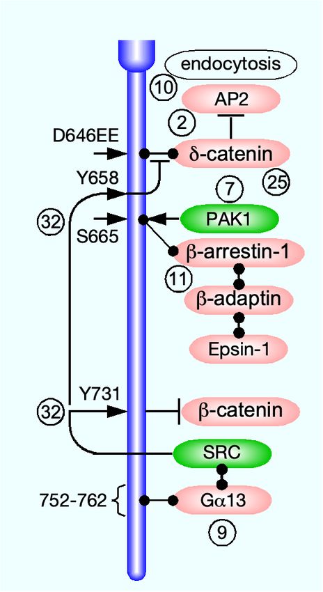
[76]. The cytoplasmic domain VE-cadherin harbors a con-
PROTEINS
served region close to the δ-catenin binding site that was
phosphorylated by p21-activated kinase (PAK)-1 on S665
3.1. CADHERINS
in a Src-dependent manner. The phosphorylation of this
Cadherin membrane trafficking is better understood site was required and sufficient for VE-cadherin endocy-
than that of any other cell junction integral protein. tosis. In contrast, a non-phosphorylatable S665V mutant
Cadherins are the largest junction transmembrane pro- remained at the cell junctions, whereas a S665D phospho-
tein family, consisting of 114 members [38]. They ad- mimetic mutant was endocytosed constitutively in the ab-
join neighboring cells by homophilic binding interactions. sence of VEGF [76]. The phosphorylation of S665 likely
This review focuses on the most abundant (though not recruited the clathrin-binding endocytic adapter arrestin-
the only) cadherins in epithelial and in ECs: epithelial β1, which bound to and collocated with endocytosed VE-
(E) and VE-cadherin, respectively. It includes relevant cadherin. Src is constitutively active in vein ECs, induc-
studies on neuronal (N) cadherin, which is expressed also ing the phosphorylation of Y658 and Y685 in the cyto-
in endothelial [186] and epithelial [190] cells. Human E- plasmic domain of VE-cadherin in HUVECs [195]. The
and VE-cadherin are 882 and 784 amino acid-long single- phosphorylation sensitized VE-cadherin to bradykinin, an
pass transmembrane proteins. They harbor five modular inflammatory cytokine that increases vessel permeability
immunoglobulin-like extracellular-cadherin domains and [103]. Bradykinin induced VE-cadherin ubiquitination
cytoplasmic domains of 151 and 163 amino acids, respec- and clathrin-mediated endocytosis, without dissociating
tively (Fig. 4). Unlike claudins, JAMs, and nectins, cad- δ-catenin. Apparently, the promotion of endocytosis by
herins do not harbor a PDZ-binding motif and do not in- ubiquitination overcame the inhibitory effect of δ-catenin
teract directly with PDZ domain-containing scaffold pro- binding to VE-cadherin.
teins. The canonical binding partners of their cytoplasmic VE-cadherin was tyrosine-phosphorylated in resting
domains are three members of the catenin protein fam- mouse tracheal vein (but not in artery) ECs [195]. The
ily, α-, β-, and δ-catenin (p120) [199]. β-catenin binds phosphorylation, which was localized to Y658 and Y685
the carboxy-terminus region of the E-cadherin cytoplas- in the VE-cadherin cytoplasmic domain, was attributed
mic domain (residues 811-882 of human E-cadherin) and to Src, having been abolished by a specific pharmacolog-
α-catenin [102], which crosslinks E-cadherin to f-actin un- ical inhibitor, AZD0530 [93]. Bradykinin, an inflamma-
der stretch [26]. δ-catenin binds the juxtamembrane re- tory cytokine that increases vessel permeability [103], in-
gion of the E-cadherin cytoplasmic domain (residues 758- duced clathrin-mediated endocytosis and ubiquitination
769) [289]. α-catenin crosslinks E-cadherin to f-actin un- of tyrosine-phosphorylated VE-cadherin. The endocy-
der stretch [26]. VE-cadherin interacts with the three tosed VE-cadherin remained bound to δ-catenin, suggest-
catenins in a similar manner [131]. There is ample evi- ing that the promotion of endocytosis by ubiquitination
dence that VE-cadherin endocytosis in response to VEGF overcame the inhibitory effect of δ-catenin binding to VE-
reduces endothelial barrier integrity and increases its per- cadherin. Both effects were blocked by Src inhibition
meability [76, 132, 195]. or by replacement of Y658 and Y685 by phenylalanine,
3.1.1. Endocytosis indicating that endocytosis and ubiquitination required
Similar to tight junction transmembrane proteins, E- phosphorylation of VE-cadherin. Exposure of HUVECs
cadherin was construed to undergo constitutive endocy- to flow produced the same tyrosine phosphorylation, sug-
tosis, induced by transferring either MDCK cells [137], or gesting that the phosphorylation of VE-cadherin in vein
MCF-7 epithelial breast cancer cells [203] from 18◦C to ECs was a hemodynamic effect.
37◦C. This conclusion was based on the inhibitory effec- A recent study reported that the proteoglycan
tiveness of K+ depletion [135] and of the pharmacological syndecan-4 collocated with VE-cadherin along HUVEC
inhibitor bafilomycin A1 [118]. The presence of a YDSLL junctions and interacted with it independently of the
motif at position 827 of human E-cadherin cytoplasmic former’s glycosaminoglycan chains [47]. VEGF admin-
domain, known to recruit the host protein to clathrin- istration induced Src-mediated phosphorylation of Y180
coated pits [261], further supported this premise [137]. in the syndecan-4 cytoplasmic domain, followed by co-
In contrast, the E-cadherin constitutive endocytosis in endocytosis with VE-cadherin. VEGF-induced endocy-
MCF-7 cells was found to be clathrin-independent. Nev- tosis of VE-cadherin in ECs devoid of syndecan-4 was
ertheless, E-cadherin was detected in clathrin-coated pits approximately 3-fold lower and the increase in their per-
by transmitted electron microscopy (EM), and, as the au- meability was around half than in syndecan-4-expressing
thors admitted, clathrin-dependent endocytosis may have ECs. The manner by which syndecan-4 facilitated VE-
11You can also read