SIMULATED SUNLIGHT DECREASES THE VIABILITY OF SARS- COV-2
←
→
Page content transcription
If your browser does not render page correctly, please read the page content below
Simulated sunlight decreases the viability of SARS-
CoV-2
Angela Sloan
National Microbiology Laboratory, Public Health Agency of Canada, Winnipeg, Manitoba, Canada
Todd Cutts
National Microbiology Laboratory, Public Health Agency of Canada, Winnipeg, Manitoba, Canada
Bryan D Gri n
National Microbiology Laboratory, Public Health Agency of Canada, Winnipeg, Manitoba, Canada
Samantha Kasloff
National Microbiology Laboratory, Public Health Agency of Canada, Winnipeg, Manitoba, Canada
Zachary Schiffman
Department of Medical Microbiology and Infectious Diseases, University of Manitoba, Winnipeg,
Manitoba, Canada
Mable Chan
National Microbiology Laboratory, Public Health Agency of Canada, Winnipeg, Manitoba, Canada
Jonathan Audet
National Microbiology Laboratory, Public Health Agency of Canada, Winnipeg, Manitoba, Canada
Anders Leung
National Microbiology Laboratory, Public Health Agency of Canada, Winnipeg, Manitoba, Canada
Darwyn Kobasa
National Microbiology Laboratory, Public Health Agency of Canada, Winnipeg, Manitoba, Canada
Derek Stein
Cadham Provincial Laboratory, Winnipeg, Manitoba, Canada
Guillaume Poliquin ( guillaume.poliquin@canada.ca )
National Microbiology Laboratory, Public Health Agency of Canada, Winnipeg, Manitoba, Canada
Research Article
Keywords: COVID-19, SARS-CoV-2, viability, arti cial sunlight, sun, solar simulator
DOI: https://doi.org/10.21203/rs.3.rs-37057/v1
License: This work is licensed under a Creative Commons Attribution 4.0 International License.
Read Full License
Page 1/10Abstract
The novel coronavirus, SARS-CoV-2, has spread into a pandemic since its emergence in Wuhan, China in
December of 2019. This has been facilitated by its high transmissibility within the human population and
its ability to remain viable on inanimate surfaces for an extended period. To address the latter, we
examined the ability of sunlight to degrade SARS-CoV-2 on stainless steel. All assays were performed
using a solar simulator at the equivalent of one air mass (i.e. equatorial sun at its Zenith). Heat-controlled
experiments were conducted at approximately 34% relative humidity (RH); otherwise, RH decreased with
sunlight exposure until a constant temperature was maintained. When initially suspended in tissue
culture medium, the virus was rendered non-viable after two hours of sunlight exposure. However, when
suspended in an organic matrix designed to mimic bodily secretions, three hours of continuous sunlight
was required for complete degradation. From this work, we demonstrate that sunlight represents an
effective decontamination method but the speed of decontamination is variable based on the underlying
matrix. This information has an important impact on the development of infection prevention and control
protocols to reduce the spread of this deadly pathogen.
Introduction
Severe acute respiratory syndrome coronavirus 2 (SARS-CoV–2) is a novel coronavirus which emerged in
the city of Wuhan, Hubei province, China in December of 20191. The resultant disease, COVID–19, has
since a icted millions and killed hundreds of thousands throughout the world2,3,4,5,6. Efforts to contain
the spread of the virus have required whole-of-society mobilization efforts, such as physical distancing
and business closures, which have led to worldwide economic devastation. The need for such measures
has been driven in part by the ability of this virus to be transmitted by asymptomatic carriers and pre-
symptomatic patients7,8,9. Furthermore, the role of fomites may also be important, as the virus can
remain infectious on some surfaces, such as plastic, glass, and steel, for up to four days10. One way to
reduce the spread of disease is to regularly disinfect contaminated surfaces with biocidal agents,
especially in high tra c areas like public transit stations and emergency rooms. However, this technique
is not practical for outdoor surfaces as, generally, there is no one employed to regularly clean them. To
address this need, we have examined the ability of sunlight to act as a natural sterilizing medium and
reduce the viability of SARS-CoV–2.
Results
Carrier Tests
All of the experiments employed control carriers that were
inoculated with virus and maintained within the same
biosafety cabinet (BSC). Importantly, all of the carriers
demonstrated a relatively stable infectious load over the
Page 2/10course of our experiment, as expected from other studies
of environmental stability10,11,12,13. Figure 1 demonstrates
that the viability of SARS-CoV–2 decreased the longer the
virus was exposed to sunlight. Inactivation occurred most
e ciently when the virus was suspended in culture
medium. Under these conditions, in heat-controlled
experiments the virus measured at 1.75 x 10³ at time-point
(TP) 0 and showed no signs of degradation after one hour
on control carriers. However, SARS-CoV–2 was rendered
inactive after 60 minutes of sunlight exposure when carrier
heat was maintained constant at 22.5°C (relative humidity
(RH) ~34%). When carrier temperature was allowed to rise
with a concomitant decrease in RH, the viability of sunlight-
exposed virus was extended to 120 minutes. Here the titre
of viable SARS-CoV–2 was reduced to below the limit of
quanti cation, while the titre of control virus fell only slightly
from 9.68 x 10³ to 1.39 x 10³.
Notably, the presence of an organic matrix extended the
survival of the virus when exposed to sunlight. Under these
conditions, sunlight exposure for three hours was necessary
to fully inactivate the virus across all biological replicates in
both heat-controlled and heat-permitted assays. The
viability of virus recovered from control carriers did not
decrease over the same time span (from 3.98 x 10³ at TP0
to 4.88 x 10³) when heat was maintained constant and fell
only slightly when carrier temperature rose and RH declined
(from 3.98 x 10³ to 1.37 x 10³).
Discussion
Page 3/10The medical, social and economic impacts of COVID–19 have raised important questions regarding how
to safely reopen society. One critical question is the risk posed by fomites in outdoor spaces. Other
studies have demonstrated that COVID–19 is able to survive on a variety of surfaces and remain
infectious10,11,12,13. Our study demonstrates that the viability of SARS-CoV–2 can be signi cantly
reduced by exposure to solar radiation. Notably, infectivity of the virus remains relatively constant in the
absence of solar rays in both experiments during the same time span. Importantly, however, we
demonstrate that there are important additional factors that affect the e cacy of sunlight in reducing
infectivity.
First, the matrix within which the virus is suspended has a demonstrable impact on the effect of sunlight
as a disinfection agent. When culture medium was used, infectious virus was no longer detected one hour
after sunlight exposure. By contrast, suspending SARS-CoV–2 in an organic matrix appeared to be
somewhat protective, with detection of very low viral titres after two hours of exposure to sunlight. This is
not surprising, as the organic matrix comprises three types of protein (high molecular weight proteins, low
molecular weight peptides, and mucous material), designed to represent bodily secretions14 which may
insulate the virus. By three hours, however, SARS-CoV–2 is no longer viable in the matrix. Given that most
shed virus is excreted within mucus, the longer exposure time appears more relevant to implementation
of these ndings.
A second important nding is that both heat and humidity impacts virus survival. When carrier heat was
kept constant (22.5°C; RH ~34%), viral viability decreased to below the limit of quantitation (LOQ) after
one hour of sunlight exposure. Interestingly, when the steel carriers were allowed to heat up from the light
exposure and a concomitant decrease in RH ensued, the period of viability was extended, requiring two
hours of exposure to achieve the same result. The same effects were observed for the control virus, as a
decrease in viability, albeit slight, was only observed in heat-controlled experiments where higher RH was
higher (~34%). The validity of these ndings is supported by observations that higher levels of humidity
lead to lower incidences of COVID–19 transmission and mortality15,16,17. Since the heat and humidity
conditions did impact survival, it is important to consider environmental conditions when determining
decontamination protocols. We would favour the latter scenario (i.e. variable heat and variable humidity)
as more representative of real-world conditions.
Our ndings are important in demonstrating that sunlight can be used to decontaminate surfaces
con rmed or suspected of having been exposed to SARS-CoV–2. However, our study has important
limitations. First, we examined sunlight conditions equivalent to a sun at equatorial latitudes in the
absence of cloud cover. As solar intensity varies geographically, it would be important to adjust exposure
times to deliver a similar solar radiation dose based on local conditions. A second limitation is the use of
a non-porous surface for these experiments. It is known that surface characteristics can also impact
survival of the virus, with non-permeable surfaces allowing the virus to persist longer than do absorbent
materials10,11,12,13. Finally, we examined simulated mucus but no other spiked or simulated bodily uids.
SARS-CoV–2 RNA has been detected routinely from patients, but recovering infectious virus appears to
Page 4/10be much less frequent18,19. In spite of this, it would be worthwhile to examine the effect of sunlight on
SARS-CoV–2 in other matrices (e.g. naso-/oro-pharyngeal uids, stool, etc.) where infectious virus has
been recovered18,19.
Overall, these ndings are important in determining plans for the maintenance and decontamination of
outdoor spaces as public health measures are relaxed. Sunlight does appear effective in reducing levels
of infectious virus following three hours of exposure when embedded within mucus. Removal of mucus
through surface cleaning would be expected to increase the e ciency of viral decontamination. Careful
attention to total solar dose and RH should be considered, since these factors affect the rate of
decontamination. Further work to explore other surfaces and environmental conditions should be
performed.
Methods
Virus Propagation
The initial virus aliquot of SARS-CoV-2 (cultured from patient sample; viral passage 1; hCoV-
19/Canada/ON-VIDO-01/2020, GISAID accession# EPI_ISL_425177) was provided by the Vaccine and
Infectious Disease Organization (VIDO; Saskatoon, Saskatchewan, Canada). Vero E6 cells were grown in
150 cm2 tissue-cultured treated asks to 80-90% con uence in Dulbeco’s Minimum Essential Medium
(DMEM) supplemented with 5% bovine calf serum (BCS). Within a biosafety level (BSL)-3 laboratory, the
medium was removed and the cells washed with DMEM containing 0.1% bovine serum albumin (BSA).
The cells were then infected with the SARS-CoV-2 aliquot (5 μl) in DMEM (5 ml) containing 0.5 μg/ml
TPCK-Trypsin and 0.1% BSA and incubated at 37°C and 5% CO2. After 30 minutes of absorption with
intermittent rocking every 5-10 minutes, additional maintenance medium (30 ml) was added and the cells
were again incubated at 37°C and 5% CO2. Any resulting cytopathic effect (CPE) was monitored daily,
with the supernatant harvested ve days post-infection (dpi). The initial virus inoculum was quanti ed by
end-point titration on Vero E6 cells and determined to be 4.6x10⁶ TCID 50/ml (i.e. 50% tissue culture
infectious dose per milliliter).
Organic Matrix
The organic matrix used in this study is the standard tripartite soil load described in ASTM E2197-17
e114. Exceptionally, the mucin suspension in 0.85% NaCl was gamma-irradiated at 2 MRads on wet ice as
an alternative to lter-sterilization to avoid clogging of the lter.
Solar Simulator
Page 5/10The arti cial sunlight used in this study was produced by the SunLite Solar Simulator Model 11002 from
Abet Technologies. Solar output was set to 1 sun, equivalent to 1 air mass or natural sunlight emitted at
the equator during peak hours on a cloudless day. An atmospheric edge lter was used to block all
wavelengths below 305 nm, as radiation below this level is absorbed by the atmosphere in the natural
environment.
Carrier Tests
Vero cells were seeded into clear, at-bottomed, tissue-culture treated 96-well plates in Minimum
Essential Medium (MEM; 100 μl) supplemented with 5% BCS and 1% L-glutamine, and grown to
approximately 90% con uence overnight at 37°C and 5% CO2. All subsequent procedures were performed
in a BSL-4 laboratory in a class II biological safety cabinet by workers wearing positive-pressure ILC Dover
suits. One stock vial of SARS-CoV-2 was thawed at room temperature and 340 μl added to 160 μl of either
maintenance medium (MEM containing 1% BCS and 1% L-glutamine) which emulates the virus in a
laboratory environment, or organic matrix which emulates the virus in its natural environment20. Positive
controls were prepared in triplicate by adding the viral suspension (10 μl) to maintenance medium (1 ml).
Carriers were prepared by adding the viral suspension (10 μl) to the centre of sterilized stainless steel
disks (1 cm in diameter and 0.7 mm thick) and allowed to dry for 45 minutes. Carriers were prepared in
triplicate for test and control conditions at each designated time point. Once dry (i.e. TP0), maintenance
medium (1 ml) was pipetted up and down on each of three carriers to re-suspend the virus, and used to
infect Vero cells (see following paragraph). In experiments designed to control for the confounding
variable of heat, half of the carriers were placed directly under the solar simulator light source set to 1 sun
in a digital block heater/cooler set to 14°C, which ensured the carriers remained at room temperature
(22.5°C). Corresponding carriers were placed in a petri dish within the BSC. For experiments where
infrared heat was permitted, control carriers were placed in the block heater/cooler and heated to the
same temperature the disks reached under the solar simulator, which was periodically measured with a
thermometer and wire probe.
Shortly after collection, each sample and positive control were used to infect the nearly con uent Vero
cells in triplicate: after removal of the growth medium, neat virus (100 μl) was added to the top row of the
96-well plate and a series of virus dilutions (10-1 to 10-6) prepared in maintenance medium were added to
the six consecutive rows below. The wells in the bottom row of the plate contained maintenance medium
only and served as negative controls. The plates were incubated at 37°C and 5% CO2 for four days, at
which time individual wells were examined for CPE. The Reed and Muench calculation21 was used to
calculate TCID 50/ml values and the LOQ. Log10 TCID 50/ml values were then calculated and plotted to
examine virus viability in all treatment groups over time.
Declarations
Author Contributions
Page 6/10A.S. conducted and analyzed carrier tests, and wrote the manuscript; T.C. conducted and analyzed carrier
tests; B.D.G. analyzed carrier tests; S.K. conducted carrier tests; Z.S. and M.C. performed cell culture and
reagent preparation; J.A. aided in analysis of results; A.L. and D.K. propagated the SARS-CoV-2 virus;
D.R.S. provided invaluable expertise and guidance; G.P. provided oversight into all functions of this study
and edited the manuscript. All authors reviewed the manuscript.
Additional Information
The authors declare that no competing interests exist.
References
[1] Lu, H., Stratton, C. W. & Tang, Y. Outbreak of pneumonia of unknown etiology in Wuhan, China: The
mystery and the miracle. J. Med. Virol. 92, 401-402 (2020).
[2] Novel coronavirus – Thailand (ex-China). Geneva: World Health Organization
https://www.who.int/csr/don/14-january-2020-novel-coronavirus-thailand/en/ (2020).
[3] Novel Coronavirus – Japan (ex-China). Geneva: World Health Organization
https://www.who.int/csr/don/16-january-2020-novel-coronavirus-japan-ex-china/en/ (2020).
[4] Update on the novel coronavirus pneumonia outbreak (Jan 24, 2020). Beijing: China National Health
Commission China National Health Commission
http://www.nhc.gov.cn/xcs/yqfkdt/202001/c5da49c4c5bf4bcfb320ec2036480627.shtml (2020).
[5] Novel coronavirus – Republic of Korea (ex-China). Geneva: World Health Organization
https://www.who.int/csr/don/21-january-2020-novel-coronavirus-republic-of-korea-ex-china/en/ (2020).
[6] First travel-related case of 2019 novel coronavirus detected in United States. Atlanta, GA: US Centers
for Disease Control and Prevention US Centers for Disease Control and Prevention
https://www.cdc.gov/media/releases/2020/p0121-novel-coronavirus-travel-case.html (2020).
[7] Bai, Y. et al. Presumed asymptomatic carrier transmission of COVID-19. JAMA. 323, 1406-1407 (2020).
[8] Huang, L. et al. Rapid asymptomatic transmission of COVID-19 during the incubation period
demonstrating strong infectivity in a cluster of youngsters aged 16-23 years outside Wuhan and
characteristics of young patients with COVID-19: A prospective contact-tracing study. J. Infect. 80, e1-e13
(2020).
[9] Rothe, C. et al. Transmission of 2019-nCoV Infection from an Asymptomatic Contact in Germany. N.
Engl. J. Med. 382, 970-971 (2020).
[10] Chin, A. W. H. et al. Stability of SARS-CoV-2 in different environmental conditions. Lancet Microbe 1,
e10 (2020).
Page 7/10[11] Pirtle, E. C. & Beran, G. W. Virus survival in the environment. Rev. Sci. Tech. 10, 733-748 (1991).
[12] Ren, S. Y. et al. Stability and infectivity of coronaviruses in inanimate environments. World. J. Clin.
Cases. 8, 1391-1399 (2020).
[13] van Doremalen, N. et al. Aerosol and surface stability of SARS-CoV-2 as compared with SARS-CoV-1.
N. Engl. J. Med. 382, 1564-1567 (2020).
[14] ASTM E2197-17e1: standard quantitative disk carrier test method for determining bactericidal,
virucidal, fungicidal, mycobactericidal, and sporicidal activities of chemicals. ASTM International, West
Conshohocken, PA; doi: 10.1520/E2197-17E01 (2017).
[15] Wang, J., Tang, K., Feng, K. & Lv, W. High temperature and high humidity reduce the transmission of
COVID-19. Preprint at http://dx.doi.org/10.2139/ssrn.3551767 (2020).
[16] Islam, N., Shabnam, S. & Erzurumluoglu, A. M. Temperature, humidity, and wind speed are associated
with lower Covid-19 incidence. Preprint at https://doi.org/10.1101/2020.03.27.20045658 (2020).
[17] Ma, Y. et al. Effects of temperature variation and humidity on the death of COVID-19 in Wuhan,
China. Sci. Total Environ. 724, 138226 (2020).
[18] Wölfel, R. et al. Virological assessment of hospitalized patients with COVID-2019. Nature 581, 465-
469 (2020).
[19] Wang, W. et al. Detection of SARS-CoV-2 in different types of clinical specimens. JAMA. 323, 1843-
1844 (2020).
[20] Sattar, S. A., Springthorpe, V. S., Adegbunrin, O., Zafer, A. A. & Busa, M. A disc-based quantitative
carrier test method to assess the virucidal activity of chemical germicides. J. Virol. Methods 112, 3-12
(2003).
[21] Reed, L. J. & Muench, H. A simple method of estimating fty per cent endpoints. Am. J. Epidemiol. 27,
493-497 (1938).
Figures
Page 8/10Page 9/10
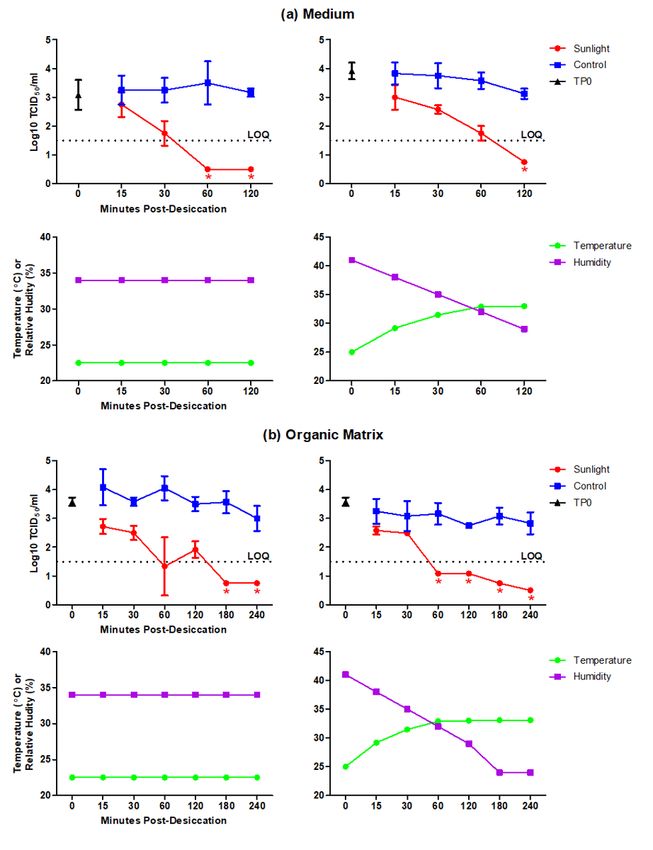
Figure 1

Viability of SARS-CoV-2 on stainless steel after exposure to simulated sunlight. SARS-CoV-2 was
suspended in (a) culture medium or (b) an organic matrix, deposited on stainless steel, desiccated
(“TP0”), and exposed to either simulated sunlight (“Sunlight”) or corresponding ambient conditions
(“Control”). Graphs in the top row of (a) and (b) show the titer of viable eluted virus, expressed as the
Log10 50% tissue culture infectious dose per milliliter (TCID50/ml), following culture in Vero cells. The
limit of quantitation (LOQ), denoted by a dashed line, is 1.5 logs or 3.16 x 101 TCID50/ml. Plots show the
mean and standard deviation of three biological replicates per time-point, with each biological replicate
representing the average of three technical replicates. Plot points denoted by an asterisk were not
quanti able and assigned values for graphing purposes only; consequently, standard deviation could not
be calculated for these data. Graphs in the bottom row of (a) and (b) show the carrier temperature and
relative humidity readings measured at each time-point, depicting heat-controlled (left) and heat-
permitted (right) assays, and correspond to the experiment represented in the graph located directly
above.

                                                 Page 10/10
You can also read