Intra- and Periarticular Ganglia (Synovial Cysts) of the Hip with Compression of the Obturator Nerve, Concomitant with Lesions of the Ligamentum ...
←
→
Page content transcription
If your browser does not render page correctly, please read the page content below
Open Journal of Orthopedics, 2021, 11, 170-182
                                                                                                https://www.scirp.org/journal/ojo
                                                                                                          ISSN Online: 2164-3016
                                                                                                            ISSN Print: 2164-3008

Intra- and Periarticular Ganglia
(Synovial Cysts) of the Hip with
Compression of the Obturator Nerve,
Concomitant with Lesions of the
Ligamentum Teres—A Report of 3 Cases

Florian Haug*, Richard Herzog

Investigation performed at Department of Orthopedics and Trauma Surgery, Lucerne Cantonal Hospital, Wolhusen, Switzerland

How to cite this paper: Haug, F. and          Abstract
Herzog, R. (2021) Intra- and Periarticular
Ganglia (Synovial Cysts) of the Hip with      Background: Synovial cysts of the hip are commonly found in patients with
Compression of the Obturator Nerve,           intra- or extraarticular pathologies of the joint. Symptoms are mostly unspe-
Concomitant with Lesions of the Ligamen-
                                              cific. To date there are no guidelines for a gold standard of treatment. Aim of
tum Teres—A Report of 3 Cases. Open
Journal of Orthopedics, 11, 170-182.          this article is to show up how lesions of the ligamentum teres (LT) might pos-
https://doi.org/10.4236/ojo.2021.115017       sibly lead to a specific formation of synovial cysts of the hip joint and how
                                              this can be treated arthroscopically. Methods: This case series included 3 pa-
Received: March 12, 2021
Accepted: May 28, 2021
                                              tients with ganglia of the hip. All patients had impingement symptoms, com-
Published: May 31, 2021                       bined with untypical location of pain. All patients qualified for joint preserv-
                                              ing surgery and underwent hip arthroscopy with pre- and postoperative MRI
Copyright © 2021 by author(s) and             imaging. The mean follow-up time was 22 months. Results: MRI imaging
Scientific Research Publishing Inc.
This work is licensed under the Creative
                                              showed extensive ganglia, presumably originating from the pelvic root of LT,
Commons Attribution International             extending to the obturator lodge. In 2 of 3 cases MRI showed lesions of the
License (CC BY 4.0).                          LT. Hip arthroscopy revealed damage of the LT in all cases, caused by chronic
http://creativecommons.org/licenses/by/4.0/
                                              instability of the joint. The postoperative MRI showed a complete regression
               Open Access
                                              of the ganglia in all patients after offset correction. After follow-up, 2 of 3 pa-
                                              tients were mostly symptom free. One patient was still suffering from a
                                              chronic weakness of the gluteus medius muscle. Conclusion: Whenever un-
                                              specific radiating pain of surrounding areas of the hip is encountered and
                                              cannot be explained by common pathologies of the hip, possible compression
                                              of nerves by ganglion cysts should be excluded. This should be done by MRI
                                              arthrography. A partial rupture of the LT can occur during FAI with consec-
                                              utive formation of ganglia in the obturator canal, compressing the obturator
                                              nerve. Primarily the articular pathology needs to be repaired. In our cases,

DOI: 10.4236/ojo.2021.115017 May 31, 2021                        170                                 Open Journal of Orthopedics
F. Haug, R. Herzog

                                 this was feasible by hip arthroscopy, as a minimally invasive and safe tech-
                                 nique.

                                 Keywords
                                 Ganglion of the Hip, Lesion of Ligamentum teres, Hip Arthroscopy

                               1. Introduction
                               Synovial cysts are commonly found in the shoulder or the knee joint. The exact
                               cause is still unknown, however the most common thesis is an excessive produc-
                               tion of synovial fluid due to an intraarticular pathology [1] [2]. Apart from ne-
                               crosis and trauma, these involve osteoarthritis, rheumatoid arthritis and labral
                               tears. In some cases cysts might be caused by inflammation of extraarticular
                               bursas [3].
                                  Most synovial cysts of the hip are located anterosuperiorly. The bursa of the
                               iliopsoas communicates with the hip joint in 15% of the cases. In patients with
                               osteoarthritis this is even more common and can be found in up to 40%. The
                               bursa lies between the iliofemoral and the pubofemoral ligament, where the joint
                               capsule has its thinnest portion [4]. This might explain why anterosuperior cysts
                               of the hip joint are more common than posterior cysts.
                                  Posteromedial cysts, mostly protrude between the ischiofemoral ligament and
                               the zona orbicularis, compressing the obturator nerve [5]. Paralabral cysts are
                               formed when synovial fluid passes paralabral tears through a check valve me-
                               chanism [6]. Labral tears are mostly found in the anterosuperior labrum. Accor-
                               dingly, 56% of the paralabral cysts are located anterosuperiorly, whereas only
                               17% are located posteriorly [7].
                                  The Ligamentum teres originates at the transverse acetabular ligament and the
                               pubic and ischial margins of the acetabular notch. The function of the Ligamen-
                               tum teres (LT) has not been fully clarified to date. According to recent studies, it
                               restricts external rotation and flexion in the hip joint, where it acts as a final sta-
                               bilizer [8] [9] [10]. Additionally, the LT seems to have mechanoreceptors which
                               help to counter micro instability by activating muscular stabilizers of the hip
                               joint [11]. Consequently, chronic instability, such as dysplasia or femoroaceta-
                               bular impingement (FAI) with hypomochlion effect, can lead to recurrent over-
                               stretching of the LT and concomitant lesions [12]. This can also be caused by
                               certain sports where a large range of motion is required [10].

                               2. Symptoms, Signs and Diagnostics
                               Lesions of the LT and synovial ganglia of the hip joint are both, often asympto-
                               matic and unspecific [1] [2], as symptoms and signs are mostly superimposed by
                               underlying pathologies of the hip joint. Partial lesions of the LT often manifest
                               as hip or groin pain, along with locking or a sensation of instability [9]. In Phys-
                               ical examination, patients with lesions of the LT often present with a positive

DOI: 10.4236/ojo.2021.115017                         171                                  Open Journal of Orthopedics
F. Haug, R. Herzog

                               anterior impingement sign. In recent studies, asymptomatic cystic lesions of the
                               hip were found in 26 out of 129 (20.2%) MRIs [13] [14]. Synovial cysts might
                               become symptomatic when compressing adjacent structures, such as nerves or
                               vessels [2].
                                 Compression of peripheral nerves
                                  As synovial cysts are mostly located anteriorly of the hip joint, symptoms are
                               mostly caused by compression of the femoral nerve. To date, 22 of such cases are
                               described in literature. Patients describe tenderness over the anterior hip and ra-
                               diating pain, down the medial thigh over the knee, down to the medial side of
                               the foot, mimicking L2-4 radiculopathy [2]. Joint related cysts, compressing the
                               obturator nerve, are to date only described in 8 cases [15]-[22]. Patients de-
                               scribed tenderness of the adductor muscles as well as pain of the groin and the
                               anteromedial thigh. In chronic cases weakness of the adductor muscles was de-
                               scribed. Other cases describe compression of the sciatic nerve, with typical scia-
                               tic pain, radiating from the buttock, down the posterior thigh, to the lateral as-
                               pect of the lower leg. Compression of arteries is associated with intermittent
                               claudicatio and coolness of the foot, whereas compression of veins where de-
                               scribed in 40 cases, resulting in swelling of the leg [2].

                               3. Pathophysiology
                               The underlying cause varies for each case, although they all have an intraarticu-
                               lar pathology in common. Those included anterior labral tears and osteoarthro-
                               sis of the hip [20]. In one case the stalk of the cyst could be followed to the ante-
                               rior labrum [16], whereas in another case the stalk was following the obturator
                               externus muscle [15]. Ganglion cysts arising from the transverse ligament have
                               been described in 3 cases [3] [19], although reviews have shown, that they might
                               as well originate from articular branches of the obturator nerve, proceeding an
                               intraneural path [21].
                                  The obturator nerve follows the linea terminalis, lying behind and medial of
                               the psoas muscle. Below the linea terminalis it runs through the obturator fora-
                               men at its anterolateral border. Here it splits into an anterior and a posterior
                               branch, divided by the adductor brevis muscle. Its motor nerves innervate the
                               adductor muscles, where its sensitive nerve innervates the distal part of the inner
                               thigh (Figure 1).

                               4. Treatment
                                 Synovial cysts of the hip
                                  To date there is no gold standard in treatment of symptomatic synovial cysts
                               of the hip. In case of asymptomatic synovial cysts, observation is possible. Con-
                               servative therapy or other approaches like synovectomy did not show any signif-
                               icant improvement of symptoms [23]. Infiltration has shown to reduce the
                               symptoms [2] [19]. Yukata et al. suggests infiltration as a possible first line ther-
                               apy for symptomatic relief. Although, 37%, recurrence has been reported, com-
                               pared to surgery, with only 5% recurrence [24]. With surgery, correction of the

DOI: 10.4236/ojo.2021.115017                        172                                 Open Journal of Orthopedics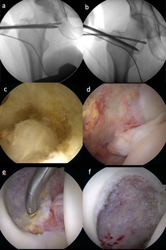
F. Haug, R. Herzog

                                            Figure 1. Transverse and frontal view of the obturator
                                            nerve, passing through the obturator foramen. More distally
                                            the anterior and posterior branches are indicated. The MRI
                                            images were taken from one of our cases, where the proxim-
                                            ity to the cyst can be seen. Source upper left and right: Es-
                                            sential Anatomy 5 (by 3D4Medical from Elsevier), lower left
                                            and right: Case Nr. 2)

                               underlying intraarticular pathology is possible. Therefore surgery seems to be
                               the only curative option [1] [21].
                                 Lesions of the LT
                                  Recent literature reviews reported good outcomes after debridement of the
                               LT. Pergaminelis et al. treated 35 Patients with isolated lesions of the LT with
                               arthroscopic radiofrequency ablation [23]. They reported significant improve-
                               ment of the symptoms for a mean period of 17.7 months (range 6 - 42 months).
                               In some cases, a persisting sensation of instability was reported. Those might
                               qualify for reconstruction of the LT, although the benefit of LT-reconstruction
                               remains unclear. Various possibilities, with different graft sources have been de-
                               scribed in a few case reports and some small case studies [9] [10] [11].

                               5. Case Reports
                               5.1. Case, Mr. F, 43 y/o
                               A 43-year-old male patient presented with typical impingement complaints of
                               the right hip, mostly on exertion. The initial MRI confirmed an offset disorder
                               with small chondrolabral tearing and extensive ganglia, originating from a mu-
                               coid degenerated, thinned out LT. These extended beyond the transverse liga-
                               ment into the obturator foramen (Figure 2(a), Figure 2(b)). Intraoperatively, it
                               was shown that one of the ganglia originated directly from the partially ruptured
                               LT (Figure 2(c)). The ligament and the intra-articular part of the ganglia were re-
                               sected, causing the ganglia to empty (Figure 2(d), Figure 2(e)). Additionally, the

DOI: 10.4236/ojo.2021.115017                         173                                  Open Journal of Orthopedics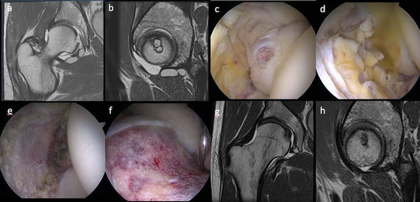
F. Haug, R. Herzog

                               Figure 2. (a) ap. and (b) transversal preoperative MRI, (c) intraoperative image of par-
                               tially ruptured LT with cyst, (d) cyst after incision, (e) acetabular fossa after resection of
                               the LT, (f) offset correction, (g) ap and (h) transversal postoperative MRI after 3 months.

                               offset was corrected (Figure 2(f)). The patient quickly became mostly symp-
                               tom-free. Due to renewed complaints, a check-up with a control MRI was car-
                               ried out, which showed a good correction of the offset (Figure 2(g), Figure
                               2(h)). The original edema in the anterolateral labrum had disappeared, however
                               a small capsule weakness, adhesions and remaining chondrolabral damage could
                               be seen. The ganglia have receded completely, although only a partial resection
                               was performed.

                               5.2. Case, Mr. S, 43 y/o
                               A 43-year-old male patient presented with chronic pain of the left hip since 1 1/2
                               years. The pain was located mostly gluteal and lateral of the hip, radiating to the
                               medial thigh with a burning sensation. The patient works in retail trade. At the
                               time of presentation, he was 100% unfit for work. MRI showed a synovial cyst of
                               the hip, extending to the obturator loge, compressing the obturator nerve, as
                               well as a slight offset disorder (Figure 3(c)). Conservative treatment like anal-
                               gesics, physiotherapy or infiltration of the sacroiliac joint showed no relief.
                               Puncture of the joint ganglion brought only short-term improvement. Although
                               the MRI did not show any significant joint pathology responsible for the com-
                               plaints, a hip arthroscopy was performed. It showed an edema of the labrum and
                               the LT (Figure 3(a), Figure 3(b), Figure 3(e)). The ligament was resected and
                               the offset corrected. There were no intraarticular ganglia visible. The radiating
                               pain to the medial thigh quickly disappeared. The remaining symptoms are pre-
                               sumably caused by muscle weakness of the gluteus medius and the piriformis
                               muscles and improved over time. 3 months postoperative an MRI was per-
                               formed, which confirmed complete regression of the ganglia that had com-
                               pressed the obturator nerve (Figure 3(d)). 3 months postoperatively the patient
                               was able to take up his work to 100%.

                               5.3. Case, Mrs. R, 47 y/o
                               A 47-year-old female patient was suffering from chronic recurrent perineal pain

DOI: 10.4236/ojo.2021.115017                          174                                     Open Journal of Orthopedics
F. Haug, R. Herzog
Figure 3. (a) ap. and (b) axial preoperative MRI, the arrows indicating
the LT, with the origin of the cysts at its base (c) preoparative MRI,
showing the proximity of the obturator nerve to the cysts (d) postopera-
tive MRI after 3 months (e) intraoperative images of the partially rup-
tured LT (f) acetabular fossa after resection of the LT.
for 5 years. The pain was exercise-related pain, also induced by long sitting. It
was described as radiating pain to the genital area, with a burning sensation. 3
years ago, after visiting different specialists a uterus myomatosus was diagnosed
and a total hysterectomy was performed. This didn’t show any relief. Also, me-
dication with Nsaids and tricyclic antidepressants didn’t help. She used to be
physically active, whereas due to the current situation she now focuses on Pilates
and physiotherapy. Still, the pain forced her to reduce work to 80%. Native MRI
showed small ganglia of the hip joint, whereas MR arthrography showed exten-
sive ganglia, reaching from the hip joint to the obturator loge, as well as a slight
cam impingement (Figure 4). Assuming a pathology of the LT we performed an
arthroscopy of the hip to resolve the intraarticular pathology. Extraarticular
preparation below the psoas muscle allowed visualization of the ganglion
through the obturator foramen, where the cyst could be opened (Figures
5(a)-(c)). Intraarticular the slight offset disorder was confirmed, and an edema
of the Labrum could be seen. The offset disorder was corrected (Figure 5(f)).
The LT showed a chronic partial rupture (Figure 5(d)). After resection of the LT
another cyst could be visualized and drained in the medial part of the acetabular fos-
sa (Figure 5(e)). Postoperatively the patient reported a sensation of relieved pressure
in the perineal and genital area. 3 months postoperatively the exercise-related pain
DOI: 10.4236/ojo.2021.115017 175 Open Journal of OrthopedicsF. Haug, R. Herzog
was gone. The burning sensation returned after stopping her tricyclic antidepres-
sants. The follow-up MRI showed complete regression of the ganglia.
Figure 4. (a) ap. and (b) axial preoperative MRI with extensive cysts
(c), (d) MRI 3 months after surgery. The cysts have disappeared.
Figure 5. (a) - (c) Arthroscopic approach to the obturator foramen. Intra-
operative imaging with x-ray and arthroscopic visualization. (c) extraarti-
cular view of the cyst, protruding through the obturator foramen (d) intra-
operative image of partially ruptured LT with signs of inflamation, (e) ace-
tabular fossa after resection of the LT, (f) offset correction.
DOI: 10.4236/ojo.2021.115017 176 Open Journal of OrthopedicsF. Haug, R. Herzog
6. Discussion
Symptoms
In our cases the pain was mostly located in different areas around the hip.
Most radicular pain that is caused by an intraspinal source is greater in the leg
than in the back [25]. The symptoms are therefore more suggestive for an intra-
articular pathology of the hip, rather than a spinal genesis. Table 1, suggests a
typical presentation of chronic recurrent pain with a burning sensation, radiat-
ing to the medial thigh or to the genitals. This was observed if the obturator
nerve was compressed by the ganglia. (The radiating pain to the genitals might
be explained by compression of the external pudendal artery and vein.) All pa-
tients described worsening of the symptoms during physical exertion, although
resting pain was common, especially after long sitting. The painful episodes of-
ten improved after long term physical rest. 2 of 3 Patients reported chronic pain,
resulting in significant limitations in work and daily activities.
Clinical findings
The clinical findings seen in Table 2 were unspecific. One patient had an im-
paired internal rotation and a positive anterior impingement sign. Other than
that, the range of motion was within normal limits. There was no significant loss
in muscle strength.
Radiological findings
As shown in Table 3 conventional radiographs did not show any significant
pathology, explaining the symptoms mentioned above. They all had a mild cam
deformity. MR Arthrography showed lesions of the LT in 2 of 3 Patients and the
above-mentioned ganglia, which were all protruding through the obturator fo-
ramen. A review from Hafezi-nejad et al. showed a sensitivity of 82.2% and spe-
cificity of 88.6% of MR Arthrography in detecting lesions of the LT, compared to
a significantly lower sensitivity in native MRI (64.7%) [26]. Consequently, le-
sions of the LT can only be safely excluded by hip arthroscopy.
Table 1. Presenting complaint.
Mr. F. Mr. S. Mrs. R.
Gluteal pain 1 0 1 0
Thigh pain 1 0 1 0
Inguinal pain 1 1 0 0
Perineal pain 1 0 0 1
Genital pain 2 0 1 1
Pain on exertion 3 1 1 1
Pain after long standing 1 0 1 0
Pain after long sitting 3 1 1 1
Pain w/o exertion 2 0 1 1
Night pain 1 0 1 0
Burning sensation 3 1 1 1
Radiating pain 2 0 1 1
DOI: 10.4236/ojo.2021.115017 177 Open Journal of OrthopedicsF. Haug, R. Herzog
Table 2. Clinical findings.
Mr. F. Mr. S. Mrs. R.
Reduced range of motion 1 0 0
Flexion/Extension path 100/0/20˚ 110/0/5˚ 110/0/20˚
Flexion/Extension phys 115/0/20˚ 115/0/10˚
ext./int. Rotation path 40/0/0˚ 35/0/30˚ 50/0/35˚
ext./int. Rotation phys 40/0/25˚ 35/0/35˚
Ab-/Adduction path 45/0/30˚ 45/0/30˚
Ab-/Adduction phys 45/0/30˚
Impingement test positive 1 0 0
Table 3. Radiologic findings.
Mr. F. Mr. S. Mrs. R.
Cam deformity 1 1 1
Alpha Angle 51 72 70
Antetorsion 6 26
CCD Angle 123 128 145
CE Angle 29 32 35
Labrum
Tear 1 0 1
Degeneration 1 0 1
Cartilage
Tear 0 0
Degeneration 1 0 0
Lig. teres edema edema 0
Acet. retroversion 1 0
Ganglia 64 × 9 × 8 mm 2.5 × 17 × 12 mm 33 × 28 × 15 mm
Crossover sign 0 0 0
Posterior wall sign 0 0 0
Pathophysiology
Briem et al. suggests that recurrent sustained micro-subluxations of the hip
joint, due to bony deformities might cause capsular stress [25]. We believe, that
in our cases, the offset and pincer disorders or rotational disorders, as primary
pathology, lead to subluxations of the hip, due to a hypomochlion effect. This
results in strain on the LT and the labrum with concomitant lesions. This leads
to extensive production of synovial fluid. With increased intraarticular pressure,
the synovial fluid may enter der LT through those microlesions, causing the
ganglia to grow in size. After physical rest the excess synovial fluid is partially
resorbed. The compressing effect of the ganglia on adjacent structures decreases.
Therefore, pain on exertion might possibly be associated with a fluctuating size
of the ganglia. In the presented cases, excessive production of synovial fluid
DOI: 10.4236/ojo.2021.115017 178 Open Journal of OrthopedicsF. Haug, R. Herzog
might also be caused by stress on the labrum due to mild FAI.
Intraoperative findings
Arthroscopically a chronic lesion of the LT was seen in all three cases, as well
as edema of the labrum. CAM disorders were mild, and the labrum didn’t re-
quire reconstruction. The ganglion cysts were directly associated with the LT in
2 out of 3 cases and could directly be drained in 2 out of 3 cases. In one case the
ganglion could be drained directly from the LT. In the other case, the stalk of the
cysts was found directly medially of the LT. This supports our suggestion.
Treatment
The presented cases had minor pathologies of the hip joint in common,
which, if asymptomatic, wouldn’t qualify for surgery. In case number 2 and 3
conservative treatments like physiotherapy and analgesics didn’t show any relief.
Hip arthroscopy is a safe method to address the intraarticular pathologies. In all
3 cases, this led to an acute relief of symptoms. Extraarticular the approach to
the obturator foramen seems a good way to reach the ganglia. The intraarticular
part seems to be addressed best in the acetabular fossa. Here the LT originates in
the very medial part of the fossa, just posterior of the transverse ligament. After
resection of the LT, the stalk of the cyst can be visualized and drained.
Follow-up images showed continuous regression of the ganglia. The
long-term follow-up MRI images showed regression of the cysts, even if they
weren’t opened intraoperatively.
Preoperative aspiration of the cysts would have been a possible option. This
has to be discussed with each patient individually, pointing out clearly, that this
doesn’t address the primary problem and recurrence is likely. An open ap-
proach, via surgical subluxation of the hip was described in a similar case, where
a synovial ganglion of the hip was thought to be causing L5 radiculopathy [25].
This is a possible option, although, in our opinion there is no benefit in open
surgery.
Figure 6. Preoperative (first row) and postoperative (second row) images, showing
complete regression of the ganglia in all cases (case number 1 - 3 from left to right).
DOI: 10.4236/ojo.2021.115017 179 Open Journal of OrthopedicsF. Haug, R. Herzog
7. Conclusions
The above-mentioned cases show that a partial rupture of the LT and formation
of ganglia can occur during hip impingement. This may cause unspecific neuro-
logical symptoms and untypical pain. Our findings were suggestive for microle-
sions of the LT causing the formation of synovial cysts. Although, the exact
cause remains uncertain. In other cases intermittent claudicatio or leg edema,
have been described [2]. Those may easily be misinterpreted as vascular or lum-
bar pathologies. When in doubt we suggest performance of an MR arthrography
in addition to diagnostics of lumbar or vascular disease. This might reveal gan-
glia formation around the hip joint, although lesions of the LT might be missed
[23] [26]. In analogy to the Baker cyst of the knee, primarily the articular dam-
age needs to be repaired to achieve permanent relief. In our cases, this included
resection of the damaged LT and, if possible, resection of the ganglia, as well as
correction of the offset. For this purpose, hip arthroscopy is a minimally invasive
and safe technique (Figure 6). Due to the incision site being further away from
the neurovascular bundle it is safer than an open approach to the ganglion. Re-
section of the ganglion can result in immediate improvement of the symptoms,
however, in case of chronic disease, the affected nerve may take some time to
recover. If the ganglion cannot be resected it may still form back on its own once
the intraarticular damage was addressed. In case of excess production of synovial
fluid on the base of rheumatic disease, injections with corticosteroids might be a
good option [1] [2].
There are further studies necessary to evaluate typical symptoms correspond-
ing to various locations of the cysts, for further differentiation from lumbar or
vascular pathologies. Furthermore, optimal arthroscopic approaches for drain-
ing the cysts need to be identified.
Conflicts of Interest
The authors declare no conflicts of interest regarding the publication of this pa-
per.
References
[1] Angelini, A., Zanotti, G., Berizzi, A., Staffa, G., Piccinini, E. and Ruggieri, P. (2017)
Synovial Cysts of the Hip. Acta Biomedica, 88, 483-490.
[2] Yukata, K., Nakai, S., Goto, T., et al. (2015) Cystic Lesion around the Hip Joint.
World Journal of Orthopedics, 6, 688-704. https://doi.org/10.5312/wjo.v6.i9.688
[3] Botchu, R., Esler, C.N., Lloyd, D.M. and Rennie, W.J. (2013) Ganglia Arising from
the Transverse Acetabular Ligament: A Report of Two Cases. Journal of Orthopae-
dic Surgery (Hong Kong), 21, 380-382.
https://doi.org/10.1177/230949901302100324
[4] Chandler, S.K. (1934) The Iliopsoas Bursa in Man. The Anatomical Record, 58,
235-240. https://doi.org/10.1002/ar.1090580304
[5] Robinson, P., White, L.M., Agur, A., Wunder, J., Bell, R.S. and Robinson, R. (2003)
Obturator Externus Bursa—Origin and MR Features of Pathology.
DOI: 10.4236/ojo.2021.115017 180 Open Journal of OrthopedicsF. Haug, R. Herzog
[6] Magee, T. and Hinson, G. (2000) Association of Paralabral Cysts with Acetabular
Disorders. American Journal of Roentgenology, 174, 1381-1384.
https://doi.org/10.2214/ajr.174.5.1741381
[7] Magerkurth, O., Jacobson, J.A., Girish, G., Brigido, M.K., Bedi, A. and Fessell, D.
(2012) Paralabral Cysts in the Hip Joint: Findings at MR Arthrography. Skeletal Ra-
diology, 41, 1279-1285. https://doi.org/10.1007/s00256-012-1395-4
[8] Jo, S., Hooke, A.W., An, K.N., Trousdale, R.T. and Sierra, R.J. (2018) Contribution
of the Ligamentum Teres to Hip Stability in the Presence of an Intact Capsule: A
Cadaveric Study. Arthroscopy: The Journal of Arthroscopic & Related Surgery, 34,
1480-1487. https://doi.org/10.1016/j.arthro.2017.12.002
[9] Kraeutler, M.J., Garabekyan, T., Pascual-Garrido, C. and Mei-Dan, O. (2016) Liga-
mentum Teres Tendinopathy and Tears. Muscle, Ligaments and Tendons Journal,
6, 337-342. https://doi.org/10.32098/mltj.03.2016.09
[10] Martin, R.L., McDonough, C., Enseki, K., Kohreiser, D. and Kivlan, B.R. (2019)
Clinical Relevance of the Ligamentum Teres: A Literature Review. International
Journal of Sports Physical Therapy, 14, 459-467.
https://doi.org/10.26603/ijspt20190459
[11] Rosinsky, P.J., Shapira, J., Lall, A.C. and Domb, B.G. (2020) All about the Ligamen-
tum Teres: From Biomechanical Role to Surgical Reconstruction. The Journal of the
American Academy of Orthopaedic Surgeons, 28, E328-E339.
https://doi.org/10.5435/JAAOS-D-19-00352
[12] Park, J., Kang, Y., Ahn, J.M., Lee, E., Lee, J.W. and Kang, H.S. (2019)
Non-Traumatic Ligamentum Teres Tears: Association with MRI Morphometry of
the Hip. Acta Radiologica, 60, 615-622. https://doi.org/10.1177/0284185118788897
[13] Schmitz, M.R., Campbell, S.E., Fajardo, R.S. and Kadrmas, W.R. (2012) Identifica-
tion of Acetabular Labral Pathological Changes in Asymptomatic Volunteers Using
Optimized, Noncontrast 1.5-T Magnetic Resonance Imaging. The American Journal
of Sports Medicine, 40, 1337-1341. https://doi.org/10.1177/0363546512439991
[14] Register, B., Pennock, A.T., Ho, C.P., Strickland, C.D., Lawand, A. and Philippon,
M.J. (2012) Prevalence of Abnormal Hip Findings in Asymptomatic Participants: A
Prospective, Blinded Study. The American Journal of Sports Medicine, 40,
2720-2724. https://doi.org/10.1177/0363546512462124
[15] Stuplich, M., Hottinger, A.F., Stoupis, C. and Sturzenegger, M. (2005) Combined
Femoral and Obturator Neuropathy Caused by Synovial Cyst of the Hip. Muscle
and Nerve, 32, 552-554. https://doi.org/10.1002/mus.20364
[16] Yukata, K., Arai, K., Yoshizumi, Y., Tamano, K., Imada, K. and Nakaima, N. (2005)
Obturator Neuropathy Caused by an Acetabular Labral Cyst: MRI Findings. AJR
American Journal of Roentgenology, 184, 112-114.
https://doi.org/10.2214/ajr.184.3_supplement.0184s112
[17] Bachar, A.I., Amar, E., Efrima, B., Kollander, Y., Rath, E. and Volaski, H. (2018)
Hip Arthroscopy as a Treatment for Obturator Neuropathy Secondary to In-
tra-Pelvic Ganglion: A Case Report. Journal of Hip Preservation Surgery, 5,
319-322. https://doi.org/10.1093/jhps/hny023
[18] Anderson, S., Ryan, J. and Baria, M.R. (2020) Obturator Internus Synovial Cyst.
American Journal of Physical Medicine & Rehabilitation, 99, e134-e135.
[19] Vidoni, A., Sankara, S.T.V., Ramana, V. and Botchu, R. (2019) Ganglion Cyst Aris-
ing from the Transverse Acetabular Ligament (TAL): A Rare Cause of Entrapment
of the Anterior Branch of the Obturator Nerve. Case Report and Review of the Lite-
rature. Skeletal Radiology, 48, 163-165. https://doi.org/10.1007/s00256-018-2992-7
DOI: 10.4236/ojo.2021.115017 181 Open Journal of OrthopedicsF. Haug, R. Herzog
[20] Kim, S.H., Seok, H., Lee, S.Y. and Park, S.W. (2014) Acetabular Paralabral Cyst as a
Rare Cause of Obturator Neuropathy: A Case Report. Annals of Rehabilitation
Medicine, 38, 427-432. https://doi.org/10.5535/arm.2014.38.3.427
[21] Jitpun, E., Howe, B., Matthew, M., Amrami, K.K., Trousdale, R.T. and Spinner, R.J.
(2019) Obturator Intraneural Ganglion Cysts: Joint Connected and Underdiag-
nosed. World Neurosurgery, 126, e259-e269.
https://doi.org/10.1016/j.wneu.2019.02.029
[22] Campeas, S. and Rafii, M. (2002) Pelvic Presentation of a Hip Joint Ganglion: A
Case Report. Bulletin of the NYU Hospital for Joint Diseases, 61, 89-92.
[23] Pergaminelis, N., Renouf, J., Fary, C., Tirosh, O. and Tran, P. (2017) Outcomes of
Arthroscopic Debridement of Isolated Ligamentum Teres Tears Using the
iHOT-33. BMC Musculoskeletal Disorders, 18, 4-9.
https://doi.org/10.1186/s12891-017-1905-6
[24] Colasanti, M., Sapienza, P., Moroni, E., Mosiello, G., Postacchini, F. and di Marzo,
L. (2006) An Unusual Case of Synovial Cyst of the Hip Joint Presenting as Femoral
Vein Compression and Severe Lower Limb Edema. European Journal of Vascular
and Endovascular Surgery, 32, 468-470. https://doi.org/10.1016/j.ejvs.2006.05.012
[25] Briem, T., Haemmerle, G., Kramers-de Quervain, I. and Leunig, M. (2016) Synovial
Ganglion of the Hip as a Rare Cause of L5 Radiculopathy: A Case Report. JBJS Case
Connect, 6, e59. https://doi.org/10.2106/JBJS.CC.15.00234
[26] Hafezi-nejad, N., Vaidya, D. and Eng, J. (2018) MR Arthrography—A Sytematic
Review an Meta-Analysis. American Journal of Roentgenology, 211, 52-63.
DOI: 10.4236/ojo.2021.115017 182 Open Journal of OrthopedicsYou can also read