COMPARATIVE STUDY OF WEANING PIGS' MUSCLE PROTEINS USING TWO-DIMENSIONAL ELECTROPHORESIS
←
→
Page content transcription
If your browser does not render page correctly, please read the page content below
Potravinarstvo Slovak Journal of Food Sciences

                                                                         Potravinarstvo Slovak Journal of Food Sciences
                                                                                                   vol. 15, 2021, p. 52-57
                                                                                            https://doi.org/10.5219/1449
                                                                   Received: 27 August 2020. Accepted: 17 January 2020.
                                                            Available online: 28 January 2021 at www.potravinarstvo.com
                                               © 2021 Potravinarstvo Slovak Journal of Food Sciences, License: CC BY 4.0
                                                                                                 ISSN 1337-0960 (online)

    COMPARATIVE STUDY OF WEANING PIGS' MUSCLE PROTEINS USING
               TWO-DIMENSIONAL ELECTROPHORESIS

                                       Anastasiya Akhremko, Liliya Fedulova

ABSTRACT
The proteostasis system of animals, including various types of protein modification during the growth stage, leads to an
almost incomprehensible number of possible forms of protein, and each can regulate numerous functions. In the presented
work, the composition of muscle tissue protein from different portions of piglets was studied to understand the main muscle
protein formation. Comparative analysis of weaned piglets' main muscle protein from l. dorsi, biceps femoris, and
brachiocephalicus were analyzed using two-dimensional electrophoresis. Changes in the staining intensity of protein
fractions inherent in different muscles were revealed. As part of this work, candidate groups of pig muscle proteins have been
selected. Eleven protein spots were revealed for the longest muscle of the back, and seven for the biceps; the muscles of the
neck are characterized by indicators of low protein fraction volume. Among the proteins found, myosin light chains,
phosphoglycerate mutase, troponins, and adenylate kinase is most likely present. The obtained results of protein identification
in muscle tissues, obtained during the intensive growth period, will allow a more detailed understanding of protein regulation,
function, and interactions in complex biological systems, which will subsequently be significantly important for
biomonitoring health and predicting farm animals productivity.
Keywords: 2-DE; muscle; protein; two-dimensional electrophoresis; pig; piglet

INTRODUCTION                                                          biological systems, which will subsequently be vitally
  The increase in demand for lean pork has required the               important for biomonitoring the health and productivity of
selection of carcasses showing increased muscle mass. The             farm animals. Also, practical applications in identifying
selection for this indicator has significantly improved the           counterfeit meat products could be based on this technique
meat qualities of most domestic and foreign pig breeds.               (Mora, Gallego and Toldrá, 2018).
However, an increase in "meat content" without taking into              As you know, with the growth and increase in animal live
account other economically useful traits led to a decrease in         weight, the amount of muscle, adipose, and bone tissue in
natural resistance and significant shifts in metabolic                their body is steadily increasing. However, the growth of
processes in the animals, which ultimately affected the               these tissues proceeds with unequal intensity. The
quality and organoleptic characteristics of pork (Fedulova            development of muscle tissue in young animals is due to a
et al., 2018; Benešová et al., 2019; Gorlov et al., 2020).            high rate of metabolic processes and protein synthesis.
  It should be noted that the selection efficiency is assessed        Moreover, the main factors that determine the rate of muscle
mainly by identifying genes that control beneficial traits, as        growth are genetic, feeding, muscle activity (exercise),
well as polymorphic DNA variants in these genes, which                stress hormones, and growth stimulants (natural or
directly affect the phenotype of the animal. In this case,            synthetic).
genome and proteome interactions are completely ignored.                As growth slows down and stops, the rates of protein
It is known that an animal's proteostasis system, including           synthesis and degradation are balanced. In conditions of
folding, modification of the primary structure, and protein           poor nutrition, diseases, including parasitic ones, stress,
degradation, are crucial for the realization of phenotypic            unfavorable environmental conditions, muscles can
traits. At the same time, the huge number of possible post-           atrophy, which means that protein degradation outweighs
translational modifications, combined with the multitude of           synthesis (Purslow, 2017).
amino acid residues, leads to an almost incomprehensible                Until recently, not enough attention has been paid to the
number of possible protein forms, and each can regulate               development rate of muscle tissue in growing animals to
numerous functions (Spoel, 2018). Thus, the use of                    predict the factors that stimulate the growth of animals and
proteomic methods in animal husbandry for muscle protein              determine meat quality.
identification will allow a more detailed understanding of
protein regulation, function, and interactions in complex

Volume 15                                                        52                                                        2021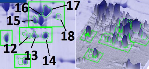
Potravinarstvo Slovak Journal of Food Sciences
Scientific Hypothesis Protein spots on two-dimensional electropherograms were
Identifying the composition of muscle proteins of various interpreted following the Swiss-Prot database (O'Donovan
parts of a growing animal - weaned piglets - will expand the et al., 2002) and the Muscle organ proteomics database
available information on the formation of muscle tissue to (Kovaleva et al., 2013).
identify quality, safety, and authenticity markers in
livestock products. Statistical analysis
The experimental data were analyzed using one-way
MATERIALS AND METHODS ANOVA (between gels obtained with a different variation
Object of IEF) by ImageMaster™ 2D Platinum software powered
The object of the study was Vietnamese pot-bellied × by Melanie 8.0 (GE Healthcare and Genebio, Switzerland).
Wiesenau weaned pigs from Krolinfo LLC laboratory A p 2, for instance, to
Materials
identify those proteins in which treatment causes at least a
Chemical reagents: Urea, Thiourea, Ditiothreitol, Sodium
two-fold spot abundance increase or decrease.
hydroxide (NaOH), Glycerol, Sodium dodecyl sulfate
(SDS), Tris, Acrylamide, Ammonium persulfate (APS), 2-
Propanol, Acetic acid, Bis-acrylamide, RESULTS AND DISCUSSION
Tetramethylethylenediamine (TEMED), Mercaptoethanol, A study was carried out on three different muscles of
Bromophenol blue, Glycine, Coomassie Brilliant Blue G- l. dorsi, biceps femoris, and brachiocephalicus by two-
250 (PanReac, Spain); Triton X-100 (Helicon Russia); dimensional electrophoresis to reveal significant
Ampholyte (Serva, Germany) and Phosphoric (V) acid differences in protein composition (Zia et al., 2020).
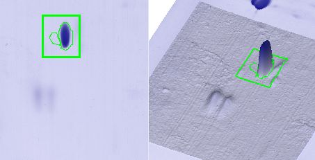
(H3PO4) (Component-reaktiv, Russia). Two-dimensional electrophoretogram fragments with the
Samples were taken within 20 minutes after slaughter and detected changes are shown in Fig. 1; a total of 18 fractions
placed in dry ice. Frozen muscle tissues (50 mg) were were selected with Fold index >2 and presented in Table. 1.
homogenized in 1 mL 7 M Urea, 2 M Thiourea, 1 % Protein spots indicated by numbers 1 and 2 in Figure 1,
Ditiothreitol, 0,4 % Triton X-100, 2% pH 3 – 10 and presumably myosin light chains fast (MLC1f)
Ampholyte. Homogenates were centrifugated with an (Murgiano et al., 2010; Yang et al., 2016) and slow
acceleration of 20 000 g for 20 minutes. Three samples, (MLC1s/v), respectively, were quite well manifested in the
obtained from different animals, were studied by two- longest muscle (Kim et al., 2017; Zou et al., 2018). In the
dimensional electrophoresis in three replicates. biceps femoris, only the slow chain was detected, and in the
brachiocephalicus, a weakly expressed fraction MLC1s/v.
Montowska and Pospiech (2012) used MLC in their
Two-dimensional gel electrophoresis (2-DE) studies as markers in the authentication of meat products
The samples described above were subjected to 2-DE. made from pork and other meats.
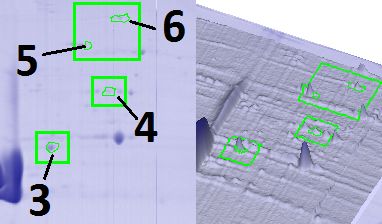
Proteins were separated by IEF (isoelectric focusing) in the A group of proteins in the range of molecular weights from
first dimension and SDS-PAGE in the second dimension. 50 kDa to 60 kDa, marked with numbers 3 – 7 in Figure 1,
This was completed as described by Matsumoto et al. were more pronounced in the muscles of the biceps, a
(2019) with slight modifications, IEF in the first dimension smaller amount identified in the longest muscle, and the
was performed at 3650 V.h-1. muscles of the neck trace amounts were detected.
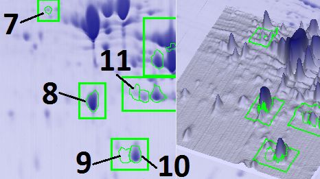
2-DE was performed according to the method of O'Farrell Protein number 8 intensity (Figure 1) decreased evenly
(O'Farrell, 1975) with isoelectric focusing on ampholine pH from l. dorsi to brachiocephalicus, the same tendency was
gradient by Glass Tube-Based IEF. The subsequent observed in fractions 13 – 17, among them troponin is
detection of the proteins was carried out by staining with presumably present. The skeletal muscle protein troponin I
Coomassie Brilliant Blue G-250 (PanReac, Spain). have already been characterized as a potential thermostable
For computerized densitometry, two-dimensional and species-specific biomarker of mammalian muscle tissue
electropherograms were used in a wet state. Their full in raw meat and meat products (Zvereva et al., 2015; Mitra
digital images and/or images of individual fragments were et al., 2018; Zuber et al., 2019), which makes it promising
obtained using a Bio-5000 plus scanner (Serva, Germany). for identifying muscles of different regions of the animal.
Scanned images were analyzed with ImageMaster™ 2D The lower spot volume in the neck muscles is possibly
Platinum software powered by Melanie 8.0 (GE Healthcare associated with the biological function of this muscle since
and Genebio, Switzerland). Spots were detected and it is less active than l. dorsi and biceps femoris.
quantified automatically with minimum thresholds: Interesting distribution of protein spots was found in
saliency – 11, min area – 5, and smooth – 3. The relative fractions 9 and 10, only protein number 9 was found in the
optical density (OD) and relative volume were computed to biceps, but fraction 10 (adenylate kinase) was present in a
correct for differences in gel staining. These measures take small amount, but its intensity in the large, flat muscle on
into account variations due to protein loading and staining, the back was greater than in the neck muscle (Lee et al.,
by considering the total OD or volume over all the spots in 2011; Scheffler, Park and Gerrard, 2011; Oliván et al.,
the gel. The digitized 2DE images of the cortex were then 2015).
compared by the matching method (Grove et al., 2006).
Volume 15 53 2021
Potravinarstvo Slovak Journal of Food Sciences

                 l. dorsi                              biceps femoris                          brachiocephalicus

 Figure 1 Fragments 2-DE gels of pig muscle tissue.

Table 1 Results of the densitometry analysis.
                                                              Muscle tissue
        No of Spot                                                                                              Fold
                                     l. dorsi            biceps femoris           brachiocephalicus
               1                9.07 ±1.19 x 10⁶        2.60 ±1.63 x 10⁶           1.46 ±0.36 x 10⁶             6.20
                                               7                        7                         7
               2                0.11 ±1.35 x 10         8.93 ±1.13 x 10            2.78 ±1.77 x 10              4.18
                                               6                        6                         6
               3                3.84 ±0.67 x 10         2.74 ±1.15 x 10            1.02 ±0.37 x 10              3.78
               4                1.15 ±0.34 x 10⁶        2.63 ±0.52 x 10⁶           1.18 ±0.57 x 10⁶             2.29
                                               5                        6                         5
               5                2.55 ±0.63 x 10         1.18 ±0.29 x 10            2.72 ±0.67 x 10              4.62
                                               5                        6                         6
               6                6.88 ±3.57 x 10         2.42 ±0.40 x 10            1.42 ±0.47 x 10              3.52
                                               6                        6                         6
               7                1.49 ±0.79 x 10         5.96 ±1.67 x 10            3.49 ±0.22 x 10              4.00
               8                6.21 ±0.29 x 10⁷        5.16 ±0.83 x 10⁷           1.75 ±1.52 x 10⁷             3.55
                                               6                        7                         6
               9                4.90 ±2.28 x 10         1.86 ±1.21 x 10            2.08 ±0.16 x 10              8.94
                                               7                        7                         6
              10                3.33 ±0.48 x 10         1.27 ±0.92 x 10            8.07 ±5.80 x 10              4.12
                                               6                        6                         6
              11                5.93 ±1.10 x 10         5.78 ±1.72 x 10            2.03 ±0.49 x 10              2.93
              12                1.77 ±0.45 x 10⁷        8.42 ±2.46 x 10⁶           1.69 ±0.54 x 10⁶             10.50
              13                4.37 ±0.32 x 10⁷        3.38 ±1.30 x 10⁷           1.24 ±0.34 x 10⁷             3.15
                                               7                        7                         7
              14                5.99 ±0.19 x 10         5.19 ±1.02 x 10            1.84 ±1.53 x 10              3.25
                                               7                        7                         7
              15                5.31 ±0.88 x 10         4.18 ±0.62 x 10            2.52 ±0.99 x 10              2.11
              16                1.56 ±0.28 x 10⁷        7.86 ±2.20 x 10⁶           3.90 ±2.56 x 10⁶             3.98
                                               8                        8                         7
              17                1.40 ±0.12 x 10         1.16 ±0.05 x 10            6.86 ±1.51 x 10              2.04
                                               7                        7                         6
              18                1.07 ±0.23 x 10         1.21 ±0.17 x 10            3.71 ±1.17 x 10              3.27
 Note: Spot Vol* were normalized by total valid spot volume and mean of value from duplicate analytical gels from three
replicates. Data represented are means ±SD of three independent experiments. *Vol: The volume of a spot is the sum of the
background-subtracted gray values of all pixels delimited by the spot.

 Volume 15                                                  54                                                      2021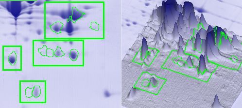
Potravinarstvo Slovak Journal of Food Sciences
  The volume of spot number 11 in brachiocephalicus was                  Bermúdez, R., Franco, D., Carballo, J., Sentandreu, M. Á.,
three times less than in l. dorsi and biceps femoris. The              Lorenzo, J. M. 2014. Influence of muscle type on the evolution
highest Fold value was in fraction 12, which is probably               of free amino acids and sarcoplasmic and myofibrillar proteins
phosphoglycerate mutase, corresponded to 10.5, and is                  through the manufacturing process of Celta dry-cured ham.
maximally expressed in l. dorsi (Welzenbach et al., 2016;              Food Research International, vol. 56, p. 226-235.
He et al., 2016; Lepczynski et al., 2019). Protein                     https://doi.org/10.1016/j.foodres.2013.12.023
number 18 is more strongly expressed in the muscles of the               Fedulova, L., Elkina, A., Vasilevskaya, E., Barysheva, E.
                                                                       2018. Identification of tissue-specific proteins of
biceps, less intensely noted in l. dorsi and a small amount in
                                                                       immunocompetent organs of Sus scrofa isolated in deuterium
the neck.
                                                                       depleted medium. Medical Science, vol. 22, no. 94, p. 509-513.
  The changes described above in the staining intensity of
                                                                         Gorlov, I., Slozhenkina, M., Mosolov, A., Baranikov, V.,
selected protein fractions may reflect the dynamics of                 Nikolaev, D., Chernyak, A., Sherstyuk, B., Krotova, O. 2020.
muscle tissue formation in growing animals (Chen et al.,               Nutritional and biological value of pork obtained from animals
2018; Li et al., 2020). For example, in l. dorsi, the most             fed with lysine and methionine. Potravinarstvo Slovak Journal
powerful muscle of the spinal column, which determines the             of      Food      Sciences,      vol.     14,   p.     112-117.
movement of the spinal column and head, the maximum                    https://doi.org/10.5219/1192
amount of intensely colored protein fractions was revealed               Grove, H., Hollung, K., Uhlen, A. K., Martens, H.,
(Liu et al., 2014; Paredi et al., 2019). Candidate markers             Faergestad, E. M. 2006. Challenges related to analysis of
for l. dorsi can be fractions 1, 3, 8, 10 – 17 (Figure 1, Table        protein spot volumes from two-dimensional gel electrophoresis
1) (Zou et al., 2017). For the biceps femoris, which                   as revealed by replicate gels. Journal of Proteome Research,
functionally exhibits actin, the hip extensor, and hock joints,        vol.         5,       no.         12,       p.       3399-3410.
and the flexor of the knee joint, muscle fractions 2, 4 – 7, 9,        https://doi.org/10.1021/pr0603250
and 18 can serve as markers (Figure 1, Table 1). The thigh               Grujić, R., Savanović, D. 2018. Analysis of myofibrillar and
muscle and its protein profile are quite well studied, since           sarcoplasmic proteins in pork meat by capillary gel
various versions of jerky ham are made from pork, such as              electrophoresis. Foods and Raw Materials, vol. 6 no. 2, p. 421-
ham and prosciutto, it is already known that the peptides              428. https://doi.org/10.21603/2308-4057-2018-2-421-4281
formed in this muscle have biological functionality (some                He, J., Yang, H., Wei, W., Chu, W., Yu, S., Tian, Y., Peng,
peptides from MLC1, CK, MYO, TNT, and MHC7 proteins                    F., Liu, H., Zhang, Z., Chen, J. 2016. The c.–360 T> C mutation
                                                                       affects PGAM2 transcription activity and is linked with the
were the most influential) (Bermúdez et al., 2014; Mora et
                                                                       water holding capacity of the longissimus lumborum muscle in
al., 2016; Chernukha et al., 2018; Zhou et al., 2020).                 pigs.     Meat      Science,     vol.    122,    p.    139-144.
  For l. dorsi composition, 11 proteins were identified (Kim           https://doi.org/10.1016/j.meatsci.2016.07.023
et al., 2008; Grujić and Savanović, 2018), and                           Chen, J., Su, W., Kang, B., Jiang, Q., Zhao, Y., Fu, C., Yao,
7 proteins for brachiocephalicus, besides, the latter showed           K. 2018. Supplementation with α-ketoglutarate to a low-
a tendency of lower intensity protein fractions, some of               protein diet enhances amino acid synthesis in tissues and
which are smaller in comparison to l. dorsi.                           improves protein metabolism in the skeletal muscle of growing
 The lowest intensity of staining of protein fractions was             pigs. Amino Acids, vol. 50, no. 11, p. 1525-1537.
noted in brachiocephalicus, which is possibly associated               https://doi.org/10.1007/s00726-018-2618-3
with low metabolic processes in this muscle, due to a low                Chernukha, I., Fedulova, L., Kotenkova, E., Akhremko, A.
functional load.                                                       2018. Hypolipidemic action of the meat product: in vivo study.
                                                                       Potravinarstvo Slovak Journal of Food Sciences, vol. 12, no.
CONCLUSION                                                             1, p. 566-569. https://doi.org/10.5219/959
  As part of this work, data were obtained on the variations             Kim, G.-D., Seo, J.-K., Yum, H.-W., Jeong, J.-Y., Yang, H.-
in muscle protein groups in the muscles of different regions           S. 2017. Protein markers for discrimination of meat species in
in piglets. For the longest muscle of the back, eleven protein         raw beef, pork and poultry and their mixtures. Food Chemistry,
spots with a pronounced intensity of staining were revealed,           vol.                217,               p.              163-170.
                                                                       https://doi.org/10.1016/j.foodchem.2016.08.100
for the biceps - seven, for the muscles of the neck, low
                                                                         Kim, J. H., Seong, P. N., Cho, S. H., Park, B. Y., Hah, K. H.,
indicators of the spot volume are characteristic. These
                                                                       Yu, L. H., Lim, D. G., Hwang, I. H., Kim, D. H., Lee, J. M.,
protein fractions vary across the entire spectrum of                   Ahn, C. N. 2008. Characterization of nutritional value for
intercellular proteins, from heat shock proteins and enzymes           twenty-one pork muscles. Asian-Australasian Journal of
of energy metabolism to structural proteins. The identified            Animal Sciences, vol. 21, no. 1, p. 138-143.
protein spots can be used as molecular markers of muscle               https://doi.org/10.5713/ajas.2008.70208
tissue development. However, much remains to be clarified                Kovaleva, M., Kovalev, L., Lisitskaya K. V., Eremina, L. S.,
on a structural and molecular basis. This study further                Ivanov, A., Krakhmaleva, I., Sadykhov E., Shishkin, S. 2013.
revealed the markers of productivity and adaptation of                 Muscle organs proteomics multi-level database. FEBS Journal,
animals, as well as the quality, safety, and authenticity of           vol. 280, no. 1, p. 488.
animal products.                                                         Lee, H. Y., Kim, J. M., Byun, M. J., Kang, K. S., Kim, T. H.,
                                                                       Hong, K. C., Lee, K. T. 2011. Structure and polymorphisms of
REFERENCES                                                             the 5′ regulatory region of porcine adenylate kinase 3-like 1
  Benešová, L., Golian, J., Martišová, P., Semjon, B., Zajác,          gene and effect on trait of meat quality. Genes & Genomics,
P., Čapla, J., Vlčko, T. 2019. Authentication and preference           vol. 33, no. 2, p. 147. https://doi.org/10.1007/s13258-010-
mapping of ham. Potravinarstvo Slovak Journal of Food                  0091-9
Sciences,      vol.    13,   no.     1,     p.   1051-1056.              Lepczynski, A., Herosimczyk, A., Ozgo, M., Barszcz, M.,
https://doi.org/10.5219/1263                                           Taciak, M., Skomial, J. 2019. Modification of ileal proteome
                                                                       in growing pigs by dietary supplementation with inulin or dried

Volume 15                                                         55                                                            2021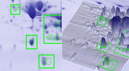
Potravinarstvo Slovak Journal of Food Sciences
chicory root. Journal of Animal and Feed Sciences, vol. 28, p.             Scheffler, T. L., Park, S., Gerrard, D. E. 2011. Lessons to
177-186. https://doi.org/10.22358/jafs/109518/2019                       learn about postmortem metabolism using the AMPKγ3R200Q
  Li, X., Xie, S., Qian, L., Cai, C., Bi, H., Cui, W. 2020.              mutation in the pig. Meat Science, vol. 89, no. 3, p. 244-250.
Identification of genes related to skeletal muscle growth and            https://doi.org/10.1016/j.meatsci.2011.04.030
development by integrated analysis of transcriptome and                    Spoel, S. H. 2018. Orchestrating the proteome with post-
proteome in myostatin-edited Meishan pigs. Journal of                    translational modifications. Journal of Experimental Botany,
Proteomics,            vol.        213,        p.        103628.         vol.        69,        no.        19,       p.        4499-4503.
https://doi.org/10.1016/j.jprot.2019.103628                              https://doi.org/10.1093/jxb/ery295
  Liu, J., He, J., Yu, J., Mao, X., Zheng, P., Huang, Z., Yu, B.,          Welzenbach, J., Neuhoff, C., Heidt, H., Cinar, M. U., Looft,
Chen, D. 2014. Birth weight alters the response to postnatal             C., Schellander, K., Tholen, E., Große-Brinkhaus, C. 2016.
high-fat diet-induced changes in meat quality traits and skeletal        Integrative analysis of metabolomic, proteomic and genomic
muscle proteome of pigs. British Journal of Nutrition, vol. 111,         data to reveal functional pathways and candidate genes for drip
no.                 10,               p.             1738-1747.          loss in pigs. International Journal of Molecular Sciences, vol.
https://doi.org/10.1017/S0007114513004431                                17, no. 9, p. 1426. https://doi.org/10.3390/ijms17091426
  Matsumoto, H., Haniu, H., Kurien, B. T., Komori, N. 2019.                Yang, H., Xu, X. L., Ma, H. M., Jiang, J. 2016. Integrative
Two-Dimensional Gel Electrophoresis by Glass Tube-Based                  analysis of transcriptomics and proteomics of skeletal muscles
IEF and SDS-PAGE. In Kurien B., Scofield R. Electrophoretic              of the Chinese indigenous Shaziling pig compared with the
Separation of Proteins. New York, USA : Humana Press, vol                Yorkshire breed. BMC genetics, vol. 17, no. 1, p. 80.
1855,       p.      107-113.      ISBN      978-1-4939-8793-1.           https://doi.org/10.1186/s12863-016-0389-y
https://doi.org/10.1007/978-1-4939-8793-1_11                               Zhou, C. Y., Tang, C. B., Wang, C., Dai, C., Bai, Y., Yu, X.
  Mitra, B., Lametsch, R., Akcan, T., Ruiz-Carrascal, J. 2018.           B., Li, C-B., Xu, X-L., Zhou, G-H., Cao, J. X. 2020. Insights
Pork proteins oxidative modifications under the influence of             into the evolution of myosin light chain isoforms and its effect
varied time-temperature thermal treatments: A chemical and               on sensory defects of dry-cured ham. Food Chemistry, vol.
redox proteomics assessment. Meat Science, vol. 140, p. 134-             315,                          p.                         126318.
144. https://doi.org/10.1016/j.meatsci.2018.03.011                       https://doi.org/10.1016/j.foodchem.2020.126318
  Montowska, M., Pospiech, E. 2012. Myosin light chain                     Zia, Q., Alawami, M., Mokhtar, N. F. K., Nhari, R. M. H. R.,
isoforms retain their species-specific electrophoretic mobility          Hanish, I. 2020. Current Analytical Methods for Porcine
after processing, which enables differentiation between six              Identification in Meat and Meat Products. Food Chemistry, vol.
species: 2DE analysis of minced meat and meat products made              324,                          p.                         126664.
from beef, pork and poultry. Proteomics, vol. 12, no. 18, p.             https://doi.org/10.1016/j.foodchem.2020.126664
2879-2889. https://doi.org/10.1002/pmic.201200043                          Zou, C., Li, J., Luo, W., Li, L., Hu, A., Fu, Y., Hou, Y., Li,
  Mora, L., Calvo, L., Escudero, E., Toldrá, F. 2016.                    C. 2017. Transcriptome analysis reveals long intergenic non-
Differences in pig genotypes influence the generation of                 coding RNAs involved in skeletal muscle growth and
peptides in dry-cured ham processing. Food Research                      development in pig. Scientific Reports, vol. 7, no. 1, p. 1-11.
International,           vol.        86,        p.        74-82.         https://doi.org/10.1038/s41598-017-07998-9
https://doi.org/10.1016/j.foodres.2016.04.023                              Zou, X., Zhou, G., Yu, X., Bai, Y., Wang, C., Xu, X., Dai,
  Mora, L., Gallego, M., Toldrá, F. 2018. New approaches                 C., Li, C. 2018. In vitro protein digestion of pork cuts differ
based on comparative proteomics for the assessment of food               with muscle type. Food Research International, vol. 106, p.
quality. Current Opinion in Food Science, vol 22, p. 22-27.              344-353. https://doi.org/10.1016/j.foodres.2017.12.070
https://doi.org/10.1016/j.cofs.2018.01.005                                 Zuber, E., Outhouse, A. C., Dekkers, J. C. M., Gabler, N. K.,
  Murgiano, L., D’Alessandro, A., Egidi, M. G., Crisa, A.,               Huff-Lonergan, E., Lonergan, S. M. 2019. Rate and extent of
Prosperini, G., Timperio, A. M., Valentini, A., Zolla, L. 2010.          troponin-T degradation in loins from pigs selected for low and
Proteomics and transcriptomics investigation on longissimus              high residual feed intake. Meat and Muscle Biology, vol. 1, no.
muscles in Large White and Casertana pig breeds. Journal of              3. https://doi.org/10.221751/rmc2017.154
Proteome Research, vol. 9, no. 12, p. 6450-6466.                           Zvereva, E. A., Kovalev, L. I., Ivanov, A. V., Kovaleva, M.
https://doi.org/10.1021/pr100693h                                        A., Zherdev, A. V., Shishkin, S. S., Lisitsyn, A. B., Chernukha,
  O'Donovan, C., Martin, M. J., Gattiker, A., Gasteiger, E.,             I. M., Dzantiev, B. B. 2015. Enzyme immunoassay and
Bairoch, A., Apweiler, R. 2002. High-quality protein                     proteomic characterization of troponin I as a marker of
knowledge resource: SWISS-PROT and TrEMBL. Briefings in                  mammalian muscle compounds in raw meat and some meat
bioinformatics,       vol.    3,     no.   3,     p.    275-284.         products.      Meat     Science,     vol.   105,     p.   46-52.
https://doi.org/10.1093/bib/3.3.275                                      https://doi.org/10.1016/j.meatsci.2015.03.001
  Oliván, M., Fernández-Suárez, V., Díaz-Martínez, F., Sierra,
V., Coto-Montes, A., de Luxán-Delgado, B., Peña, R., Bassols,            Funds:
A., Fàbrega, E., Dalmau, A., Velarde, A. 2015. Identification              The reported study was funded by RFBR, project number 19-
of biomarkers of stress in meat of pigs managed under different          316-90056.
mixing treatments. Biotechnology Journal International, vol.
11, no. 1, p. 1-13. https://doi.org/10.9734/BBJ/2016/22402               Conflict of Interest:
  Paredi, G., Mori, F., de Marino, M. G., Raboni, S., Marchi,             The authors declare no conflict of interest.
L., Galati, S., Buschini, A., Fiego, D. P., Mozzarelli, A. 2019.
Is the protein profile of pig Longissimus dorsi affected by              Ethical Statement:
gender and diet? Journal of Proteomics, vol. 206, p. 103437.              The research was approved by the bioethical commission
https://doi.org/10.1016/j.jprot.2019.103437
                                                                         of the V.M. Gorbatov Federal Research Centre for Food
  Purslow, P. P. 2017. The structure and growth of muscle. In
Toldra, F. Lawrie´s Meat Science. Cambridge, UK : Woodhead               Systems of the Russian Academy of Sciences (protocol
Publishing, p. 49-97. ISBN-13 978-0-08-100697-9.                         #08/2019, dated November 14, 2019).
https://doi.org/10.1016/B978-0-08-100694-8.00003-0

Volume 15                                                           56                                                            2021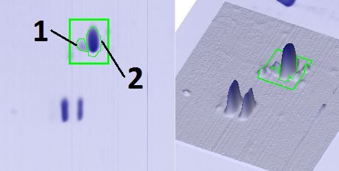
Potravinarstvo Slovak Journal of Food Sciences
Contact address:                                                       Liliya Fedulova, V. M. Gorbatov Federal Research Center
  *Anastasiya Akhremko, V. M. Gorbatov Federal Research              for Food Systems of RAS, Experimental-clinical research
Center for Food Systems of RAS, Experimental-clinical                laboratory of bioactive substances of animal origin,
research laboratory of bioactive substances of animal origin,        Talalikhina st., 26, 109316, Moscow, Russia, Tel.:
Talalikhina st., 26, 109316, Moscow, Russia, Tel.:                   +74956769211,
+79152379497,                                                        E-mail: l.fedulova@fncps.ru
E-mail: a.ahremko@fncps.ru                                           ORCID: http://orcid.org/0000-0003-3573-930X
ORCID: https://orcid.org/0000-0002-0211-8171
                                                                     Corresponding author: *

Volume 15                                                       57                                                      2021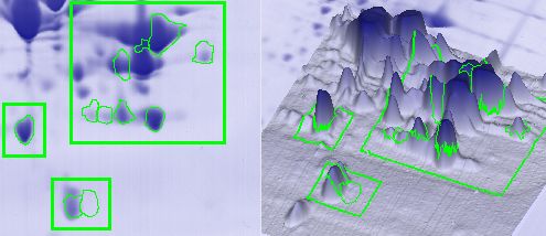
You can also read