Three New Species and Six Newly Recorded Species of Jumping Spiders (Araneae: Salticidae) in Taiwan - Scientific Research Publishing
←
→
Page content transcription
If your browser does not render page correctly, please read the page content below
Natural Resources, 2021, 12, 290-320
                                                                                                  https://www.scirp.org/journal/nr
                                                                                                           ISSN Online: 2158-7086
                                                                                                             ISSN Print: 2158-706X

Three New Species and Six Newly Recorded
Species of Jumping Spiders (Araneae: Salticidae)
in Taiwan

Kuo-Ming Chen1,2, Tai-Yu Lin1,3, Yih-Tsong Ueng1*
1
 Department of Environmental Engineering, Kun Shan University, Taiwan
2
 Tainan Municipal Yongkang Junior High School, Taiwan
3
 Tainan Municipal Dujia Elementary School, Taiwan

How to cite this paper: Chen, K.-M., Lin,       Abstract
T.-Y. and Ueng, Y.-T. (2021) Three New
Species and Six Newly Recorded Species of       From August 2013 to August 2014 at Aogu Wetland of Chiayi County, Tai-
Jumping Spiders (Araneae: Salticidae) in        wan, a total of 32 adult spiders of Salticidae were identified, comprising 15
Taiwan. Natural Resources, 12, 290-320.
                                                species from 13 genera. This paper describes 3 new species and 6 newly rec-
https://doi.org/10.4236/nr.2021.129021
                                                orded species as follows: Microbianor formosana sp. nov., Euophrys taiwanus
Received: August 5, 2021                        sp. nov., and Evarcha chiayiensis sp. nov.; Carrhotus tristis, Evarcha bulbosa,
Accepted: September 27, 2021                    Menemerus bivittatus, Plexippus petersi, Rhene rubrigera, and Synagelides
Published: September 30, 2021
                                                zhilcovae. The males distinguish them from other congeners. In addition, the
Copyright © 2021 by author(s) and               female Sibianor pullus is described for the first time. Species morphologies
Scientific Research Publishing Inc.             and detailed structures are depicted in micrographs to compensate for the
This work is licensed under the Creative
                                                lack of textual description.
Commons Attribution International
License (CC BY 4.0).
http://creativecommons.org/licenses/by/4.0/     Keywords
               Open Access
                                                Salticidae, Taxonomy, New Species, Newly Recorded Species, Taiwan

                                              1. Introduction
                                              The earliest record of the jumping spider family (Araneae: Salticidae) in Taiwan
                                              was in an article on the beneficial insects among Taiwan pests, which mentioned
                                              the spider Pyroderses formosanus (Matsumura, 1910) [1]. Subsequently, species
                                              were recorded by scholars revising the list of spiders in Taiwan, with a total of 10
                                              genera and 18 species recorded [2]-[9].
                                                As of June 2021, Academia Sinica’s Taiwan Species List contained 32 genera
                                              and 50 species with the scientific name Salticidae [8]. We collect and report on
                                              Salticidae spiders to increase the biodiversity of Taiwan spiders.

DOI: 10.4236/nr.2021.129021        Sep. 30, 2021                   290                                             Natural Resources
K.-M. Chen et al.

                              2. Materials and Methods
                              All specimens were collected from Aogu Wetland (23˚30'19"N, 120˚07'03"E) in
                              Chiayi County, Taiwan, from August 2013 to August 2014 each monthly [10]. A
                              sweep net (diameter: 38 cm) was used to catch spiders [11]. The purpose of that
                              research was to find the clustering and diversity of the spiders.
                                The environment in Aogu Wetland includes grassland, terrestrial shrubs, wa-
                              terside shrubs, and casuarina forest [12]. In total, 15,467 spiders were collected,
                              of which 4576 were adult spiders, belonging to 14 families and 141 morphologi-
                              cal species. Among them, 251 specimens of Salticidae were examined [10]. All
                              collected specimens were preserved in 40% alcohol and brought to a Kun Shan
                              University laboratory, where they were washed and preserved in 75% alcohol for
                              subsequent identification.
                                 First, determine whether 251 specimens of Salticidae were adult spiders based
                              on the characteristics of the spiders [13]. If they were juvenile and immature
                              spiders, they will not be processed and stored at the Kun-Shan University labor-
                              atory. If they were adult spiders, they will continue to be examined using the
                              following references: Huang (2004) [7], Chen and Chen (2013) [8], Davies and
                              Zabka (1989) [13], Simon (1903) [14], Ono et al. (2009) [15], Kim et al. (2000)
                              [16], Kim and Kim (2009) [17], Kim and Lee (2014) [18], Żabka (1985) [19],
                              Żabka and Waldock (2012) [20], Prószynski (2008) [21], World Spider Catalog
                              (2021) [22], and Jumping spiders (2021) [23].
                                Except for one specimen of the Pseudicius sp. was lost during the photo-
                              graphing process, the remaining 31 adult specimens were stored at the National
                              Museum of Natural Science (NMNS), Taichung (NMNS-8370-001—NMNS-
                              8370-014). The other 219 were young spiders, the characteristics of the species
                              are no obvious and it is difficult to distinguish.
                                Color images of specimens were captured using a stereo zoom microscope
                              (SZ-60, OLYMPUS) and a Nikon CoolPix 4500 camera. The images of the male
                              genitalia were captured under a microscope (Eclipse 50i, NIKON). The mea-
                              surement accuracy of the body length and each part of each specimen was 0.01
                              mm.
                                Abbreviations
                                Abbreviations used in the paper are as follows: AER, anterior eye row; ALE,
                              anterior lateral eye; AME, anterior median eye; PLE, posterior lateral eye; PME,
                              posterior median eye; MOA, median ocular area (area between promarginal
                              teeth and retromarginal teeth). Eye size was measured as the length of its long
                              axis, but for the PME, it was the length of its horizontal plane.
                                Leg length is femur + patella + tibia + metatarsus + tarsus lengths; pedipalp
                              length is femur + patella + tibia + cymbium lengths; MOA-L, median ocular area
                              length (from AME to PME); MOA-AW, anterior width of the AME lenses cal-
                              culated from their external edges; MOA-PW, posterior width of the PME lenses
                              calculated from their external edges; AMI, anterior median eye interval; AMLI,
                              anterior median and lateral eye interval; PMI, posterior median eye interval;

DOI: 10.4236/nr.2021.129021                         291                                           Natural Resources
K.-M. Chen et al.

                              PMLI, posterior median and lateral eye interval [8].

                              3. Results
                              From August 2013 to August 2014 at Aogu Wetland, in total 251 specimens Sal-
                              ticidae were identified: 219 specimens were juvenile or immature, and 32 speci-
                              mens were adult spider (Appendix 1). These adult spiders with 13 genera and 15
                              species identified, including 3 new species and 6 newly recorded species as fol-
                              lows: Microbianor formosana sp. nov., Euophrys taiwanus sp. nov., and Evarcha
                              chiayiensis sp. nov.; Carrhotus tristis, Evarcha bulbosa, Menemerus bivittatus,
                              Plexippus petersi, Rhene rubrigera, and Synagelides zhilcovae, respectively.
                              These are distinguishable from other congeners of the male spider. Additionally,
                              the female Sibianor pullus is also described herein for the first time. Species
                              morphology and detailed structures are shown in micrographs to compensate
                              for the lack of textual description.

                              3.1. Key for the Identification Salticidae Species at Aogu Wetland
                                   of Taiwan
                                1) Body shape resembles that of an ant (Figure 1, Figure 32)………………...2
                                Body shape unlike that of an ant………………………………………………..3
                                2) Promarginal teeth (2 teeth), cervical groove not notable (Figure 32)—
                              Synagelides zhilcovae (new record).
                                Promarginal teeth (more than 2 teeth), cervical groove notable (Figure 1)—
                              Myrmarachne formosicola.
                                3) The ocular area is longer than one-half the length of the cephalothorax…..4
                                The ocular area is shorter than one-half the length of the cephalothorax…….7
                                4) The length and width of the cephalothorax are almost equal (Figure
                              30(B))—Rhene rubrigera (new record).
                                The length of the cephalothorax is much greater than the width (Figure
                              22(A))……………………………………………………………………………….5
                                5) The posterior middle eye is closer to the anterior eye, but farther away from
                              the posterior eye (Figure 2)—Bianor angulosus.
                                The posterior middle eye is equidistant from the anterior and posterior eyes
                              (Figure 3(A), Figure 22(A))……………………………………………………....6
                                6) Body length less than 2 mm, radial furrow not notable (Figure 25(A))—
                              Microbianor formosana sp. nov.
                                Body length greater than 2 mm, radial furrow notable (Figure 3(A))—Sibianor
                              pullus.
                                7) Body length less than 3 mm (Figure 10)—Euophrys taiwanus sp. nov.
                                Body length greater than 3 mm…………………………………………………8
                                8) Male spider……………………………………………………………………9
                                Female spider…………………………………………………………………...16
                                9) Pdipalpal tibia longer than the cymbium (Figure 4(B))—Hasarius adansoni
                                Pdipalpal tibia shorter than the cymbium……………………………………..10

DOI: 10.4236/nr.2021.129021                        292                                         Natural Resources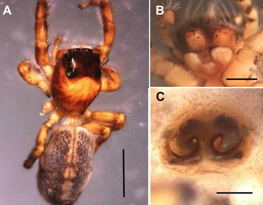
K.-M. Chen et al.

                              Figure 1. Myrmarachne formosicola (NMNS-8370-010). (A-D) Male; (A) dorsal view; (B)
                              cephalothorax; (C) chelicera and fangs; (D) palpal organ; (E) female promarginal teeth,
                              retromarginal teeth, and fang. Scale bars: A = 3 mm, B-C, E = 1 mm, D = 0.5 mm.

                              Figure 2. Bianor angulosus (NMNS-8370-005). (A) Male dorsal view; (B) male pedipalp
                              and palpal organ ventral view; (C) female cephalothorax and ocular area dorsal view.
                              Scale bars: A = 2 mm, B-C = 1 mm.

                                10) Bulbus shape is almost round……………………………………………...11
                                Bulbus shape is not round; some species have a protuberance……………….13
                                11) Three tibia protuberances (Figure 5)—Cosmophasis lami.
                                Fewer than 3 tibia protuberances (Figure 16)………………………………...12

DOI: 10.4236/nr.2021.129021                         293                                            Natural Resources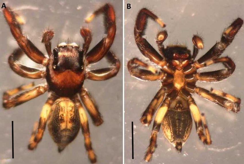
K.-M. Chen et al.

                              Figure 3. Female Sibianor pullus (NMNS-8370-013). (A) Dorsal view; (B) lateral view;
                              (C) chelicera and sternum; (D) epigynum. Scale bars: A-C = 1 mm, D = 0.3 mm.

                              Figure 4. Hasarius adansoni (collected from Tainan City of Taiwan). (A) Female dorsal
                              view; (B) male pedipalpal tibia longer than the cymbium. Scale bars: A = 2 mm, B = 0.3 mm.

                                12) Tibial protrusion ends with bifurcate apophysis (Figure 15(A))—Evarcha
                              bulbosa (new record).
                                Tibia protruding end does not bifurcate (Figure 16(D))—Evarcha chiayiensis
                              sp. nov.
                                13) Embolus slender (Figure 8(B), Figure 26(C))…………………………....14
                                Embolus beak (Figure 6(D), Figure 20(A))…………………………………..15
                                14) The length of the genitalia bulb is greater than the width (Figure 8(B))—
                              Carrhotus tristis (new record).
                                The length and width of the genitalia bulb are the approximately same—
                              Plexippus petersi (new record).

DOI: 10.4236/nr.2021.129021                         294                                              Natural Resources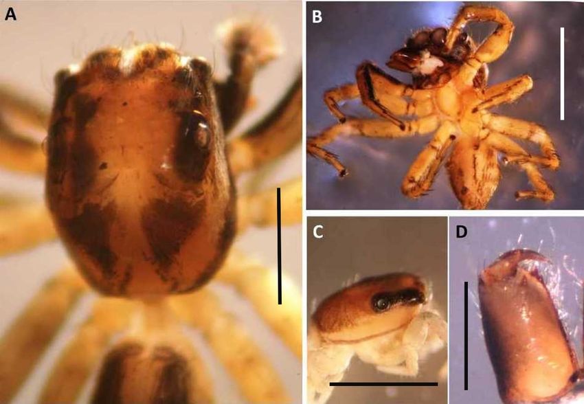
K.-M. Chen et al.

                              Figure 5. Cosmophasis lami (collected from Tainan City of Taiwan). (A) Male dorsal
                              view; (B) tibia and protuberance; (C) embolus; (D) female lateral view. Scale bars: A, D =
                              2 mm, B-C = 0.5 mm.

                              Figure 6. Male Phintella versicolor (NMNS-8370-015). (A) Dorsal view; (B) promarginal
                              teeth, retromarginal teeth and fang; (C) tibia and protuberance. Scale bars: A = 2 mm, B =
                              0.1 mm, C = 0.5 mm.

                                15) Seminal receptacle channel is clearly visible (Figure 6(C))—Phintella ver-
                              sicolor.
                                Seminal receptacle channel is not obvious (Figure 20(A))—Menemerus bi-
                              vittatus (new record).
                                16) Under the posterior median eye (PME) and posterior lateral eye (PLE) is a
                              row of microspines (stridulatory spines) (Figure 29)—genus Pseudicius.
                                No row of microspines under the PME or PLE……………………………….17

DOI: 10.4236/nr.2021.129021                         295                                              Natural Resources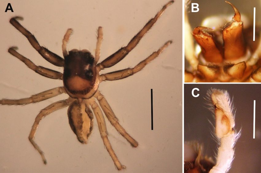
K.-M. Chen et al.

                                17) Epigynum dark brown (Figure 7(C))—Evarcha flavocincta.
                                Epigynum pink (Figure 4, Figure 5, Figure 21)……………………………...18
                                18) No light spots behind the ocular area (Figure 5(D))—Cosmophasis lami.
                                Light spots behind the ocular area……………………………………………..19
                                19) The length of the light spot behind the ocular area is greater than its width
                              (Figure 28(A))—Plexippuspetersi (new record).
                                The width of the light spot behind the ocular area is greater than its length
                              (Figure 4(A), Figure 21(A))……………………………………………………..20
                                20) The abdomen ventral surface is densely covered with brown spots (Figure
                              4(A))—Hasarius adansoni.
                                The abdomen has no stains on the ventral surface (Figure 20)—Menemerus
                              bivittatus (new record).

                              3.2. Salticidae Spiders at Aogu Wetland of Taiwan
                                Carrhotus tristis Thorell, 1895 (new record) (Figure 8 & Figure 9)
                                Carrhotus tristis Thorell, 1895; Prószyński, 1984; Prószyński, 1992b: 168, f.
                              8-11.
                                Type Material
                                Male total length (mm) 4.70. Carapace length 2.30, width 1.60; abdomen length
                              2.10, width 1.50. MOA ratio, MOA-L:MOA-AW:MOA-PW = 0.73:0.97:1.40.
                              AMI:AMLI:PMI:PMLI            =        0.10:0.13:1.27:0.23.   AME:ALE:PME:PLE           =
                              0.43:016:0.07:0.17. Leg I 3.65 (1.20, 1.25, 0.60, 0.60); leg II 3.65 (1.20, 1.25, 0.60,
                              0.60); leg III 3.60 (1.10, 1.25, 0.50, 0.75); leg IV 3.65 (1.25, 1.10, 0.60, 0.70); pedi-
                              palp 1.65 (0.50, 0.25, 0.25, 0.65).

                              Figure 7. Female Evarcha flavocincta. (A) Dorsal view; (B) chelicera and retromarginal
                              teeth; (C) epigynum. Scale bars: A = 2 mm, B-C = 0.3 mm.

DOI: 10.4236/nr.2021.129021                         296                                             Natural Resources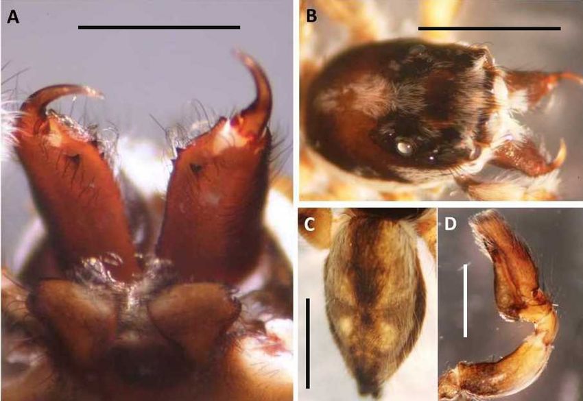
K.-M. Chen et al.

                              Figure 8. Male Carrhotus tristis. (A) Dorsal view; (B) ventral view. Scale bars: A-B = 2
                              mm.

                              Figure 9. Carrhotus tristis. (A) Chelicerae and marginal teeth; (B-D) pedipalp and palpal
                              organ; (B) front view; (C) lateral view; (D) dorsal view. Scale bars: A = 0.3 mm, B-D = 0.5
                              mm.

                                Characteristics
                                Male
                                Cephalothorax brown, ocular area black, thoracic groove obvious. AER re-
                              curve. Chelicera and fangs are reddish brown. Two promarginal teeth, 1 retro-
                              marginal tooth, labium brown. Sternum brown, oval and front truncated. Ab-
                              domen long oval, dorsal yellow brown and with hairs; dorsal view dark brown
                              and with small yellow spots; 3 arcs near the end of abdomen; ventral view dark
                              brown, lateral view light brown. Spinnerets are yellowish brown. The length of

DOI: 10.4236/nr.2021.129021                          297                                              Natural Resources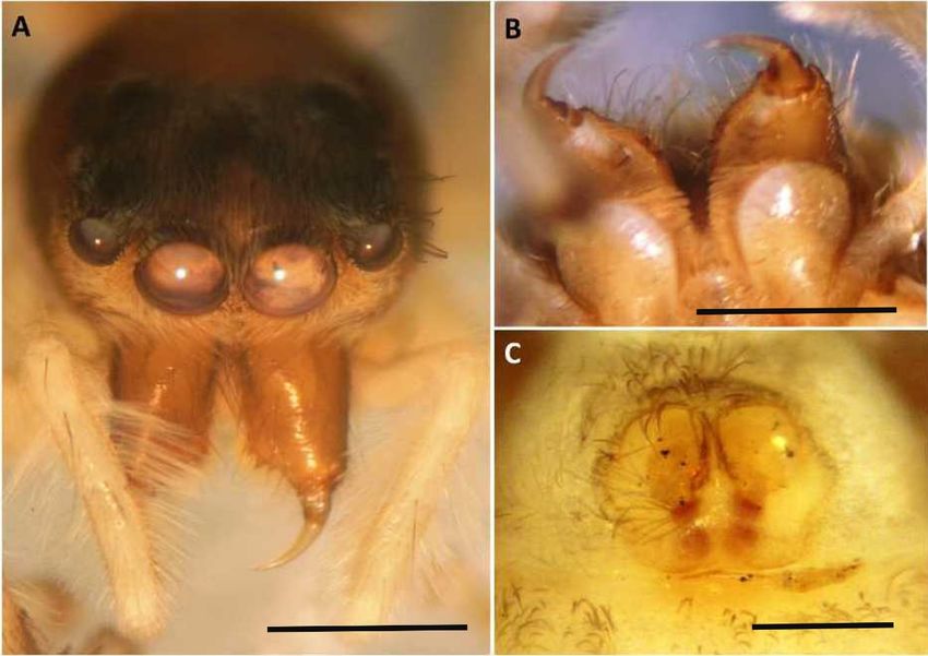
K.-M. Chen et al.

                              each leg is approximately the same, with each leg coxa and trochanter yellow,
                              femur front two-thirds yellow and end dark brown, tibia dark brown, metatarsus
                              and tarsus yellow with black rings. Genitalia bulb length is greater than its width,
                              embolus slender, base wraps around the top of the spheroid and extends along
                              the cymbium, tibial apophysis ends with a single sharp process, the cymbium
                              longer than the tibia.
                                Ecology
                                Low grassland of Aogu Wetland, Taiwan.
                                Distribution
                                India, Myanmar, Taiwan area.

                                Euophrys taiwanus sp. nov. Chen, Lin and Ueng, 2021 (Figures 10-13)
                                Type Material
                                Holotype: A male specimen collected from Aogu Wetland, Chiayi County, by
                              Tai-Yu Lin (NMNS-8370-001) on February 3, 2014.
                                Paratypes: Two female specimens collected from the locality on same date as
                              holotype by Tai-Yu Lin (NMNS-8370-002) on February 3, 2014.
                                Male
                                Male total length (mm) 2.45. Carapace length 1.10, width 0.80; abdomen
                              length 1.35, width 0.70. MOA ratio, MOA-L:MOA-AW:MOA-PW =
                              0.40:0.530.76. AMI:AMLI:PMI:PMLI = 0.07:0.04:0.67:0.20. AME:ALE:PME:PLE
                              = 0.22:011: 0.04:0.11. Leg I 1.85 (0.60, 0.80, 0.25, 0.20); leg II 1.35 (0.45, 0.50,
                              0.20, 0.20); leg III 1.65 (0.55, 0.50, 0.25, 0.30); leg IV 1.80 (0.60, 0.70, 0.25, 0.25).
                              Pedipalp 1.65 (0.33, 0.17, 0.13, 0.33).
                                Female
                                Female total length (mm) 2.90; carapace length 1.40, width 0.90; abdomen

                              Figure 10. Male Euophrys taiwanus sp. nov. (A) Dorsal view; (B-D) pedipalp and palpal
                              organ; (B) lateral view; (C) dorsal view; (D) ventral view. Scale bars: A = 1 mm, B-D = 0.5
                              mm.

DOI: 10.4236/nr.2021.129021                          298                                              Natural Resources
K.-M. Chen et al.

                              Figure 11. Male Euophrys taiwanus sp. nov. (A) Ventral view; (B) right lateral view; (C)
                              AER; (D) promarginal and retromarginal teeth. Scale bars: A-B = 1 mm, C = 0.5 mm, D =
                              0.1 mm.

                              Figure 12. Female Euophrys taiwanus sp. nov. (A) Dorsal view; (B) ventral view. Scale
                              bars: A-B = 1 mm.

                              length 1.50, width 1.00. MOA ratio, MOA-L:MOA-AW:MOA-PW =
                              0.40:0.53:0.80. AMI:AMLI: PMI:PMLI = 0.04:0.04:0.67:0.12. AME:ALE:PME:PLE
                              = 0.24:011: 0.06:0.13. Leg I 1.95 (0.75, 0.75, 0.20, 0.25); leg II 1.55 (0.50, 0.60,
                              0.20, 0.25); leg III 1.65 (0.60, 0.60, 0.20, 0.25); leg IV 1.75 (0.50, 0.75, 0.25, 0.25).
                                Characteristics
                                Male
                                Cephalothorax brown and flat, ocular area blackish brown, white hairs,
                              fan-shaped radial groove behind the thoracic groove. AER recurve. Chelicera

DOI: 10.4236/nr.2021.129021                         299                                               Natural Resources
K.-M. Chen et al.
Figure 13. Female Euophrys taiwanus sp. nov. (A) cephalothorax; (B) AER; (C) first leg;
(D) first leg ventral view; (E) promarginal and retromarginal teeth; (F) epigynum. Scale
bars: A-D = 0.5 mm, E-F = 0.1 mm.
and fangs are yellowish brown. Two promarginal teeth, 1 retromarginal tooth, la-
bium brown. Sternum black, shield-shaped, front truncated. Abdomen long
ovoid, hairy, dorsal yellow, on the front part of abdomen with large black
patches, and 9 - 12 inverted V-shaped black patterns on the second part of ab-
domen. Abdomen ventral is light yellow. The black longitudinal band extends to
the end of the abdomen. Spinnerets are dark brown. Leg lengths are in the order
I > IV > III > II. First leg: femur, patella, and tibia brown; metatarsus and tarsus
yellow. The second, third, and fourth legs are light yellow, patella and tibia with
obvious black belts. Palpal organ brown, tibia shorter than cymbium, tibial apo-
physis bifurcated. The bifurcation on the side near the spheroid is sharper, and
the bifurcation on the other side is blunt. The genitalia bulb protruding, and its
length is greater than width. If the genitalia bulb is viewed from the side, the
lower end of the bulb is most prominen, and genitalia bulb cover part of the ti-
bia, embolus sickle shaped.
Female
Cephalothorax brown and flat, ocular area blackish brown, white hairs,
fan-shaped radial groove behind the thoracic groove, AER recurve. Chelicere
and fangs are yellowish brown. Two promarginal teeth, 1 retromarginal tooth,
labium brown. Sternum black, shield-shaped, front truncated. Abdomen long
ovoid, hairy, dorsal yellow, on the front part of abdomen with large black
patches and black pattern, and 4 - 5 inverted V-shaped black patterns on the
second part; 2 pairs of black dots on each side of the black of abdomen vertical
band. Abdomen ventral is light yellow. The black longitudinal band extends to
the end of the abdomen. Spinnerets are dark brown. Leg lengths are in the order
DOI: 10.4236/nr.2021.129021 300 Natural ResourcesK.-M. Chen et al.
I > IV > III > II. Legs light yellow, lateral view of femur, patella, and tibia with
obvious black belt; metatarsus and tarsus yellow. The second, third, and fourth
legs are light yellow, patella and tibia with obvious black belts, ventral side of the
first has 3 pairs of spines. Epigynum is reddish brown, and spermatheca is round
and faintly visible.
Ecology
Aogu Wetland casuarina forest, Euophrys taiwanus sp. nov. is the dominant
species of casuarina forest.
Distribution
Taiwan area.
Evarcha bulbosa Zabka, 1985 (new record) (Figure 14, Figure 15)
Evarcha bulbosa Zabka, 1985: 222, f. 173-175; Peng, 1989: 159, f. 6A-C; Zhang,
Song, and Zhu, 1992: 1, f. 1.1-3; Peng et al., 1993: 64, f. 175-178; Song, Zhu, and
Chen, 1999: 510, f. 293J-K, 325L; Yin et al., 2012: 1359, f. 736a-d
Type Material
Male total length (mm) 5.70. Carapace length 2.80, width 2.10; abdomen
length 2.90, width 1.60. MOA ratio, MOA-L:MOA-AW:MOA-PW = 0.90:1.07:1.57.
AMI:AMLI:PMI:PMLI = 0.07:0.10:1.43:0.33. AME:ALE:PME:PLE =
0.50:023:0.06:0.17. Leg I 5.00 (1.55, 2.05, 0.85, 0.55); leg II 4.25 (1.50, 1.50, 0.75,
0.50); leg III 4.39 (1.50, 1.60, 0.74, 0.55); leg IV 4.65 (1.70, 1.50, 0.75, 0.70). Pe-
dipalp 1.95 (0.80, 0.20, 0.20, 0.75).
Characteristics
Male
Cephalothorax black-brown, orange-red bands on both sides and back of
ocular area, white hairs on both sides of the band, and black at the end of the
cephalothorax, AER recurve. Chelicera and fangs are reddish brown, One pro-
marginal tooth, 1 retromarginal tooth, labium reddish brown. Yellowish brown
Figure 14. Male Evarcha bulbosa. (A) Dorsal view; (B) ventral view. Scale bars: A-B = 2
mm.
DOI: 10.4236/nr.2021.129021 301 Natural ResourcesK.-M. Chen et al.
Figure 15. Male Evarcha bulbosa. (A-C) Palpal organ; (A) front view; (B) lateral view; (C)
dorsal view; (D) tibial apophysis; (E) chelicerae, promarginal and retromarginal teeth.
Scale bars: A = 0.3 mm, B-D = 0.5 mm, E = 0.1 mm.
with darker central color. Abdomen long ovoid, dorsal view black, 2 yellow belts
in the center extend to two-thirds of the abdomen, inside with black patches,
and the black parts on both sides are densely covered with small yellow spots;
ventral view center black, 4 vertical lines composed of yellow spots, book lung
yellowish brown. Spinnerets are dark brown. First leg is the longest and thickest,
the first and second legs are dark brown, and the third and fourth legs are yellow
and black. Genitalia bulb round, sperm duct visible, embolus elongated, and end
flagellated. Tibial apophysis is single and with bifurcations at the end, and the
cymbium is longer than the tibia. This species and Evarcha pococki [19] can be
distinguished by whether the end of the tibial apophysis is bifurcated.
Ecology
Low grassland and terrestrial shrubs of Aogu Wetland, Taiwan.
Distribution
Chinese Mainland, Vietnam, Taiwan area.
Evarcha chiayiensis sp. nov. Chen, Lin and Ueng, 2021 (Figure 16, Figure
17)
Holotype: A male specimen collected from Aogu Wetland, Chiayi County, 4
April 2014 by Tai-Yu Lin (NMNS-8370-003).
Type Material
Male total length (mm) 3.80. Carapace length 2.00, width 1.60; abdomen
length 1.80, width 1.20. MOA ratio, MOA-L:MOA-AW:MOA-PW = 073:0.76:1.35.
AMI:AMLI:PMI:PMLI = 0.02:0.06:1.20:0.27. AME:ALE:PME:PLE =
0.38:0.17:007:0.17. Leg I 2.96 (1.00, 1.05, 0.45, 0.46); leg II 2.17 (0.82, 0.75, 0.35,
0.25); leg III 3.30 (1.00, 1.20, 0.60, 0.50); leg IV 3.20 (1.05, 1.05, 0.60, 0.50). Pe-
dipalp 1.65 (0.80, 0.20, 0.20, 0.75).
DOI: 10.4236/nr.2021.129021 302 Natural ResourcesK.-M. Chen et al.
Figure 16. Male Evarcha chiayiensis sp. nov. (A) Dorsal view; (B) pedipalp; (C-D) long
apophysis; (C) dorsal view; (D) ventral view. Scale bars: A = 2 mm, B-D = 0.5 mm.
Figure 17. Evarcha chiayiensis sp. nov. (A) Cephalothorax dorsal view; (B) ventral view;
(C) lateral cephalothorax; (D) chelicerae, promarginal and retromarginal teeth. Scale bars:
A-C = 2 mm, D = 0.1 mm.
Characteristics
Male
Cephalothorax yellowish brown, flat, black around the PME and PLE, forming
a black area. A black area is present behind the PLE. The 2 black areas form 2
black vertical bands from a distance, but they are slightly cut off behind the PLE.
On observation, the cephalothorax\is light yellow near the ventral surface. AER
recurve. Chelicera and fangs are yellowish brown. Two promarginal teeth, 1 re-
tromarginal tooth. Labium yellowish brown. Sternum yellow, and ovoid. Abdo-
DOI: 10.4236/nr.2021.129021 303 Natural ResourcesK.-M. Chen et al.
men long oval, dorsal view orange with a wide black longitudinal band on each
side near the edge, and the longitudinal width is approximately one-third of the
abdomen’s width. Abdomen’s ventral surface yellow, with 1 black thin longitu-
dinal line in the center, and on both sides 1 lighter black thin line. Spinnerets are
yellowish brown. First leg yellowish brown, and from leg coxa to tarsus with
black lines. Second, third, and fourth legs yellow, and trochanter, femur, and
patella have black rings on the ventral contact. Palpal organ reddish brown, tibial
apophysis monoprocessed, and its end straight and short. Genitalia bulb nearly
round, sperm duct faintly visible, embolus elongated and linear and protrude
along the genitalia bulb. Cymbium covered with many white hairs and longer
than the tibia. This species can be distinguished from Evarcha bulbosa by no bi-
furcation at the end of the tibial apophysis. Additionally, this species and Evar-
cha pococki can be distinguished by the palpal organ, detailed description is
shown in Table 1 and Figure 16 [18].
Ecology
Waterside shrubs of Aogu Wetland, Taiwan.
Distribution
Taiwan area.
Menemerus bivittatus (Dufour, 1831) (new record) (Figures 18-21)
Salticus bivittatus Dufour, 1831: 369, pl. 11, f. 5.
Attus cinctus Walckenaer, 1837: 430.
Marpissa balteata C. L. Koch, 1846: 68, f. 1133.
Salticus convergens Doleschall, 1859: 15, pl. 9, f. 4.
Dendryphantes balteata Simon, 1864: 314.
Menemerus vittatus Simon, 1877b: 59.
Icius convergens Thorell, 1878b: 232.
Menemerus bivittatus Peckham and Peckham, 1886: 292; Petrunkevitch,
1925a: 241, f. 152-153; Davies and Zabka, 1989: 250, pl. 55.
Type Material
Male total length (mm) 5.20. Carapace length 3.10, width 2.00; abdomen
length 2.10, width 1.85. MOA ratio, MOA-L:MOA-AW:MOA-PW= 0.91:1.02:1.62.
Table 1. Comparison of the palpal organ of male Evarcha chiayiensis sp. nov. and E. po-
cocki.
Species Embolus Genitalia bulb Tibial apophysis
end straight, long, and
nearly round shape, sperm
thin line shape; knife-shaped; more than half
Evarcha duct is visible and long,
cut out along the length of the genitalia bulb;
chiayiensis narrow at the top and wide
the genitalia bulb bifurcation near the inner
at the bottom
side of the cymbium
thin line shape, slight curvature at the end, about
nearly round shape, visible
after touching the half the length of the genitalia
E. pococki and short, wide at the top
genitalia bulb, it rises bulb, no bifurcation near the
and narrow at the bottom
nearly vertically inner side of the cymbium
DOI: 10.4236/nr.2021.129021 304 Natural ResourcesK.-M. Chen et al.
Figure 18. Male Menemerus bivittatus. (A) Dorsal view; (B) ventral view. Scale bars: A-B
= 2 mm.
Figure 19. Male Menemerus bivittatus. (A) Chelicera, retromarginal teeth; (B) cephalo-
thorax; (C) abdomen dorsal view; (D) pedipalp. Scale bars: A-C = 1 mm, D = 0.5 mm.
AMI:AMLI: PMI:PMLI = 0.13:0.06:1.44:0.33. AME:ALE:PME:PLE =
0.44:024:0.09:0.22. Leg Iblack; leg II 3.75 (1.25, 1.65, 0.50, 0.35); leg III 3.55 (1.15,
1.60, 0.50, 0.30); leg IV 4.35 (1.50, 1.75, 0.60, 0.50). Pedipalp 2.10 (0.95, 0.30,
0.25, 0.60).
Female total length (mm) 6.60. Carapace-length 2.30, width 1.90; abdomen-
length 4.30, width 2.30. MOA ratio, MOA-L:MOA-AW:MOA-PW =
0.97:1.10:1.43. AMI:AMLI: PMI:PMLI = 0.10:0.13:1.30:0.23. AME:ALE:PME:PLE
= 0.50:027:0.07:0.20. Leg I 3.75 (1.25, 1.50, 0.50, 0.50); leg II 3.70
DOI: 10.4236/nr.2021.129021 305 Natural ResourcesK.-M. Chen et al.
Figure 20. Female Menemerus bivittatus. (A) Dorsal view, (B) ventral view. Scale bars:
A-B = 2 mm.
Figure 21. Female Menemerus bivittatus. (A) AER; (B) chelicera; (C) epigynum. Scale
bars: A-B = 1 mm, C = 0.2 mm.
(1.25, 1.50, 0.50, 0.45); leg III 3.70 (1.15, 1.55, 0.50, 0.50); leg IV 4.45 (1.45, 2.00,
0.50, 0.50).
Characteristics
Male
Cephalothorax brown, ocular area dark brown; thoracic groove and behind
AER with white hairs AER recurve. Chelicera and fangs reddish brown. Two
promarginal teeth, 1 retromarginal tooth. Labium brown. Sternum dark brown,
oval, with white hairs on the bottom edge. Abdomen oval, with gray hairs. Wide
DOI: 10.4236/nr.2021.129021 306 Natural ResourcesK.-M. Chen et al.
dark brown vertical strip in the center of the back, with or-ange-yellow edges on
both sides of the vertical strip, and 2 blackish gray tri-angular stains extending to
the sides at three-fifths of the vertical line. The dark-brown vertical strips behind
the stain are covered by 2 yellow herringbone stripes and the herringbone stripes
are divided abdomen into 3 blocks. Abdomen’s ventral surface grayish yellow.
Several yellow stripes behind at the extragastric sulcus, and the book lung is yel-
lowish brown. Spinnerets are dark brown. Legs with white hairs, ventral yello-
wish brown; femur, patella, tibia, metatarsus, and tarsus with black markings.
Pedipalp, femur, and tibia are with white hairs tufts on the outside. Genitalia
bulb front view is with a vertical line. Protuberance under the genitalia bulb
covering approximately half the tibia, embolus base large, base with a
hook-shaped protrusion at the end. Cymbium with black hairs with a small pro-
trusion from the base of the dorsal side.
Female
Cephalothorax brown, between the PME and PLE with a butterfly-shaped
dark brown block. Cephalothorax has dark-brown stripes on both sides of the
edge, a thoracic groove of the middle fossa is in the shape of the number “l”, and
a horizontal band of white hairs is present on the forehead. AER recurve. Cheli-
cera and fangs are brown. Two promarginal teeth, 1 retromarginal tooth. La-
bium is brown, and length is greater than width. Sternum grayish black, densely
covered with small yellow spots, ovoid, front truncated, and white hairs on the
edges. Abdomen oval, covered with scattered black hairs, dorsal view yellow with
a brown longitudinal band on each side edge that extends to the end of the ab-
domen and finally meets; ventral view yellow, without a pattern, and some black
hairs are present on the extragastric groove. Spinnerets are yellow. Leg yellow,
femur covered with 4 - 5 black hairs on the dorsal side of the leg. Pedipalp
densely covered with white hairs; genitalia obvious, pink.
Ecology
Waterside shrubs and casuarina forest of Aogu Wetland, Taiwan.
Distribution
Chinese Mainland, Vietnam, Taiwan area.
Microbianor formosana sp. nov. Chen, Lin and Ueng, 2021 (Figures
22-24)
Holotype: A male specimen collected from Aogu Wetland, Chiayi County, on
August 2, 2014, by Tai-Yu Lin (NMNS-8370-004).
Type Material
Male total length (mm) 1.56. Carapace length 0.67, width 0.70; abdomen
length 0.89, width 0.67. MOA ratio, MOA-L:MOA-AW:MOA-PW =
058:0.58:0.74. AMI:AMLI: PMI:PMLI = 0.01:0.01:0.63:0.25. AME:ALE:PME:PLE
= 0.16:0.08:0.04:0.11. Leg I 1.52 (0.53, 0.60, 0.20, 0.19); leg II 1.21 (0.41, 0.41,
0.20, 0.19); leg III 1.33 (0.41, 0.53, 0.20, 0.19); leg IV 1.46 (0.45, 0.57, 0.23, 0.21).
Pedipalp 1.08 (0.40, 0.20, 0.07, 0.41).
DOI: 10.4236/nr.2021.129021 307 Natural ResourcesK.-M. Chen et al.
Figure 22. Male Microbianor formosana sp. nov. (A) Dorsal view; (B) ventral view. Scale
bars: A-B = 1 mm.
Figure 23. Microbianor formosana sp. nov. (A) AER and chelicerae; (B) first leg; (C-D)
lateral palpal organ. Scale bars: A-D = 0.5 mm.
Characteristics
Male
Body size small, less than 2 mm, resembles a beetle with a metallic luster. Ce-
phalothorax is dark brown, and ocular area is darker than the chest. Ocular area is
widest at the PLE, radial furrow obvious, and ventral surface yellow. AER recurve,
and a row of white hairs on the forehead. Chelicera and fangs brown, labium dark
brown. Sternum brown, shield-shaped, and with yellow dots. Abdomen dorsal
view is brown; 2 pairs muscle points are at front, and ventral view is yellow. First
leg is much thicker than other legs, and the color is darker and brown. Rows of
DOI: 10.4236/nr.2021.129021 308 Natural ResourcesK.-M. Chen et al.
Figure 24. Microbianor formosana sp. nov. (A-B) Front pedipalp cymbium; (B) dorsal
pedipalp cymbium; (C-D) lateral pedipalp cymbium and tibial apophysis. Scale bars: A-D
= 0.2 mm.
Figure 25. Male Plexippus petersi. (A) Dorsal view; (B) ventral view. Scale bars: A-B = 3
mm.
black shag are present on both sides of the femur, and the outer side black shag
contains more than the inner side. After the midpoint of the patella approaching
the tibia, thick, tassel-like, coarse, black hairs are observed. The second, third,
and fourth legs are yellowish brown, with less hairs than on the first leg. Palpal
organ dark brown, tibial apophysis single and spiked at the end, slightly curved
outward toward the cymbium. Genitalia bulb oval, approximately one-fifth of its
lower end is most prominent, and its lower edge does not cover the palpal organ
tibia. Embolus short, straight, knife-shaped, and surround a quarter turn of the
genitalia bulb. The position extends to the top. Cymbium contains more hairs,
DOI: 10.4236/nr.2021.129021 309 Natural ResourcesK.-M. Chen et al.
and the hairs are longer than those of the tibia. Although Microbianor formosa-
na sp. nov. and M. furcatus (Haddad & Wesolowska, 2013) are similar [24], dif-
ferences exist in structures such as the first leg and the palpal organ, as shown in
Table 2; thus, they are identified as different species.
Ecology
Casuarina forest of Aogu Wetland, Taiwan.
Distribution
Taiwan area.
Plexippus petersi (Karsch, 1878) (new record) (Figures 25-28)
Euophrys petersii Karsch, 1878a: 332, pl. 2, f. 7.
Plexippus petersi Simon, 1903a: 728; Zabka, 1985: 433, f. 464-470; Prószyński,
1987: 80; Próchniewicz, 1989: 219, f. 39-43; Zabka, 1990b: 172, f. 26-27; Song and
Chai, 1991: 21, f. 12A-D; Xie, 1993: 359, f. 11-15; Peng et al., 1993: 183, f. 639-645;
Barrion and Litsinger, 1995: 83, f. 41a-g; Song, Zhu, and Chen, 1999: 541, f.
310Q, 312C, 328M; Peng and Li, 2003b: 752, f. 3A-F; Jang, Choe, and Kim, 2007:
101, f. 3-4 (f); Yin et al., 2012: 1443, f. 787a-f.
Table 2. Comparison of first leg and palpal organ of male Microbianor formosana sp.
nov. and M. furcatus.
Species First leg Palpal organ
patella slender, length:width = 3:1,
single tibial apophysis, embolus
M. formosana tassel-like hair from the inner side
straight, short, and knife-shaped.
of the patella joint only after halfway.
patella stubby, length:width = 2:1,
tibial apophysis bifurcated, embolus
M. furcatus tassel-like hair from the inner side
straight, short, and knife-shaped.
of the femur junction.
Figure 26. Male Plexippus petersi. (A) Cephalothorax dorsal view; (B) chelicera and pe-
dipalp; (C-D) palpal organ. Scale bars: A = 3 mm, B-D = 1 mm.
DOI: 10.4236/nr.2021.129021 310 Natural ResourcesK.-M. Chen et al.
Figure 27. Female Plexippus petersi. (A) Dorsal view; (B) ventral view. Scale bars: A-B =
3 mm.
Figure 28. Female Plexippus petersi. (A) Cephalothorax dorsal view; (B) chelicera and
retromarginal teeth; (C) abdomen ventral view; (D) epigynum. Scale bars: A = 1 mm, (B)
D = 0.5 mm, C = 2 mm.
Type material
Male total length (mm) 5.45. Carapace length 2.65, width 2.20; abdomen
length 2.80, width 1.96. MOA ratio, MOA-L:MOA-AW:MOA-PW =
1.02:1.17:1.22. AMI:AMLI: PMI:PMLI = 0.10:0.06:1.10:0.22. AME:ALE:PME:PLE
= 0.60:0.21:0.08:0.18. Leg I 3.26 (1.23, 0.58, 0.77, 0.68); leg II 3.26 (0.96, 0.57,
1.23, 0.50); leg III 3.81 (1.16, 1.30, 0.80, 0.55); leg IV 3.31 (1.05, 1.35, 0.45, 0.46).
Pedipalp 2.03 (0.83, 0.33, 0.20, 0.67).
DOI: 10.4236/nr.2021.129021 311 Natural ResourcesK.-M. Chen et al.
Female total length (mm) 7.02. Carapace length 3.20, width 2.20; abdomen
length 3.82, width 2.00. MOA ratio, MOA-L:MOA-AW:MOA-PW =
0.77:1.17:1.53. AMI:AMLI: PMI:PMLI = 0.10:0.07:1.33:0.21. AME:ALE:PME:PLE
= 0.53:0.20:0.10:0.27. Leg I 4.53 (1.50, 1.83, 0.67, 0.53); leg II 3.37 (1.50, 1.67,
0.67, 053); leg III 4.84 (1.67, 1.67, 0.67, 0.83); leg IV 5.73 (1.83, 1.83, 1.00, 1.07).
Characteristics
Male
Cephalothorax brown, ocular area black-brown, covered with black hairs,
from the PLE to the carapace with a wide black longitudinal band on the left and
right sides. AER recurve. Chelicera and fangs are brown. Two promarginal teeth,
1 retromarginal tooth. Labium brown. Sternum yellow, with a light U-shaped at
the front end, and the rear end does not extend to the fourth leg coxa. Abdomen
long ovoid, yellow, with several black, long hairs in the center, and 2 dark brown
longitudinal bands on the 2 sides, separated by a yellow horizontal strip at
two-thirds the abdomen. Several brown mountain-shaped patterns can be seen
at the end of the center of the dorsal of abdomen. Abdomen ventral is yellowish
brown, with a large dark brown wedge-shaped spot under the epigastric furrow.
Spinnerets are grayish yellow. The legs yellow, hairy, with dark brown longitu-
dinal spots on both sides. Genitalia bulb noncircular and with a protuberance
underneath. Sperm duct visible, extending upward along the side of the genitalia
bulb. Embolus has a slender tip. The front edge of the genitalia bulb is higher
than the base of the embolus. Tibia has thick clusters of white hairs and tibial
apophysis monoliths. Close to the cymbium, the end of the tibial apophysis ex-
ceeds the base of the embolus, and the dorsal side of the cymbium is covered
with numerous white hairs. The palpal organ structure of this species is similar
to that of P. paykulli (Audouin, 1826). In this species, the embolus is relatively
slender, and the front edge of the tip of the tibial apophysis exceeds the embolus.
Female
Cephalothorax brown, with black hairs, black around the eyes, ocular area less
than half the length of the cephalothorax. Ocular area has a yellow longi-tudinal
band behind the center. AER recurve. Chelicera and fangs are are brown and
fangs are reddish brown. Two promarginal teeth, 1 retromarginal tooth. Labium
yellow, width is greater than length. Sternum light yellow, with white burrs on
the rear end. Abdomen oval, dorsal view blackish brown with a thick grayish
white longitudinal band in the center. This central longitudinal band protrudes
to the left and right at the center and two-thirds of the abdo-men, forming 2
pairs of small white spots. Several thin, arc-shaped, horizontal bands at the end
of the central longitudinal band. Abdomen ventral view yel-low. Many black
spots are evenly distributed under the epigastric furrow. Spots directly above the
spinnerets are largest and are grayish yellow. Pedi-palp yellow and black, with
hairs. Leg brown with white hairs, and with many dark spots in the dorsal view.
Black bristles on the dorsal side of the femur, 3 pairs of spines in the tibia ventral
view, 2 pairs of spines in the metatarsus ventral view. Epigynum long, its center
DOI: 10.4236/nr.2021.129021 312 Natural ResourcesK.-M. Chen et al.
having a gutter-shaped ditch. Front positioned hood. The copulatory openings
on both sides are longitudinal, long, and crack-like. Spermatheca round, and 2
are visible. The main difference of this species from P. paykulli lies is in the posi-
tion of the hood; specifically, it has a front position in this species and a middle
position in P. paykulli.
The top front edge of the tibial apophysis of the male Plexippus petersi ex-
ceeds the base of the embolus (Figure 25). The position of the epigynum of the
female is higher than that of P. paykulli (Figure 28). The 2 species can be dis-
tinguished.
Ecology
Terrestrial shrubs of Aogu Wetland, Taiwan.
Distribution
Chinese Mainland, Japan, Vietnam, Malay Islands, New Guinea, Taiwan area.
Pseudicius sp. (Figure 29)
Characteristics
Female
Cephalothorax flat, dark brown, ocular area black, the edges of the cephalotho-
rax and the ocular area covered with white hairs. The ocular area is less than half
the length of the cephalothorax. PME located in the middle of ALE and PLE; un-
der the PME and PLE is a row of stridulatory spines [21]. AER recurve. Chelicera
and fangs are reddish brown. Two promarginal teeth, 1 retromarginal tooth. La-
bium reddish brown. Sternum width is greater than length, brown and with white
hairs on the edges. Abdomen oval, dorsal view black and yellow, and the black part
is covered with numerous black hairs. A short white longitudinal band is in the
Figure 29. Female Pseudicius sp. (A) Dorsal view; (B) cephalothorax and microspines
(stridulatory spines); (C) chelicerae; (D) epigynum. Scale bars: A = 3 mm, B = 1 mm, C =
2 mm, D = 0.2 mm.
DOI: 10.4236/nr.2021.129021 313 Natural ResourcesK.-M. Chen et al.
center. The end of the longitudinal band is faint with 2 horizontal rings. Ventral
view is yellow. Rows of horizontal short black hairs are present along the epigas-
tric furrow. Two vertical gray bands are present under the epigastric furrow.
Spinnerets are grayish yellow. Pedipalp yellow, with many long white hairs on
the knee joints, the outer side of the tibia, and the entire tarsus. The first leg is
the thickest, and its femur is thickest and reddish brown with sporadic long
black hairs on the outside of each section. The second, third, and fourth legs are
lighter in color and yellowish brown. The epigynum is keratinized.
Rhene rubrigera (Thorell, 1887) (new record) (Figure 30, Figure 31)
Homalattus rubriger Thorell, 1887: 347.
Homalattus phoeniceus Simon, 1888b: 203.
Rhene phoenicea Simon, 1901a: 635, 638, f. 750.
Rhene rubrigera Simon, 1903g: 733; Prószyński, 1984a: 121; Zabka, 1985: 444,
f. 544-562; Peng et al., 1993: 203, f. 715-722; Song, Zhu, and Chen, 1999: 538, f.
314O-P, 315C-D, 329F; Yin et al., 2012: 1463, f. 797a-h; Sen et al., 2015: 30, f.
67-71, pl. 12.
Type Material
Male total length (mm) 5.55. Carapace length 2.50, width 2.45; abdomen
length 3.05, width 2.10. MOA ratio, MOA-L:MOA-AW:MOA-PW = 0.90:0.96:1.63.
AMI:AMLI: PMI:PMLI = 0.03:0.10:1.50:0.86. AME:ALE:PME:PLE =
0.47:024:0.06:0.10. Leg I 5.13 (1.80, 2.10, 0.72, 0.51); leg II 3.45 (1.25, 1.20, 0.55,
0.45); leg III 2.65 (0.60, 0.95, 0.50, 0.60); leg IV 2.80 (1.00, 1.00, 0.40, 0.40). Pe-
dipalp 2.10 (0.80, 0.25, 0.25, 0.80).
Figure 30. Male Rhene rubrigera. (A) Dorsal view; (B) cephalothorax; (C) palpal organ;
(D) promarginal and retromarginal teeth. Scale bars: A = 2 mm, B, D = 1 mm, C = 0.5
mm.
DOI: 10.4236/nr.2021.129021 314 Natural ResourcesK.-M. Chen et al.
Figure 31. Female Rhene rubrigera. (A) Dorsal view; (B) cephalothorax; (C) promarginal
and retromarginal teeth; (D) epigynum. Scale bars: A = 2 mm, B = 1 mm, C = 0.5 mm, D
= 0.2 mm.
Female total length 5.85. Carapace length 2.20, width 2.20; abdomen length
3.65, width 2.30. MOA ratio, MOA-L:MOA-AW:MOA-PW = 0.73:0.82:1.43.
AMI:AMLI:PMI:PMLI = 0.12:0.10:1.30:0.83. AME:ALE:PME:PLE =
0.37:0.27:0.07:0.17. Leg I 3.98 (1.25, 1.68, 0.50, 0.55); leg II 3.25 (1.00, 1.20, 0.45,
0.60); leg III 2.70 (0.80, 0.95, 0.40, 0.55); leg IV 2.60 (0.85, 1.05, 0.30, 0.40).
Characteristics
Male
Cephalothorax flat, dark brown, and ocular area black. Ocular area and its
edges are covered with white hairs. The ocular area is less than half the length of
the cephalothorax. PME located midpoint between the anterior and posterior
eyes. AER recurve. Chelicera and fangs are reddish brown. Two promarginal
teeth, 1 retromarginal tooth. Labium brown. Sternum yellowish brown, darker
center. Abdomen long oval, dorsal view yellowish brown, edge dark brown with
white hairs, 3 pairs of muscle marks. The abdomen dorsal has 3 pairs of hori-
zontal yellow pinstripes extending from the center to the sides of the abdomen.
Yellow ventral surface of the abdomen, with a large black area in the center and
its width is approximately half the width of the abdomen, and the book lung is
dark brown. Spinnerets are dark brown. First leg longest, thickest, dark brown.
Femur, patella, and tibia with thick burrs on the inside. Second, third, and fourth
legs brown. Genitalia bulb length is greater than width, kidney-shaped. Sperm
duct visible, embolus upright and short needle-shaped, carried above the genita-
lia bulb, tibial apophysis mono-protruding, single helix-shaped, curved toward
the cymbium, cymbium longer than tibia, hairy. The main difference between
this species and R. atrata (Karsch, 1881) is the shape of the embolus. In this spe-
cies, the embolus is upright and short needle-shaped, and that of R. rubrigera
has a broad base and a curved tip.
DOI: 10.4236/nr.2021.129021 315 Natural ResourcesK.-M. Chen et al.
Female
The appearance characteristics are identical to those of the male. Abdomen
ventral view yellow, and the large black patch in the middle is lighter, with yel-
low spots scattered within it. The abdomen dorsal has 3 pairs of horizontal
lightly yellow pinstripes, which was less obvious than that of male spider. Spin-
nerets are yellowish brown, genital groove black, epigynum light red. The mat-
ing hole is horizontal. The embolus of R. rubrigera is upright and short
needle-shaped (Figure 30(C)), and it can be clearly distinguished from the em-
bolus of R. atrata, with a broad base and a curved tip.
Ecology
Waterside shrubs of Aogu Wetland, Taiwan.
Distribution
Chinese Mainland, India to Sumatra, Mexico, Hawaii, Taiwan area.
Synagelides zhilcova Prószyński, 1979 (new record) (Figure 32)
Synagelides zhilcovae Prószyński, 1979: 319, f. 316-317; Dunin, 1984b: 139, f.
65 (f); Peng et al., 1993: 229, f. 816-818 (f); Song, Zhu, and Chen, 1999: 561, f.
320H, 321A, 329R (f); Kim and Kim, 2000a: 186, f. 2A-E (f); Ono, Ikeda, and
Kono, 2009: 588, f. 345-346 (f); Kim and Lee, 2014: 141, f. 100A-B, pl. 29 (f).
Type Material
Female total length (mm) 4.00. Carapace length 1.50, width 1.00; abdomen
length 2.50, width 1.05. MOA ratio, MOA-L:MOA-AW:MOA-PW =
0.37:0.67:0.93. AMI:AMLI:PMI:PMLI = 0.03:0.03:0.83:0.40. AME:ALE:PME:PLE
= 0.30:0.13:0.05:0.13. Leg I 1.75 (0.60, 0.65, 0.30, 0.20); leg II 1.40 (0.45, 0.50,
0.30, 0.15); leg III 1.35 (0.45, 0.45, 0.30, 0.15); leg IV 1.85 (0.60, 0.75, 0.30, 0.20).
Figure 32. Female Synagelides zhilcovae. (A) Dorsal view; (B) chelicera; promarginal and
retromarginal teeth; (C) cephalothorax; (D) epigynum. Scale bars: A = 2 mm, B, C = 1
mm, C = 0.2 mm.
DOI: 10.4236/nr.2021.129021 316 Natural ResourcesK.-M. Chen et al.
Characteristics
Female
Body slender and long. Cephalothorax brown, front narrow and back wide.
From ALE to PLE a black longitudinal band is formed, covered with black hairs.
PLE is black around, ocular area has 2 large black elliptical spots, and thoracic
groove witha yellow spot block; the lower radial groove is obvious. AER recurve.
Chelicera yellow, fangs brown. Two promarginal teeth, 1 retro-marginal tooth.
Labium light yellow. Sternum light yellow, shield-shaped, front truncated, rear
pointed tail.
Abdomen spindle-shaped, dorsal view brown, with 1 pair of light-yellow stripes
on the front edge and 2 pairs of light-yellow, oblique spots on the back, forming
2 “handstand Vs,” with 2 - 3 thick, light-yellow horizontal stripes at the end.
Ventral view is yellow. Spinnerets are light yellow. Leg lengths are in the order of
IV > I > II > III. Legs yellow. The first leg has a black longitudinal band from the
inner side of the femur. The tibia has 2 pairs of acupuncture-like needles on the
ventral side. Epigynum is red, with a pair of mating tubes clearly visible.
Ecology
Waterside shrubs of Aogu Wetland, Taiwan.
Distribution
Russia, Chinese Mainland, Japan, South Korea, Taiwan area.
4. Discussion
The Euophrys taiwanus sp. nov. described in this study belongs to the subfamily
Euophryinae. This subfamily includes Euophrys, Pseudeuophrys, Talavera, Neon,
and 4 other genera of spiders in China and neighboring countries. Their com-
mon morphological characteristics are small size, with a body length of less than
4 mm, and an appearance dissimilar to ants [21].
The embolus of Euophrys is smaller than that of Pseudeuophrys and larger
than that of Talavera. The embolus of Euophrys is wrapped around the top of
the genital bulb. The embolus of Pseudeuophrys is looped around the recess or
recess at the top of the genitalia bulb. The embolus of Talavera is needle-like or
thorn-like, and that of Euophrys bends.
For the subfamily Pelleninae within the family Salticidae, 3 genera have been
recorded (Bianor, Harmochirus, Sibanor), with 4 species recorded in Taiwan [8].
This subfamily is characterized by its diamond-shaped cephalothorax, which is
used in this study to report a newly recorded genus Microbianor.
The PME of Bianor is closer to the ALE, whereas in the 3 other genera, it is at
the midpoint between the ALE and the PLE. Only the side of the genitalia bulb
of Sibanor has papillary protrusions, and the 3 other genera (Bianor, Harmo-
chirus, Microbianor) do not papillary protrusion [25].
Acknowledgements
We are grateful to Mr. Keng-Yau Chang, Mr. Po-Jaing Shie, Mr. Shir-Zu Lin,
DOI: 10.4236/nr.2021.129021 317 Natural ResourcesK.-M. Chen et al.
Mr. Meng-Han Shie, Mr. Wu-Chanl Chang, Mr. Yu-An Lin, Mr. Ya-Su Luo, Mr.
Han-Chang Chen, and Mrs. Jun-Fang Chang for their help with fieldwork and
data collection. This study was supported by Chiayi County Government and by
the Construction and Planning Agency of the Ministry of the Interior.
Conflicts of Interest
The authors have no conflicts of interest to declare.
References
[1] Matsumura, S. (1910) Die schädlichen und nützlichen insekten: Vom zuckerrohr
Formosas. The Keiseisha, Tokyo. 288 p. https://doi.org/10.5962/bhl.title.35666
[2] Lee, C.-L. (1964) The Spiders of Taiwan. Da-Jian Bank Quarterly, 15, 254-290.
[3] Zhum, Y.-I. and Okuma, C. (1974) A Checklist of Spiders in Taiwan. Annual of
Taiwan Museum, 17, 29-49.
[4] Zhum, Y.-I. and Okuma, C. (1975) A Checklist of Spiders in Taiwan, Continued.
Annual of Taiwan Museum, 18, 101-119.
[5] Chen, S.-H. (1996) A Checklist of Spiders in Taiwan. Annual of Taiwan Museum,
39, 123-155.
[6] Bao, Y.H. and Peng, X.J. (2002) Six New Species of Jumping Spiders (Araneae: Sal-
ticidae) from Hui-Sun Experimental Forest Station, Taiwan. Zoological Studies, 41,
403-411. http://zoolstud.sinica.edu.tw/Journals/41.4/403.pdf
[7] Huang, J.-W. (2004) Taxonomic Study of Myrmarachne (Araneae: Salticidae) from
Taiwan. Master’s Thesis, Department of Life Sciences, Sun Yat-sen University,
Kaohsiung, 87 p.
[8] Chen, S.-H. and Chen, C.-C. (2013) A Taxonomic Study on the Spiders of Plexip-
poida (Araneae: Salticidae) of Taiwan. Master’s Thesis, Department of Life Science,
Taiwan Normal University, Taipei, 205 p.
http://rportal.lib.ntnu.edu.tw:80/handle/20.500.12235/104392
[9] Yamasaki, T. (2013) A New Species of the Genus Myrmarachne (Araneae: Saltici-
dae) from Tamsui, Taiwan. Acta Arachnologica, 62, 29-31.
https://doi.org/10.2476/asjaa.62.29
[10] Lin, T.-Y. (2015) A Study on the Spider Cluster of Aogu Wetland, Chiayi County.
Master’s Thesis, Department of Environmental Engineering, Kun-Shan University,
Tainan, 97 p.
[11] Lin, Y.-C., Chen, K.-N., Chen, K.-X. and Ueng, Y.-T. (2019) Population Fluctuation
and Life History Traits of Tiny Leafhopper Singapora nigropunctata Mahmood,
1967 (Hemiptera: Cicadellidae) in Southwestern Taiwan. Agricultural Science Re-
search Journal, 9, 151-159.
[12] Yang, J.Y.-C. and Ueng, Y.-T. (2011) Taiwan’s Wetlands of Importance. Urban and
Rural Development Branch, Construction and Planning Agency, Ministry of the
Interior, Taipei, 192 p.
[13] Davies, V.T. and Zabka, M. (1989) Illustrated Keys to the Genera of Jumping Spid-
ers (Araneae: Salticidae) in Australia. Memoirs of the Queensland Museum, 27,
189-266.
[14] Simon, E. (1903) Histoire naturelle des araignées. Paris, 2, 669-1080.
https://www.biodiversitylibrary.org/item/119058#page/6/mode/1up
DOI: 10.4236/nr.2021.129021 318 Natural ResourcesK.-M. Chen et al.
[15] Ono, H., Ikeda, H. and Kono, R. (2009) Salticidae. In: Ono, H., Ed., The Spiders of
Japan with Keys to the Families and Genera and Illustrations of the Species, Tokai
University Press, Kanagawa, 558-588.
[16] Kim, B.W. and Kim, J.P. (2000) A Revision of the Genus Synagelides Strand, 1906
(Araneae, Salticidae) in Korea. Korean Journal of Systematic Zoology, 16, 183-190.
[17] Kim, B.W., Kim, J.P. and Cho, J.H. (2003) A Revision of the Genera Euophrys,
Pseudeuophrys, and Talavera (Araneae: Salticiadae) from Korea. Korean Arachnol-
ogy, 19, 89-102.
[18] Kim, S.T. and Lee, S.Y. (2014) Arthropoda: Arachnida: Araneae: Clubionidae, Co-
rinnidae, Salticidae, Segestriidae. Spiders. Invertebrate Fauna of Korea, 21, 1-186.
[19] Zabka, M. (1985) Systematic and Zoogeographic Study on the Family Salticidae
(Araneae) from Viet-Nam. Annales Zoologici, Warszawa, 39, 197-485.
[20] Zabka, M. and Waldock, J. (2012) Salticidae (Arachnida: Araneae) from Oriental,
Australian and Pacific Regions. Genus Cosmophasis Simon, 1901. Annales Zoologi-
ci, 62, 115-198. https://doi.org/10.3161/000345412X633694
[21] Prószynski, J. (2008) Key to Identification of Genera of Salticidae (Araneae) Known
from China and Adjacent Countries.
[22] World Spider Catalog (2021) World Spider Catalog Version 22.0. Natural History
Museum Bern. https://wsc.nmbe.ch/
[23] Jumping-Spiders Company (2021) Jumping Spiders (Arachnida: Araneae: Saltici-
dae) of the World. http://www.jumping-spiders.com/
[24] Haddad, C.R. and Wesołowska, W. (2013) Additions to the Jumping Spider Fauna
of South Africa (Araneae: Salticidae). Genus, 24, 459-501.
[25] Logunov, D.V. (2001) A Redefinition of the Genera Bianor Peckham & Peckham,
1885 and Harmochirus Simon, 1885, with the Establishment of a New Genus Sibia-
nor Gen. n. (Aranei: Salticidae). Arthropoda Selecta, 9, 221-286.
DOI: 10.4236/nr.2021.129021 319 Natural ResourcesK.-M. Chen et al.
Appendix 1
All species of Salticidae spiders were collected from Aogu Wetland in Chiayi County, Taiwan (from August 2013 to
August 2014, N = 32).
Species Specimen number Collection date and quantity State Note
Bianor angulosus NMNS-8370-005 2014/6/7 = 1♀, 7/10 = 2♀, 9/7 = 1♂ Figure 2
Carrhotus tristi NMNS-8370-006 2013/12/1 = 1♂, 2014/5/3 = 1♂ New record Figure 8, Figure 9
Euophrys taiwanus NMNS-8370-001 2014/2/3 = 1♂, Holotype New Species Figure 10, Figure 11
Euophrys taiwanus NMNS-8370-002 2014/2/3 = 2♀, Paratypes New Species Figure 12, Figure 13
Evarcha bulbosa NMNS-8370-007 2013/9/7 = 2♀, 2014/5/3 = 1♂ New record Figure 14, Figure 15
Evarcha flavocincta NMNS-8370-008 2013/9/7 = 1♀, 12/2 = 1♀ Figure 7
Evarcha chiayiensis NMNS-8370-003 2014/4/4 = 1♂, Holotype New Species Figure 16, Figure 17
Menemerus bivittatus NMNS-8370-009 2013/9/7 = 1♂ & 1♀ New record Figures 22-26
Microbianor formosana NMNS-8370-004 2014/8/2 = 1♂, Holotype New Species Figures 22-24
Myrmarachne formosicola NMNS-8370-010 2014/8/2 = 1♂ & 1♀ Figure 10
Phintella versicolor NMNS-8370-015 2014/7/10 = 1♂ Figure 6
Plexippus petersi NMNS-8370-011 2014/3/8 = 1♂ & 1♀ New record Figures 25-28
Rhene rubrigera NMNS-8370-012 2014/1/4 = 2♀, 2/3 = 1♀, 3/8 = 1♂ New record Figure 30, Figure 31
Sibianor pullus NMNS-8370-013 2014/5/3 = 1♀ Figure 3
Synagelides zhilcovae NMNS-8370-014 2014/6/23 = 2♀, 7/10 = 1♀ New record Figure 32
Pseudicius sp Missing specimen 1♀ Figure 29
DOI: 10.4236/nr.2021.129021 320 Natural ResourcesYou can also read