Comparison of electromyogram reaction time at the onset of motion in badminton players at different competitive levels
←
→
Page content transcription
If your browser does not render page correctly, please read the page content below
Original Article

Comparison of electromyogram reaction time at
the onset of motion in badminton players at
different competitive levels
YUJIRO MASU 1 , ATSUYA OTSUKA
Department of Physical Therapy, Faculty of Health Science, Health Science University, Yamanashi, Japan

                                                    ABSTRACT

In this study, we evaluated the electromyogram reaction time of femoral muscles at the onset of motion in
college badminton players at different competitive levels, with the aim of clarifying the characteristics of the
motor reaction processing in response to stimulation. The participants were seven male players from the
team that won second place in the All Japan College Badminton Championship (high-performance group)
and college badminton players with no experience in participating in national championship games (low-
performance group). In both the 1-direction and 2-direction tasks, the action time was shorter, and the pre-
motor time of femoral muscle activity was significantly shorter in the high-performance than in the low-
performance group. In both tasks, significant differences were observed in the rate of increase in absolute
EMG amplitude of the swing-out and push-out legs, and the values were higher in the high-performance than
in the low-performance group. Results indicate that high-performance badminton players are able to move
quickly by synchronizing the motor units of the rectus femoris muscle at the onset of motion and perform
actions by exerting large joint torques.
Keywords: Championship; Sports performance; Shuttlecock.

    Cite this article as:
    Masu, Y., & Otsuka, A. (2021). Comparison of electromyogram reaction time at the onset of motion in
       badminton players at different competitive levels. Journal of Human Sport and Exercise, in press.
       doi:https://doi.org/10.14198/jhse.2022.174.12

1
 Corresponding author. Department of Physical Therapy, Faculty of Health Science, Health Science University, Yamanashi,
Japan.
   E-mail: y-masu@kenkoudai.ac.jp
   Submitted for publication December 08, 2020
   Accepted for publication February 09, 2021
   Published in press March 09, 2021
   JOURNAL OF HUMAN SPORT & EXERCISE ISSN 1988-5202
   © Faculty of Education. University of Alicante
   doi:10.14198/jhse.2022.174.12
                                                                            VOLUME -- | ISSUE - | 2021 |         1
Masu, et al. / Electromyogram reaction time at the onset of motion in badminton players JOURNAL OF HUMAN SPORT & EXERCISE
INTRODUCTION
In various sports, anticipating the next development by watching the opponent’s movement is an important
ability that affects competitive performance. It has been shown that excellent ability of anticipation is acquired
by sensory learning and that the amount of information concerning the opponent’s motion patterns makes
accurate anticipation possible (Farrow et al., 2002; Jackson et al., 2007). In badminton, shots of different
velocities are hit in all directions, and it is necessary to observe the opponent’s motion patterns,
instantaneously judge the point where the shuttlecock will fall, and quickly start moving. In a previous study,
in which the task of anticipating the trajectory of the shuttlecock by viewing the video of a striker before
striking was imposed, errors of anticipation were smaller, suggesting better ability of anticipation in
experienced players than beginners (Abernethy et al., 2007). In addition, according to a report that compared
changes in brain potential in subjects with and without experience of playing badminton during the task of
anticipation and judgment, the subjects with experience showed a larger amplitude of P300, which reflects
attention and concentration, than those with no experience (Jin et al., 2011). Moreover, it is known that
experienced badminton players can selectively use useful information obtained from the form of the opponent
but that inexperienced players also direct attention to unnecessary information (Abernethy, 1990; Kellman &
Garrigan, 2009). If one can anticipate where the shuttlecock will come earlier by watching the opponent’s
form, more time can be used for preparing the return shot, and coping is facilitated. With more experience,
information concerning the opponent’s form and trajectory of the shuttlecock that has been hit is accumulated,
and the player becomes more able to anticipate where the shuttlecock will fly.
When muscles are stimulated, α motor neurons are activated, and the stimulated muscles contract (so-called
stretch reflex). H wave evoked by electric stimulation is used as an index for the evaluation of muscle
contraction at the spinal cord level in such a stretch reflex. H wave is an evoked myoelectric potential that
occurs as a result of monosynaptic excitation of α motor neurons induced by Ia afferent fibres excited by
electric stimulation of peripheral nerve bundles and is an index that reflects the excitability of spinal cord
motor neurons (Nakazawa et al., 2004; Phadke et al., 2010; Nielsen et al., 1993). When badminton players
were told to hold a racket and be poised to receive, the amplitude of the H wave of the soleus muscle was
reported to be significantly reduced in college students who began playing badminton in their junior high
school days (Masu & Muramatsu, 2015). Especially, training over many years using a particular tool leads to
functional reconstitution of the motion-related cortical network. In badminton players, the motion-related
areas are activated by holding a racket, and stretch reflex is suppressed (Masu et al., 2019). Although the
functional significance of the decrease in spinal cord excitability is unclear, when the spinal cord is in a highly
excited state, the membrane potential of motor neurons elevates. This is advantageous for exciting a large
number of motor neurons but is considered to increase the susceptibility to the effects of spinal reflexes and
make it difficult to perform movements delicately controlled by the brain. On the other hand, if the spinal
excitability is appropriately suppressed, the membrane potential of motor neurons is reduced, the effects of
spinal reflex are controlled, and movements faithful to orders from the brain are facilitated. Moreover, in a
study in which college badminton players were instructed to stand still and relax with both arms stretched
along the sides and to raise one hand above the head at the moment when the light flashes, the results of
measurement of femoral muscle activities during the task suggested that the racket-holding arm can be raised
more quickly by enhancing femoral muscle activities of the leg contralateral to the racket-holding hand as
well as accelerating the switch of femoral muscle activities from the ipsilateral to contralateral leg (Masu &
Nagai, 2016). Both studies, in which the subjects were college badminton players who started playing
badminton in their elementary school or junior high school days, suggest that, by continuing to play
badminton, players more readily reflect orders from the brain in muscle movements and instantaneously
move the body. However, these studies evaluated femoral muscle activities in a stationary position for
2 | 2021 | ISSUE - | VOLUME -- © 2021 University of AlicanteMasu, et al. / Electromyogram reaction time at the onset of motion in badminton players JOURNAL OF HUMAN SPORT & EXERCISE
receiving or with the upper limbs raised, and we have encountered no report evaluating the characteristics
of leg muscle activities while players are actually moving on their legs. Also, while it has been clarified by
research on information processing in the brain from presentation of stimulation to implementation of reactive
movements that the visual motor reaction time is shorter (Hülsdünker et al., 2016) and that the amplitude of
P300, the brain potential related to the anticipation ability, is larger (Jin et al., 2011) in badminton players
compared with non-athletes. In these reports, however, the subjects were non-athletes or relatively unskilled
athletes. Therefore, these studies are considered to have been inadequate for clarifying differences in the
degree of development of the motor reaction ability among elite athletes related to the competitive level.
In this study, therefore, we evaluated the electromyogram reaction time of femoral muscles at the onset of
motion in college badminton players at different competitive levels, aiming to clarify the characteristics of
motor reaction processing in response to stimulation.
MATERIALS AND METHODS
Subjects
The subjects were seven male players who belonged to the team that won second place in the team
competition in the All Japan College Badminton Championship (high-performance group) and college
badminton players with no experience of participating in national championship games (low-performance
group) (Table 1). The high-performance and low-performance groups belonged to different teams. Both
groups practiced 6 days a week, 3 hours a day. The players in the high-performance group placed in the top
8 in national-level individual matches in high school and entered college by athlete quotas. All subjects were
given an explanation about the objective and safety of the measurement, and voluntary consent to participate
in the study was obtained. This study was carried out with approval by the research ethics committee of the
Health Science University.
Table 1. Participants' Age, Badminton playing experience, and Physical Characteristics.
Group Age (years) Badminton playing experience (years) Height (cm) Weight (kg)
Low-performance (n = 7) 19.3 ± 0.9 7.3 ± 0.9 * 170.6 ± 4.4 61.0 ± 5.2
High-performance (n = 7) 19.7 ± 0.9 12.9 ± 1.2 * 168.6 ± 7.0 64.9 ± 6.3
Note. *p < .05.
Measurement of the onset of motion
Each subject stood 1 m behind the intersection of the short service line and centre line, instantaneously
moved when the light flashed, and touched the top of the net inside the court 50 cm from the singles side-
line with a racket (Figure 1). First, the light on the forehand side was flashed, and the subjects moved to the
forehand side alone (1-direction task). Next, the lights on the forehand and backhand sides were flashed
randomly, and the subjects moved to the side of the flashed light (2-direction task). The movements during
the trials were video-recorded with a high-speed camera (Sports Coachingcam; 4Assist, Inc.; film speed 240
frames/sec.; shutter speed 1/1,000 sec) (Figure 1). In addition, electrodes were attached to the rectus femoris
and biceps femoris muscles, and muscle activities were recorded using a wireless electromyograph (4Assist,
Inc.) at 1,000 Hz.
In this study, also, the legs ipsilateral and contralateral to the hand holding the racket were defined as the
racket-side and non-racket-side legs, respectively. Moreover, in movements to the forehand side, the racket-
side and non-racket-side legs were defined as the swing-out and push-out legs, respectively. In movements
to the backhand side, the non-racket-side and racket-side legs were defined as the swing-out and push-out
VOLUME -- | ISSUE - | 2021 | 3Masu, et al. / Electromyogram reaction time at the onset of motion in badminton players JOURNAL OF HUMAN SPORT & EXERCISE
legs, respectively. Traveling to the forehand side was made in 3 strides by grounding the racket-side leg
(swing-out leg) (1st step), stepping the non-racket-side leg (push-out leg) toward the traveling direction (2nd
step), and finally grounding the racket-side leg and touching the net with the racket. Traveling to the backhand
side was made in 2 strides by grounding the non-racket-side leg (swing-out leg) and stepping the racket-side
leg toward the traveling direction and touching the net with the racket.
Figure 1. Schematic drawing of experimental setup.
Measurement using a motion capture system
By selecting 1 subject each from the high-performance and low-performance groups, 1-direction and 2-
direction tasks were video-recorded using 8 units of MAC3D System (Motion Analysis Corporation; film speed
240 frames/sec.; shutter speed 1/1,500 sec.) and 1 high-speed camera (Sports Coachingcam; 4Assist, Inc.;
film speed 240 frames/sec.; shutter speed 1/1,000 sec.) (Figure 1). Regarding the 3D coordinates, the X axis
was set as the direction of the centre line, Y axis as the direction parallel to the net, and Z axis as the direction
perpendicular to the floor.
The subjects were stripped to the waist and wore half tights and badminton shoes for the measurements.
Reflective markers were attached at a total of 29 points on the body according to the Helen Hayes marker
set: Head (1, 2, 3), acromia (shoulder joints: 4, 5), inferior angle of the right scapula (6), ulnar sides of the
elbows (7, 8), wrists (9, 10), anterior superior iliac spines (11, 12), 5th lumbar vertebra (13), femurs (14, 15),
lateral epicondyles of femur (16,17), medial epicondyles of femur (18, 19), tibias (20, 21), medial malleoli (22,
23), lateral malleoli (24, 25), second metatarsal bones (26, 27), and heels (28, 29) (Figure 2).
Adjustment of the musculoskeletal model of each subject was made using the coordinate data of the whole
body according to the human body size and shape database (National Institute of Advanced Industrial
Science and Technology) and estimates of tension against the maximum muscular tension in the present
posture were calculated using Stoeve’s method (1999). In addition, joint torques were calculated by inverse
4 | 2021 | ISSUE - | VOLUME -- © 2021 University of AlicanteMasu, et al. / Electromyogram reaction time at the onset of motion in badminton players JOURNAL OF HUMAN SPORT & EXERCISE
dynamic analysis. These analyses were performed using musculoskeletal model kinetic analysis software
(nMotion musculous).
Figure 2. Body marker placement.
Both the 1-direction and 2-direction tasks were performed until data from 3 trials in the forehand direction
could be obtained from each participant, and the mean of the 3 trials was adopted as the representative value
of the participant.
EMG analysis
After the EMG data obtained were integrated with a time constant decay of 0.02 (integrated EMG (IEMG)),
the latency from flashing of the light to the onset of motion of each muscle (pre-motor time (PMT)) was
calculated. The moment at which 4 times the standard deviation of the EMG value during stationary standing
was exceeded was defined as the time of the onset of muscle activity (Figure 3). The rates of increase in
absolute EMG amplitude (RIE) of the rectus femoris and biceps femoris muscles at the onset of motion were
determined according to the method of Kamimura et al. (2009).11) After performing full-wave rectification of
the EMG waveform first (Figure 4b), smoothing was made using a Gaussian filter with a cut-off frequency of
4 Hz (Figure 4c), the slope was calculated by temporal differentiation (Figure 4d), and the first peak amplitude
was used as RIE. RIE serves as an index reflecting the instantaneous synchronization rate of motion units
at the onset of motion. Data analysis was performed using Labchart (ADInstuents Incorporated) and
KyPlot5.0 (KyensLab Incorporated).
VOLUME -- | ISSUE - | 2021 | 5Masu, et al. / Electromyogram reaction time at the onset of motion in badminton players JOURNAL OF HUMAN SPORT & EXERCISE
Figure 3. Time from turning on the light to muscle activity. EMG: electromyogram.
Figure 4. Calculation of rate of increase in absolute EMG amplitude. (a) Raw waveform; (b) Rectification; (c)
Smoothing; (d) Temporal differentiation. EMG: electromyogram.
6 | 2021 | ISSUE - | VOLUME -- © 2021 University of AlicanteMasu, et al. / Electromyogram reaction time at the onset of motion in badminton players JOURNAL OF HUMAN SPORT & EXERCISE
Data processing
Both the 1-direction and 2-direction tasks were performed until data of 3 trials in the forehand direction could
be obtained from each subject, and the mean of the 3 trials was adopted as the representative value of the
subject. The high-performance and low-performance groups were compared by unpaired Student t-test after
testing the normality by the Kolmogorov-Smirnov test, and the t value and Cohen’s d were calculated. All
tests were performed at a significance level of p < .05. These analyses were carried out using the statistical
analysis software EZR (EasyR) (Kanda, 2013).
RESULTS
Table 2 shows the action time. In both the 1-direction and 2-direction tasks, the action time was significantly
shorter in the high-performance than low-performance players (p < .05). No significant difference was
observed in the difference between the results of the 1-direction and 2-direction tasks between the two
groups.
Table 2. Comparisons of the action time.
Action time Low-performance High-performance t value Cohen's d
1-direction task (sec) 5.762 ± 0.219 4.504 ± 0.397 6.794 4.084 *
2-direction task (sec) 6.047 ± 0.540 4.808 ± 0.324 4.821 2.869 *
Differences between 1-direction and
0.285 ± 0.595 0.304 ± 0.395 0.065 0.038
2-direction tasks (sec)
Note. *p < .05.
Table 3. Time of onset of activity of the rectus femoris and biceps femoris muscles.
1-direction task Low-performance High-performance t value Cohen's d
Rectus femoris (sec) 0.381 ± 0.045 0.349 ± 0.057 -2.044 0.632
Swing-out leg
Biceps femoris (sec) 0.357 ± 0.068 0.425 ± 0.161 -1.466 -0.595
Rectus femoris (sec) 0.350 ± 0.070 0.218 ± 0.018 0.884 3.396 *
Push-out leg
Biceps femoris (sec) 0.267 ± 0.038 0.259 ± 0.025 0.424 0.251
2-direction task
Rectus femoris (sec) 0.643 ± 0.055 0.399 ± 0.141 3.303 2.485 *
Swing-out leg
Biceps femoris (sec) 0.698 ± 0.071 0.557 ± 0.223 1.211 0.955
Rectus femoris (sec) 0.623 ± 0.067 0.305 ± 0.140 3.059 3.059 *
Push-out leg
Biceps femoris (sec) 0.667 ± 0.104 0.441 ± 0.154 2.251 1.752 *
Differences between 1-direction and 2-direction tasks
Rectus femoris (sec) 0.261 ± 0.089 0.050 ± 0.154 2.911 1.74 *
Swing-out leg
Biceps femoris (sec) 0.341 ± 0.062 0.132 ± 0.137 3.409 2.103 *
Rectus femoris (sec) 0.273 ± 0.095 0.110 ± 0.116 2.668 1.547 *
Push-out leg
Biceps femoris (sec) 0.400 ± 0.107 0.182 ± 0.163 2.741 1.617 *
Note. *p < .05.
Table 3 shows the time of onset of activity of the rectus femoris and biceps femoris muscles of the swing-out
and push-out legs. In the 1-direction task, the time of onset of activity of the rectus femoris muscle of the
push-out leg differed significantly, being shorter in the high-performance than low-performance group (p <
.05). In the 2-direction task, significant differences were observed in the time of the onset of activity in the
rectus femoris muscle of the swing-out leg and rectus femoris and biceps femoris muscles of the push-out
leg, all being shorter in the high-performance group (p < .05). In both the 1-direction and 2-direction tasks,
VOLUME -- | ISSUE - | 2021 | 7Masu, et al. / Electromyogram reaction time at the onset of motion in badminton players JOURNAL OF HUMAN SPORT & EXERCISE
significant differences were observed in all muscles, and the values were smaller in the high-performance
than low-performance group (p < .05).
Table 4. Rate of increase in absolute EMG amplitude (RIE)on rectus femoris.
1-direction task Low-performance High-performance t value Cohen's d
Swing-out leg (mV/sec) 1.958 ± 1.269 5.427 ± 3.117 2.525 1.582 *
Push-out leg (mV/sec) 2.370 ± 0.810 7.476 ± 2.315 5.099 3.267 *
2-direction task
Swing-out leg (mV/sec) 1.054 ± 0.202 3.885 ± 1.860 3.706 2.745 *
Push-out leg (mV/sec) 1.487 ± 0.647 3.358 ± 1.711 2.505 1.587 *
Differences between 1-direction and 2-direction tasks
Swing-out leg (mV/sec) 0.904 ± 1.361 1.542 ± 3.025 0.471 0.291
Push-out leg (mV/sec) 0.883 ± 0.883 4.118 ± 2.220 3.316 2.085 *
Note. *p < .05.
Table 4 shows the rate of increase in electromyographic action potential (RIE) of the rectus femoris muscle.
In both the 1-direction and 2-direction tasks, significant differences were observed in the swing-out and push-
out legs, and the values were higher in the high-performance group (p < .05). Concerning the differences
between the 1-direction and 2-direction tasks, significant differences were observed in the push-out leg, and
the values were larger in the high-performance than low-performance group (p < .05).
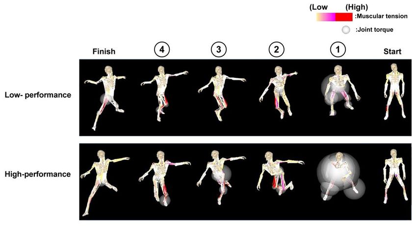
Figure 5 shows video images that reflect the estimated muscle tensions and joint torques at the onset of
motion to the forehand side in representative subjects (high-performance: a player ranked in the top 20 in
Japan, low-performance: a player with a 7-year history of playing badminton). The joint torques of the whole
body at the onset of motion were larger in the high-performance than low-performance player. However, no
marked difference was observed in the action pattern of estimated muscle tension.
Figure 5. Images of muscular tension and joint torque.
8 | 2021 | ISSUE - | VOLUME -- © 2021 University of AlicanteMasu, et al. / Electromyogram reaction time at the onset of motion in badminton players JOURNAL OF HUMAN SPORT & EXERCISE
DISCUSSION
To quickly react to the flash of light and start moving, sensory and perceptive information processing is
important. According to a report that evaluated the reaction time (RT), which reflects the steps of information
processing in the brain from presentation of a stimulus to the onset of reaction, RT after presentation of a
stimulus was suggested to be significantly shorter in soccer players than in average students (Ando et al.,
2001). However, since RT is the time of output of motion/reaction processing, it is difficult to evaluate
information processing related to decision-making about what action should be taken. Therefore, the
electromyogram reaction time (EMG-RT), which is RT measured by EMG, is used. The latency from
stimulation to the onset of muscle contraction in EMG-RT (pre-motor time (PMT)) is the time from
presentation of a stimulus to the appearance of motion/reaction processing and serves as an index for the
estimation of the degree of development of the brain and nervous system. In the present study, the action
time was shorter, and PMT of femoral muscle activity was significantly shorter, in the high-performance than
low-performance group in both the 1-direction and 2-direction tasks. EMG-RT has been suggested to be
affected by the quality and quantity of professional competitive training (Nishihara et al., 1991). Therefore,
the shorter PMT in the high-performance group is considered to be a result of differences in the period and
environment of training in the past.
Directions from the motor area are conducted to the target muscles of the leg via the spinal nerves, locomotor
neurons of the spinal cord, and motor nerves. These processes of conduction of electric signals are
performed through neurons and can be achieved more quickly as electric signals flow more rapidly from the
brain to the muscle. The time from a flash of light to touching of the net is affected by the reaction at the onset
of motion (EMG-RT) and the speed of movement. PMT is an index that reflects the motion/reaction
processing of instantaneously moving the legs, and the muscle power output of the legs is important as a
factor involved in the speed of movement. The muscle power output is related to the number and
synchronization rate of the mobilized motor units (Halliday et al., 1999). In both the 1-direction and 2-direction
tasks, RIE of the swing-out and push-out legs showed significant differences, and they were higher in the
high-performance than low-performance group. Moreover, in the action pattern of the high-performance
group, the joint torques of the whole body at the onset of action were higher compared with the low-
performance group. These results suggest that the high-performance players can move more quickly by
instantaneously synchronizing motor units of the rectus femoris muscle at the onset of action and perform
the action with larger joint torques.
A method for the assessment of information processing related to decision-making is event-related potentials
(ERPs). Badminton players who viewed videos of plays showed a larger amplitude of P300 of ERPs than
non-badminton players (Jin et al., 2011). Liu et al. (2017) performed an experiment in which, after having
subjects with no experience in badminton practice badminton for 12 weeks, they were asked to anticipate
the trajectory of the shuttlecock by viewing videos. ERPs measured during this task showed large P300
components, suggesting improvement in the anticipation ability, in those who practiced badminton compared
with the controls. P300 reflects components including elements of context updating (Picton, 1992). These
observations suggest that badminton, which requires repetition of selection of plays and motion/reaction in a
limited time, induces the development of the neurological system involved in information processing in the
brain. In addition, if presented stimuli can be assessed quickly, PMT, which indicates the output of motor
command, is also considered to be shortened. The shorter PMT in high-performance than low-performance
players in the present study suggests a difference in the development of the neurological system involved in
information processing in the brain. However, as PMT showed no significant difference between the 1-
VOLUME -- | ISSUE - | 2021 | 9Masu, et al. / Electromyogram reaction time at the onset of motion in badminton players JOURNAL OF HUMAN SPORT & EXERCISE
direction and 2-direction tasks and was delayed similarly, no difference is considered to develop in the
process of judgment of the direction of movement.
CONCLUSIONS
In this study, the electromyogram reaction time of the femoral muscles at the onset of motion was evaluated
in college badminton players at different performance levels. In both 1-direction and 2-direction tasks, the
action time was shorter, and PMT of femoral muscle activity was significantly shorter, in the high-performance
than low-performance group. In both tasks, significant differences were observed in the RIE of the swing-out
and push-out legs, and the values were higher in the high-performance than low-performance group.
From these results, high-performance badminton players are considered to be able to move quickly by
synchronizing motor units of the rectus femoris muscle at the onset of motion and perform actions by exerting
large joint torques.
AUTHOR CONTRIBUTIONS
Yujiro Masu: Research conception or design, data collection, analysis and interpretation of data, writing a
paper. Atsuya Otsuka: Supervision (writing a paper).
SUPPORTING AGENCIES
This work was supported by a Research Grant of the Health Science University.
DISCLOSURE STATEMENT
No potential conflict of interest was reported by the authors.
REFERENCES
Abernethy, B. (1990). Expertise, visual search, and information pick-up in squash. Perception, 19(1), 63-
77. https://doi.org/10.1068/p190063
Abernethy, B., & Zawi, K. (2007). Pickup of essential kinematics underpins expert perception of
movement patterns. J Mot Behav, 39(5), 353-367. https://doi.org/10.3200/JMBR.39.5.353-368
Ando. S., Kida. N., & Oda. S. (2001). Central and peripheral visual reaction time of soccer players and
nonathletes. Percept Mot Skills, 92(3), 786-794. https://doi.org/10.2466/pms.2001.92.3.786
Farrow, D., & Abernethy, B. (2002). Can anticipatory skills be learned through implicit video-based
perceptual training? J Sports Sci, 20(6), 471-485. https://doi.org/10.1080/02640410252925143
Halliday. D.M., Conway, B.A., Farmer. S.F., & Rosenberg. J.R. (1999). Load-independent contributions
from motor-unit synchronization to human physiological tremor. J Neurophysiol, 82(2), 664-675.
https://doi.org/10.1152/jn.1999.82.2.664
Hülsdünker, T., Strüder, H.K., & Mierau, A. (2016). Neural correlates of expert visuomotor performance
in badminton players. Med Sci Sports Exerc, 48(11), 2125-2134.
https://doi.org/10.1249/MSS.0000000000001010
Jackson, R.C., & Mogan, P. (2007). Advance visual information, awareness, and anticipation skill. J Mot
Behav, 39(5), 341-351. https://doi.org/10.3200/JMBR.39.5.341-352
10 | 2021 | ISSUE - | VOLUME -- © 2021 University of AlicanteMasu, et al. / Electromyogram reaction time at the onset of motion in badminton players JOURNAL OF HUMAN SPORT & EXERCISE
Jin, H., Xu, G., Zhang, J.X., Gao, H., Ye, Z., Wang, P., Lin, H., Mo, L., & Lin, C.D. (2011). Event-related
potential effects of superior action anticipation in professional badminton players. Neurosci Lett, 492,
139-144. https://doi.org/10.1016/j.neulet.2011.01.074
Kamimura. T., Yoshioka. K., Ito. S., & Kusakabe. T. (2009). Increased rate of force development of elbow
flexors by antagonist conditioning contraction. Human Movement Science, 28(4), 407-414.
https://doi.org/10.1016/j.humov.2009.02.005
Kanda, Y. (2013). Investigation of the freely available easy-to-use software 'EZR' for medical statistics.
Bone Marrow Transplantation, 48(3), 452-458. https://doi.org/10.1038/bmt.2012.244
Kellman, P.J., & Garrigan, P. (2009). Perceptual learning and human expertise. Phys Life Rev, 6(2), 53-
84. https://doi.org/10.1016/j.plrev.2008.12.001
Liu. T., Shao. M., Yin. D., Li. Y., Yang. N., Yin. R., Leng. Y., Jin. H., & Hong. H. (2017). The effect of
badminton training on the ability of same-domain action anticipation for adult novices: Evidence from
behavior and ERPs. Neurosci Lett, 660: 6-11. https://doi.org/10.1016/j.neulet.2017.08.038
Masu, Y., & Muramatsu, K. (2015). Soleus H-reflex modulation during receive stance in badminton
players in the receive stance. J Phys Ther Sci, 27(1), 123-125. https://doi.org/10.1589/jpts.27.123
Masu, Y., & Nagai, M. (2016). Characteristics of lower limb muscle activity during upper limb elevation in
badminton players. J Phys Ther Sci, 28(9), 2510-2514. https://doi.org/10.1589/jpts.28.2510
Masu, Y., Udaka, R., & Muramatsu, K. (2019). Alteration of F waves by motor imagery with and without
hitting in badminton. Int J Phys Ther Rehab, 5, 158. https://doi.org/10.15344/2455-7498/2019/158
Nakazawa, K., Miyoshi, T., Sekiguchi, H., Nozaki, D., Akai, M., & Yano, H. (2004). Effects of loading and
unloading of lower limb joints on the soleus H-reflex in standing humans. Clin Neurophysiol, 115(6),
1296-1304. https://doi.org/10.1016/j.clinph.2004.01.016
Nielsen, J., Crone, C., & Hultborn, H. (1993). H-reflexes are smaller in dancers from the Royal Danish
Ballet than in well-trained athletes. Eur J Appl Physiol Occup Physiol, 66(2), 116-121.
https://doi.org/10.1007 / BF01427051
Nishihira. Y., Araki. H., & Ishihara. A. (1991). Attenuation of somatosensory evoked potentials
immediately following rapid reaction movement. Electromyogr Clin Neurophysiol, 31(1), 15-20.
Phadke, C.P., Klimstra, M., Zehr, E.P., Thompson, F.J., & Behrman, A. L. (2010). Soleus H-reflex
modulation during stance phase of walking with altered arm swing patterns. Motor Control, 14(1),
116-125. https://doi.org/10.1123/mcj.14.1.116
Picton. T. W. (1992). The P300 wave of the human event-related potential. J Clin Neurophysiol, 9(4),
456-479. https://doi.org/10.1097/00004691-199210000-00002
Stroeve. S. (1999). Impedance characteristics of a neuromusculoskeletal model of the human arm I.
Posture control. Biol Cybern, 81(5-6), 475-494. https://doi.org/10.1007/s004220050577
This work is licensed under a Attribution-NonCommercial-NoDerivatives 4.0 International (CC BY-NC-ND 4.0).
VOLUME -- | ISSUE - | 2021 | 11You can also read