HOW THE CORONAVIRUS INFECTS OUR CELLS - Nature

Page created by Walter Wagner
 
CONTINUE READING
HOW THE CORONAVIRUS INFECTS OUR CELLS - Nature
Feature

                                                                        JANET IWASA, UNIV. UTAH

A computer simulation of the structure of the coronavirus SARS-CoV-2.

HOW THE CORONAVIRUS
INFECTS OUR CELLS
Scientists are unpicking SARS-CoV-2’s life cycle. By Megan Scudellari

640 | Nature | Vol 595 | 29 July 2021
                                                 ©
                                                 2
                                                 0
                                                 2
                                                 1
                                                 S
                                                 p
                                                 r
                                                 i
                                                 n
                                                 g
                                                 e
                                                 r
                                                 N
                                                 a
                                                 t
                                                 u
                                                 r
                                                 e
                                                 L
                                                 i
                                                 m
                                                 i
                                                 t
                                                 e
                                                 d
                                                 .
                                                 A
                                                 l
                                                 l
                                                 r
                                                 i
                                                 g
                                                 h
                                                 t
                                                 s
                                                 r
                                                 e
                                                 s
                                                 e
                                                 r
                                                 v
                                                 e
                                                 d
                                                 .
HOW THE CORONAVIRUS INFECTS OUR CELLS - Nature
T
                                                                         he coronavirus sports a luxurious         tools that have enabled the virus to spread so       2–4 times more strongly4, because several
                                                                         sugar coat. “It’s striking,” thought      quickly and claim millions of lives. “That’s why     changes in the RBD stabilize its virus-binding
                                                                         Rommie Amaro, staring at her com-         it’s so difficult to control,” says Wendy Barclay,   hotspots5.
                                                                         puter simulation of one of the trade-     a virologist at Imperial College London.                Worrying variants of SARS-CoV-2 tend to
                                                                         mark spike proteins of SARS-CoV-2,                                                             have mutations in the S1 subunit of the spike
                                                                         which stick out from the virus’s          Barbed and ready                                     protein, which hosts the RBDs and is responsi-
                                                                         surface. It was swathed in sugar          It starts with the spikes. Each SARS-CoV-2           ble for binding to the ACE2 receptor. (A second
                                                                         molecules, known as glycans.              virion (virus particle) has an outer surface         spike subunit, S2, prompts viral fusion with the
                                                                 “When you see it with all the glycans, it’s       peppered with 24–40 haphazardly arranged             host cell’s membrane.)
                                                              almost unrecognizable,” says Amaro, a compu-         spike proteins that are its key to fusing with          The Alpha variant, for example, includes
                                                              tational biophysical chemist at the University       human cells2. For other types of virus, such         ten changes in the spike-protein sequence,
                                                              of California, San Diego.                            as influenza, external fusion proteins are           which result in RBDs being more likely to stay
                                                                 Many viruses have glycans covering their          relatively rigid. SARS-CoV-2 spikes, however,        in the ‘up’ position6. “It is helping the virus
                                                              outer proteins, camouflaging them from the           are wildly flexible and hinge at three points,       along by making it easier to enter into cells,”
                                                              human immune system like a wolf in sheep’s           according to work published in August 2020           says Priyamvada Acharya, a structural biolo-
                                                              clothing. But last year, Amaro’s laboratory          by biochemist Martin Beck at the Max Planck          gist at the Duke Human Vaccine Institute in
                                                              group and collaborators created the most             Institute of Biophysics in Frankfurt, Germany,       Durham, North Carolina, who is studying the
                                                              detailed visualization yet of this coat, based       and his colleagues3.                                 spike mutations.
                                                              on structural and genetic data and rendered             That allows the spikes to flop around, sway          The Delta variant, which is now spreading
                                                              atom-by-atom by a supercomputer. On                  and rotate, which could make it easier for them      around the world, hosts multiple mutations
                                                              22 March 2020, she posted the simulation to          to scan the cell surface and for multiple spikes     in the S1 subunit, including three in the RBD
                                                              Twitter. Within an hour, one researcher asked        to bind to a human cell. There are no similar        that seem to improve the RBD’s ability to bind
                                                              in a comment: what was the naked, uncoated           experimental data for other coronaviruses,           to ACE2 and evade the immune system7.
                                                              loop sticking out of the top of the protein?         but because spike-protein sequences are
                                                                 Amaro had no idea. But ten minutes later,         highly evolutionarily conserved, it is fair to       Restricted entry
                                                              structural biologist Jason McLellan at the           assume the trait is shared, says Beck.               Once the viral spikes bind to ACE2, other
                                                              University of Texas at Austin chimed in: the            Early in the pandemic, researchers con-           proteins on the host cell’s surface initiate a
                                                              uncoated loop was a receptor binding domain          firmed that the RBDs of SARS-CoV-2 spike             process that leads to the merging of viral and
                                                              (RBD), one of three sections of the spike that       proteins attach to a familiar protein called the     cell membranes.
                                                              bind to receptors on human cells (see ‘A             ACE2 receptor, which adorns the outside of              The virus that causes SARS, SARS-CoV, uses
                                                              hidden spike’).                                      most human throat and lung cells. This recep-        either of two host protease enzymes to break
                                                                 In Amaro’s simulation, when the RBD               tor is also the docking point for SARS-CoV, the      in: TMPRSS2 (pronounced ‘tempress two’) or
                                                              lifted up above the glycan cloud, two glycans        virus that causes severe acute respiratory           cathepsin L. TMPRSS2 is the faster route in,
                                                              swooped in to lock it into place, like a kickstand   syndrome (SARS). But compared with SARS-             but SARS-CoV often enters instead through
                                                              on a bicycle. When Amaro mutated the glycans         CoV, SARS-CoV-2 binds to ACE2 an estimated           an endosome — a lipid-surrounded bubble —
                                                              in the computer model, the RBD collapsed.                                                                 which relies on cathepsin L. When virions enter
                                                              McLellan’s team built a way to try the same
                                                              experiment in the lab, and by June 2020, the
                                                                                                                   A HIDDEN SPIKE                                       cells by this route, however, antiviral proteins
                                                                                                                                                                        can trap them.
                                                                                                                   The spike protein of SARS-CoV-2 is coated in
                                                              collaborators had reported that mutating the         sugar molecules, or glycans, which disguise it          SARS-CoV-2 differs from SARS-CoV because
                                                              two glycans reduced the ability of the spike         from the immune system. It can hinge at three        it efficiently uses TMPRSS2, an enzyme found
                                                                                                                   points on the stalk, giving it flexibility.
                                                              protein to bind to a human cell receptor1 — a                                                             in high amounts on the outside of respiratory
                                                              role that no one has previously recognized in                          Receptor-binding
                                                                                                                                                                        cells. First, TMPRSS2 cuts a site on the spike’s
                                                              coronaviruses, McLellan says. It’s possible that                       domain                             S2 subunit8. That cut exposes a run of hydro-
                                                              snipping out those two sugars could reduce                                                                phobic amino acids that rapidly buries itself in
                                                              the virus’s infectivity, says Amaro, although                                                Glycan       the closest membrane — that of the host cell.
                                                              researchers don’t yet have a way to do this.                                                              Next, the extended spike folds back onto itself,
                                                                 Since the start of the COVID-19 pandemic,             S1                                               like a zipper, forcing the viral and cell mem-
                                                              scientists have been developing a detailed           subunit                                              branes to fuse (see ‘Viral entry up close’).
                                                              understanding of how SARS-CoV-2 infects                                                                      The virus then ejects its genome directly
                                                              cells. By picking apart the infection process,                                                            into the cell. By invading in this spring-
                                                              they hope to find better ways to interrupt it                                                             loaded manner, SARS-CoV-2 infects faster
CALIFORNIA, SAN DIEGO (REF. 1); GRAPHIC: NIK SPENCER/NATURE

                                                              through improved treatments and vaccines,                                                                 than SARS-CoV and avoids being trapped in
SOURCE: STRUCTURAL IMAGE FROM LORENZO CASALINO, UNIV.

                                                              and learn why the latest strains, such as the                                                             endosomes, according to work published in
                                                              Delta variant, are more transmissible.                                                                    April by Barclay and her colleagues at Imperial
                                                                 What has emerged from 19 months of work,              S2                            Hip                College London9.
                                                              backed by decades of coronavirus research, is        subunit                                                 The virus’s speedy entry using TMPRSS2
                                                              a blow-by-blow account of how SARS-CoV-2                                               Knee               explains why the malaria drug chloroquine
                                                              invades human cells (see ‘Life cycle of the pan-                  Stalk                                   didn’t work in clinical trials as a COVID-19 treat-
                                                              demic coronavirus’). Scientists have discov-                                                              ment, despite early promising studies in the
                                                              ered key adaptations that help the virus to grab                                                          lab10. Those turned out to have used cells that
                                                                                                                                                     Ankle
                                                              on to human cells with surprising strength and                                                            rely exclusively on cathepsins for endosomal
                                                              then hide itself once inside. Later, as it leaves                                                         entry. “When the virus transmits and replicates
                                                              cells, SARS-CoV-2 executes a crucial process-                                                             in the human airway, it doesn’t use endosomes,
                                                              ing step to prepare its particles for infecting                                                           so chloroquine, which is an endosomal disrupt-
                                                              even more human cells. These are some of the                                                              ing drug, is not effective in real life,” says Barclay.

                                                                                                                                                                                 Nature | Vol 595 | 29 July 2021 | 641
                                                                                                                     ©
                                                                                                                     2
                                                                                                                     0
                                                                                                                     2
                                                                                                                     1
                                                                                                                     S
                                                                                                                     p
                                                                                                                     r
                                                                                                                     i
                                                                                                                     n
                                                                                                                     g
                                                                                                                     e
                                                                                                                     r
                                                                                                                     N
                                                                                                                     a
                                                                                                                     t
                                                                                                                     u
                                                                                                                     r
                                                                                                                     e
                                                                                                                     L
                                                                                                                     i
                                                                                                                     m
                                                                                                                     i
                                                                                                                     t
                                                                                                                     e
                                                                                                                     d
                                                                                                                     .
                                                                                                                     A
                                                                                                                     l
                                                                                                                     l
                                                                                                                     r
                                                                                                                     i
                                                                                                                     g
                                                                                                                     h
                                                                                                                     t
                                                                                                                     s
                                                                                                                     r
                                                                                                                     e
                                                                                                                     s
                                                                                                                     e
                                                                                                                     r
                                                                                                                     v
                                                                                                                     e
                                                                                                                     d
                                                                                                                     .
HOW THE CORONAVIRUS INFECTS OUR CELLS - Nature
Feature
LIFE CYCLE OF THE PANDEMIC CORONAVIRUS                                                                                      The discovery also points to protease
                                                                                                                         inhibitors as a promising therapeutic option to
                                                                                                                         prevent a virus from using TMPRSS2, cathep-
A simplified account of how SARS-CoV-2 enters and exits cells.
                                                                                                                         sin L or other proteases to enter host cells.
                                                                                                                         One TMPRSS2 inhibitor, camostat mesylate,
              Stage 1 : Viral entry
                                                                                                                         which is approved in Japan to treat pancrea-
              The virus’s spike protein binds to a                                                                       titis, blocked viral entry into lung cells8, but
              receptor on the host cell called ACE2.                    Nucleocapsid
                                                                                                                         the drug did not improve patients’ outcomes
              Then, the host molecule TMPRSS2
              cleaves the spike protein, exposing                                  Spike protein                         in an initial clinical trial11.
              parts that fuse the viral membrane           RNA                                                              “From my perspective, we should have such
              with that of the host.                                               M protein
                                                                                                                         protease inhibitors as broad antivirals availa-
                                                                                                                         ble to fight new disease outbreaks and prevent
                                                                                                                         future pandemics at the very beginning,” says
              TMPRSS2
                               ACE2
                                                                                                                         Stefan Pöhlmann, director of the Infection
                                                                                                                         Biology Unit at the German Primate Center
 Stage 2: Inside the cell                                                                                                in Göttingen, who has led research on ACE2
 Viral RNA is translated into                                                                                            binding and the TMPRSS2 pathway.
 non-structural proteins (NSPs) that                                TMPRSS2 cuts
 quickly suppress the translation of                                the spike protein
 host messenger RNAs in favour of                                                                                        Deadly competition
 those belonging to the virus.                                                                                           The next steps of infection are murkier. “There
                                                                                                                         are a lot more black boxes once you are inside
 Endoplasmic                                                                                                             the cell,” says chemist Janet Iwasa at the Univer-
 reticulum (ER)                     Viral proteins                                                                       sity of Utah in Salt Lake City, who is developing
                                    (NSPs)
                                                                                                                         an annotated animation of the viral life cycle.
                                                                             The spikes unravel and pull                 “There’s more uncertainty, and competing
     NSPs                                                                    the membrane of the virus                   hypotheses.”
                                                       Viral RNA             and host cell together
                           Ribosome                                                                                         After the virus shoots its RNA genome into
                                                                                                                         the cell, ribosomes in the cytoplasm translate
        ER remodelling
                                                                                                                         two sections of viral RNA into long strings of
                                                                                                                         amino acids, which are then snipped into
                                                                                                                         16 proteins, including many involved in RNA
                      DMVs                  Stage 3: Remodelling the cell
                                            The virus transforms the cell’s ER —                                         synthesis. Later, more RNAs are generated
                                            an internal membrane network —                                               that code for a total of 26 known viral proteins,
                                            into bubble-like structures called                                           including structural ones used to make new
                                            double-membrane vesicles (DMVs).
                                            These might provide a safe haven                                             virus particles, such as the spike, and other
                                            for more viral RNA to be replicated                                          accessory proteins. In this way, the virus
                                            and translated.
                                                                                                                         begins churning out copies of its own mes-
                                                                                                                         senger RNA. But it needs the cell’s machinery
                                                                                                                         to translate those mRNAs into proteins.
                                                                                                                            Coronaviruses take over that machinery in
                                                            Furin cut                                                    many ways. Virologist Noam Stern-Ginossar
                                             Golgi
                                             apparatus                                                                   and her team at the Weizmann Institute of Sci-
                                                                                                                         ence in Rehovot, Israel, zoomed in on three
                                                                                        Stage 5: The last slice
                                                                                        A host enzyme named furin
                                                                                                                         mechanisms by which SARS-CoV-2 suppresses
                                                                                        makes a crucial cut at a site    the translation of host mRNA in favour of its
                                                                                        of five amino acids on the       own. None are exclusive to this virus, but the
                                                                                        spike protein. This prepares
                                                                                        the virus to strike another      combination, speed and magnitude of the
                                                                                        cell. Variants have a higher     effects seem unique, says Stern-Ginossar.
                                                                                                                                                                              SOURCE: HUI (ANN) LIU, UNIV. UTAH (HTTPS://ANIMATIONLAB.UTAH.EDU/COVA);
                                                                                        proportion of snipped spike         First, the virus eliminates the competition:
                                                                                        proteins, helping them to
                                                                                        infect cells more efficiently.   viral protein Nsp1, one of the first proteins
                                                                                                                         translated when the virus arrives, recruits host
                                                                                                                         proteins to systematically chop up all cellu-
                  Stage 4: Exit                                                                                          lar mRNAs that don’t have a viral tag. When
                  Once the newly made molecules                                                                          Stern-Ginossar’s team put that same tag on
                  assemble into a complete virus
                  particle, this leaves the cell through                                                                 the end of a host mRNA, the mRNA was not
                  an organelle called the Golgi                                                                          chopped up12.
                  apparatus, or perhaps through
                  lysosomes, which are cellular
                                                                                                                            Second, infection reduces overall protein
                  rubbish bins.                                                                                          translation in the cell by 70%. Nsp1 is again the
                                                                                                                                                                              GRAPHIC: NIK SPENCER/NATURE

                                                                                                                         main culprit, this time physically blocking the
                                                                                                                         entry channel of ribosomes so mRNA can’t get
                                                                                                                         inside, according to work from two research
                                                                                                                         teams 13,14. The little translation capacity
                                                                                                                         that remains is dedicated to viral RNAs, says
                                                                                                                         Stern-Ginossar.
                                                                                                                            Finally, the virus shuts down the cell’s alarm

642 | Nature | Vol 595 | 29 July 2021
                                                              ©
                                                              2
                                                              0
                                                              2
                                                              1
                                                              S
                                                              p
                                                              r
                                                              i
                                                              n
                                                              g
                                                              e
                                                              r
                                                              N
                                                              a
                                                              t
                                                              u
                                                              r
                                                              e
                                                              L
                                                              i
                                                              m
                                                              i
                                                              t
                                                              e
                                                              d
                                                              .
                                                              A
                                                              l
                                                              l
                                                              r
                                                              i
                                                              g
                                                              h
                                                              t
                                                              s
                                                              r
                                                              e
                                                              s
                                                              e
                                                              r
                                                              v
                                                              e
                                                              d
                                                              .
system. This happens in numerous ways, but
                                                                  Stern-Ginossar’s team identified one clear
                                                                                                                       VIRAL ENTRY UP CLOSE
                                                                                                                       Virus and host-cell membranes fuse after the TMPRSS2 enzyme cuts a SARS-CoV-2 spike protein.
                                                                  mechanism for SARS-CoV-2: the virus prevents         This exposes hydrophobic amino acids in the spike that rapidly embed themselves into the
                                                                                                                       nearest membrane — that of the host cell.
                                                                  cellular mRNA from getting out of the nucleus,
                                                                  including instructions for proteins meant to
                                                                  alert the immune system to infection. A sec-                                 Virus
                                                                  ond team confirmed this finding, and again
SOURCE: JANET IWASA, UNIV. UTAH (HTTPS://ANIMATIONLAB.UTAH.EDU/

                                                                  pointed to Nsp1: the protein seems to jam up
                                                                  exit channels in the nucleus so nothing can               Spike
                                                                  escape15.                                                 protein
                                                                    Because gene transcripts can’t get out of the
                                                                  nucleus, the infected cells don’t release many
                                                                                                                       TMPRSS2     Cut
                                                                  interferons — these are signalling proteins that
                                                                  alert the immune system to the presence of a
COVA); GRAPHIC: NIK SPENCER/NATURE

                                                                  virus. SARS-Cov-2 is particularly efficient at
                                                                  shutting down this alarm system: compared                                                                                     Hydrophobic
                                                                  with other respiratory viruses, including                                                                                     amino acids
                                                                                                                                                                                                attach to nearest
                                                                  SARS-CoV and respiratory syncytial virus,                                                                                     membrane
                                                                                                                                                ACE2
                                                                  SARS-CoV-2 infection induces significantly
                                                                  lower levels of interferons16. And this June,
                                                                                                                                             Host cell
                                                                  researchers reported mutations in the Alpha
                                                                  variant that seem to enable it to subdue inter-
                                                                  feron production even more efficiently17.            A second team, led by researcher Qiang Sun at          newly made viral RNA21.
                                                                    “It’s clear that SARS-CoV-2 is a very fast         the Chinese Academy of Medical Sciences in                Most viruses that have an outer wrapping,
                                                                  virus that has a unique ability to prevent           Beijing, found that some COVID-19-infected             known as an envelope, form this feature by
                                                                  our immune system from recognizing and               cells even form syncytia with lymphocytes —            assembly directly at the edge of the cell,
                                                                  combating infection in the first stages,” says       one of the body’s own immune cells19. This is          co-opting some of the cell’s own plasma
                                                                  Stern-Ginossar. By the time the immune sys-          a known mechanism of immune evasion by                 membrane on their way out. But newly made
                                                                  tem does realize there is a virus, there is so       tumour cells, but not by viruses. It suggests          coronavirus proteins take a different path.
                                                                  much of it that immune-response proteins             that infected cells avoid immune detection                For years, evidence has suggested that
                                                                  sometimes flood the bloodstream at a faster          by simply grabbing on to and merging with              coronaviruses are transported out of the cell
                                                                  rate than normal — which can cause damage.           nearby immune scouts.                                  through the Golgi complex, an organelle that
                                                                  Doctors saw early in the pandemic that some             On the inside of the cell, even more change         works like a post office, packaging molecules
                                                                  people with COVID-19 who become very ill are         is occurring. Like other coronaviruses,                in membranes and sending them off to other
                                                                  harmed by an overactive immune response to           SARS-CoV-2 transforms the long, thin endo-             parts of the cell. There, the virus forms a lipid
                                                                  SARS-CoV-2, as well as by the virus itself. Some     plasmic reticulum (ER), a network of flat              envelope from the Golgi complex’s membrane;
                                                                  proven treatments work by dampening down             membranes involved in protein synthesis and            newly formed virions are then carried inside
                                                                  this immune response.                                transport, into double-membrane spheres,               Golgi vesicles to the cell surface, where they
                                                                                                                                                                              are spat out of the cell, says virologist and cell
                                                                  Renovation station
                                                                                                                       “There are a lot more                                  biologist Carolyn Machamer at Johns Hopkins
                                                                  Once the virus has taken over host transla-                                                                 University in Baltimore, Maryland, who has
                                                                  tion, it starts a home makeover, extensively
                                                                                                                       black boxes once you                                   studied coronaviruses for 30 years.
                                                                  remodelling the interior and exterior of the         are inside the cell.”                                     But in December, cell biologist Nihal
                                                                  cell to its needs.                                                                                          Altan-Bonnet at the US National Heart, Lung,
                                                                     First, some of the newly made viral spike                                                                and Blood Institute in Bethesda, Maryland,
                                                                  proteins travel to the surface of the cell and       as if the ER were blowing bubbles. These dou-          and her colleagues reported that they had
                                                                  poke out of the host-cell membrane. There,           ble-membrane vesicles (DMVs) might provide             detected coronaviruses leaving the cell
                                                                  they activate a host calcium-ion channel,            a safe place for viral RNA to be replicated and        through lysosomes — cellular rubbish bins
                                                                  which expels a fatty coating onto the outside        translated, shielding it from innate immune            full of enzymes that break down cell parts22.
                                                                  of the cell — the same coating found on cells        sensors in the cell, but that hypothesis is still      Blocking the Golgi-based secretory pathway
                                                                  that naturally fuse together, such as muscle         being investigated.                                    didn’t seem to affect the amount of infectious
                                                                  cells. At this point, the infected cell fuses to        Proteins involved in making DMVs could be           virus being released, says Altan-Bonnet. Her
                                                                  neighbouring cells expressing ACE2, devel-           good drug targets, because they seem to be             team’s evidence22 suggests that viral proteins
                                                                  oping into massive individual respiratory cells      necessary for viral replication. For instance,         form an envelope by budding into the ER, then
                                                                  filled with up to 20 nuclei.                         a host protein, TMEM41B, is needed to mobi-            take over lysosomes to get out of the cell. The
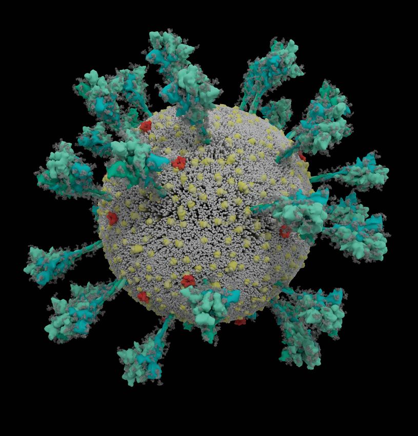
                                                                     These fused structures, called syncytia,          lize cholesterol and other lipids to expand            researchers are currently testing inhibitors
                                                                  are induced by viral infections such as HIV          the ER membranes so that all the virus parts           that block the lysosomal exit process as poten-
                                                                  and herpes simplex virus, but not by the             will fit inside20. “When you take TMEM41B              tial antiviral candidates.
                                                                  SARS virus, says molecular biologist Mauro           out, it has a major impact on infection,” says            Leaving a cell through either the Golgi or
                                                                  Giacca at King’s College London, who led the         Vineet Menachery, a coronavirus researcher             lysosomes is slow and inefficient compared
                                                                  team that published the finding in April18. He       at the University of Texas Medical Branch in           with budding out of a plasma membrane, so
                                                                  hypothesizes that forming syncytia allows            Galveston, who was involved in the research.           scientists don’t know why SARS-CoV-2 does it.
                                                                  infected cells to thrive for long periods of time,   The coronavirus transmembrane protein Nsp3             Machamer suspects that the lipid composition
                                                                  churning out more and more virions. “This is         could also be a target: it creates a crown-like        of a Golgi- or lysosome-derived envelope is
                                                                  not a hit-and-run virus,” he says. “It persists.”    pore in the walls of the DMVs to shuttle out           somehow more beneficial to the virus than one

                                                                                                                                                                                      Nature | Vol 595 | 29 July 2021 | 643
                                                                                                                         ©
                                                                                                                         2
                                                                                                                         0
                                                                                                                         2
                                                                                                                         1
                                                                                                                         S
                                                                                                                         p
                                                                                                                         r
                                                                                                                         i
                                                                                                                         n
                                                                                                                         g
                                                                                                                         e
                                                                                                                         r
                                                                                                                         N
                                                                                                                         a
                                                                                                                         t
                                                                                                                         u
                                                                                                                         r
                                                                                                                         e
                                                                                                                         L
                                                                                                                         i
                                                                                                                         m
                                                                                                                         i
                                                                                                                         t
                                                                                                                         e
                                                                                                                         d
                                                                                                                         .
                                                                                                                         A
                                                                                                                         l
                                                                                                                         l
                                                                                                                         r
                                                                                                                         i
                                                                                                                         g
                                                                                                                         h
                                                                                                                         t
                                                                                                                         s
                                                                                                                         r
                                                                                                                         e
                                                                                                                         s
                                                                                                                         e
                                                                                                                         r
                                                                                                                         v
                                                                                                                         e
                                                                                                                         d
                                                                                                                         .
Feature
                                                                                                             More furin cuts mean more spike proteins
                                                                                                          primed to enter human cells. In SARS-CoV, less
                                                                                                          than 10% of spike proteins are primed, says
                                                                                                          Menachery, whose lab group has been quan-
                                                                                                          tifying the primed spike proteins but is yet to
                                                                                                          publish this work. In SARS-CoV-2, that percent-
                                                                                                          age rises to 50%. In the Alpha variant, it’s more
                                                                                                          than 50%. In the highly transmissible Delta var-
                                                                                                          iant, the group has found, greater than 75% of
                                                                                                          spikes are primed to infect a human cell.

                                                                                                          Known unknowns
                                                                                                          The scientific community is still scratching the
                                                                                                          surface of its understanding of SARS-CoV-2.
                                                                                                          Key unknowns include the number of ACE2
                                                                                                          receptors needed to bind to each spike pro-
                                                                                                          tein; when exactly the S2 site is cleaved by
                                                                                                          TMPRSS2; and the number of spikes needed
                                                                                                          for virus–cell membrane fusion, says McLellan
                                                                                                          — and that’s just for entry. In April 2020, a team
                                                                                                          at the University of California, San Francisco,
                                                                                                          identified at least 332 interactions between

                                                                                                                                                                             MAURO GIACCA
                                                                                                          SARS-CoV-2 and human proteins25.
                                                                                                             It is not easy to keep pace with the quickly
                                                                                                          mutating virus. Most mutations so far are asso-
Fused cell structures (syncytia) seen in cells expressing the SARS-CoV-2 spike protein                    ciated with how effectively the virus spreads,
(green). Nuclei are in blue and the cell skeleton is in red.                                              not with how much the virus damages the host,
                                                                                                          experts agree. This month, a study reported
from the plasma membrane. “If we understood            those without it24.                                that the Delta variant grew more rapidly and at
this part a little bit better, there would be great       Furin is suspected to cut the site at some      higher levels inside people’s lungs and throats
opportunities for novel antiviral therapeutics,”       point during virion assembly, or just before       than did earlier versions of the virus26.
she says.                                              release. The timing might explain why the virus       But it is not yet certain how Delta’s muta-
                                                       exits through the Golgi or lysosomes, says Tom     tions have supercharged the variant in this
Last slice                                             Gallagher, a virologist at Loyola University       way, says Stern-Ginossar. “This is something
On the way out of the cell, one more event             Chicago in Illinois. “The virus, once assembled,   many labs are trying to figure out.”
makes this virus into an infectious juggernaut:        moves into an organelle where it can be bathed
a quick snip at a site of five amino acids pre-        in the presence of the furin protease.”            Megan Scudellari is a science journalist in
pares the virus to strike its next target.                By snipping the bond between the S1 and S2      Boston, Massachusetts.
   Where other coronaviruses have a single             subunits, the furin cut loosens up virion spike
arginine amino acid at the junction of the S1          proteins so that during cell entry they respond
                                                                                                          1.  Casalino, L. et al. ACS Cent. Sci. 6, 1722–1734 (2020).
and S2 subunits of the spike, SARS-CoV-2 has a         to a second cut by TMPRSS2, which exposes
                                                                                                          2.  Ke, Z. et al. Nature 588, 498–502 (2020).
line of five amino acids: proline, arginine, argi-     the hydrophobic area that rapidly buries itself    3.  Turoňová, B. et al. Science 370, 203–208 (2020).
nine, alanine and arginine. “Because the site                                                             4.  Nguyen, H. L. et al. J. Phys. Chem. B 124, 7336–7347
                                                                                                              (2020).
was unusual, we focused on it, and it turned           “We would hypothesize that                         5. Shang, J. et al. Nature 581, 221–224 (2020).
out that, yes, the site is essential for invasion of
lung cells,” says Pöhlmann. In May 2020, he and
                                                       this is the virus getting even                     6. Gobeil, S. M.-C. et al. Science https://doi.org/10.1126/
                                                                                                              science.abi6226 (2021).
his colleagues reported that a host-cell protein       better at transmitting.”                           7. Khateeb, J., Li, Y. & Zhang, H. Crit. Care 25, 244 (2021).
                                                                                                          8. Hoffmann, M. et al. Cell 181, 271–280 (2020).
called furin recognizes and clips that string of                                                          9. Peacock, T. P. et al. Nature Microbiol. 6, 899–909 (2021).
amino acids — and the cut is “essential” for the                                                          10. Wang, M. et al. Cell Res. 30, 269–271 (2020).
virus to enter human lung cells efficiently23.         in a host-cell membrane, says Gallagher. If        11. Gunst, J. D. et al. EClinicalMedicine 35, 100894 (2021).
                                                                                                          12. Finkel, Y. et al. Nature 594, 240–245 (2021).
   It’s not the first time that researchers have       spikes are not pre-clipped by furin —and they      13. Schubert, K. et al. Nature Struct. Mol. Biol. 27, 959–966
identified a furin cleavage site on a virus;           aren’t always — they bypass TMPRSS2, and               (2020).
highly pathogenic avian influenza viruses also         enter through the slower endosomal path-           14. Thoms, M. et al. Science 369, 1249–1255 (2020).
                                                                                                          15. Zhang, K. et al. Sci. Adv. 7, eabe7386 (2021).
have it, says Barclay. When a colleague sent           way, if at all.                                    16. Blanco-Melo, D. et al. Cell 181, 1036–1045 (2020).
Barclay a strain of SARS-CoV-2 in culture that           Two coronavirus variants, Alpha and Delta,       17. Thorne, L. G. et al. Preprint at bioRxiv https://doi.
had spontaneously lost the furin cleavage site,        have altered furin cleavage sites. In the Alpha        org/10.1101/2021.06.06.446826 (2021).
                                                                                                          18. Braga, L. et al. Nature 594, 88–93 (2021).
her team found that ferrets infected with this         variant, the initial proline amino acid is         19. Zhang, Z. et al. Cell Death Differ. https://doi.org/10.1038/
strain shed viral particles in lower amounts           changed to a histidine (P681H) ; in the Delta          s41418-021-00782-3 (2021).
than did those infected with the pandemic              variant, it is changed to an arginine (P681R).     20. Trimarco, J. D. et al. PLoS Pathog. 17, e1009599 (2021).
                                                                                                          21. Wolff, G. et al. Science 369, 1395–1398 (2020).
strain, and did not transmit the infection to          Both changes make the sequence less acidic,        22. Ghosh, S. et al. Cell 183, 1520–1535 (2020).
nearby animals9. At the same time as Barclay’s         and the more basic the string of amino acids,      23. Hoffmann, M., Kleine-Weber, H. & Pöhlmann, S. Mol. Cell
team reported its results in a September 2020          the more effectively furin recognizes and              78, 779–784 (2020).
                                                                                                          24. Mykytyn, A. Z. et al. eLife 10, e64508 (2021).
preprint, a study in the Netherlands also found        cuts it, says Barclay. “We would hypothesize
                                                                                                          25. Gordon, D. E. et al. Nature 583, 459–468 (2020).
that coronavirus with an intact furin cleavage         that this is the virus getting even better at      26. Li, B. et al. Preprint at medRxiv https://doi.
site enters human airway cells faster than do          transmitting.”                                         org/10.1101/2021.07.07.21260122 (2021).

644 | Nature | Vol 595 | 29 July 2021
                                                         ©
                                                         2
                                                         0
                                                         2
                                                         1
                                                         S
                                                         p
                                                         r
                                                         i
                                                         n
                                                         g
                                                         e
                                                         r
                                                         N
                                                         a
                                                         t
                                                         u
                                                         r
                                                         e
                                                         L
                                                         i
                                                         m
                                                         i
                                                         t
                                                         e
                                                         d
                                                         .
                                                         A
                                                         l
                                                         l
                                                         r
                                                         i
                                                         g
                                                         h
                                                         t
                                                         s
                                                         r
                                                         e
                                                         s
                                                         e
                                                         r
                                                         v
                                                         e
                                                         d
                                                         .
You can also read