How innovative technology leads to a faster diagnosis and more targeted treatment of UTI
←
→
Page content transcription
If your browser does not render page correctly, please read the page content below

URINALYSIS WHITE PAPER | May 2020

 Urinary Tract Infection

How innovative technology leads to a faster
diagnosis and more targeted treatment of UTI
Introduction

Globally, urinary tract infections (UTI) belong to the most frequent    obstructions of the urinary tract, urinary retention, urinary calculi,
bacterial infections, affecting around 150 million people each year     pregnancy and immunosuppression [2].
[1]. This not only results in millions of medical consultations in
both inpatient and outpatient settings, but also in high healthcare     Since urinary tract infections can follow different symptomatic
expenditures and social costs [2].                                      courses or even be asymptomatic, proper diagnosis of UTI combines
                                                                        patient history, urinary symptoms and laboratory diagnostics. Lower
The pathogenic pathway can be either extraluminal by microbial          UTI manifests with alguria, pollakiuria, dysuria, acute suprapubic
contamination of the periurethral zone and subsequent colonisation      or abdominal pain, a general feeling of illness and occasionally
towards the bladder or intraluminal by colonisation of the urinary      haematuria, cloudy or foul-smelling urine. Upper UTI shows a more
tract via urinary catheters. Urinary tract infections are thus one      severe and systemic presentation, and in addition to the symptoms
of the major nosocomial infections [2].                                 of lower UTI include costovertebral angle tenderness, fever and
                                                                        chills. In addition, non-specific symptoms such as tiredness, fatigue,
Clinically, urinary tract infections are categorised as uncomplicated   chronic headache, persistent loss of appetite, nausea, vomiting,
or complicated, depending on the absence or presence of underlying      intermittent temperature increases and change of mental status
structural or functional abnormalities of the urinary tract, respec-    can indicate a urinary tract infection [2]. A diagnosis solely based
tively [3]. Further, urinary tract infections are differentiated into   on the patient’s history and present symptoms is still common in
lower UTI (cystitis, urethritis) or upper UTI (pyelonephritis) [4].     many countries but often inaccurate [5].

Besides the female gender, a recent urinary tract infection, sexual     Although uropathogenic Escherichia coli is the most common
activity, diabetes, obesity and a certain genetic susceptibility are    pathogen associated with both complicated and uncomplicated
common risk factors associated with lower urinary tract infections.     urinary tract infections, various microorganisms, including
Complicated urinary tract infections are related to renal diseases      Gram-negative and Gram-positive bacteria and various fungal
(e.g. chronic kidney disease, renal failure, renal transplantation),    species, can cause urinary tract infections (Fig. 1; [3]).
Urinary Tract Infection: How innovative technology leads to a faster diagnosis and more targeted treatment of UTI                                           2
Urinalysis white paper | May 2020

                                                                            the sensitivity of urine microscopy is highly reliable for samples
                                                                            with ≥ 105 CFU/mL, reported insensitivities for lower bacterial
                                                                            concentrations limit its clinical utility, especially for uncomplicated
                                                                            UTI in outpatient settings [8].

                                                                            Urine culture remains an important test in the context of UTI
                                                                            diagnostics, particularly for identifying the infectious microorganism.
                                                                            The common gold standard definition of bacteriuria is the presence
                                                                            of ≥ 105 CFU/mL, which was established for women with acute
       Uropathogenic E. coli     Enterococcus spp.          P. aeruginosa   pyelonephritis or asymptomatic UTI but was adapted for other patient
       K. pneumoniae             Group B Streptococcus      S. aureus       groups [8]. Since many UTI patients show bacteriuria with
       S. saprophyticus          P. mirabilis               Candida spp.
                                                                            ≤ 105 CFU/mL, many laboratories already apply lower colony counts
                                                                            as cut-off values to increase the sensitivity of urine culture.
Fig. 1 Epidemiology of complicated (left diagram) and uncomplicated
(right diagram) urinary tract infections. Adapted from [3].
                                                                            Positive urine cultures finally result in antibiotic susceptibility
                                                                            testing (antibiogram) to identify a suitable and specific antibiotic
                                                                            for the targeted treatment of a present microbial infection. The
Suspected urinary tract infections contribute to high laboratory            susceptibility testing by agar diffusion [9] is still the reliable gold
workloads, although in the end up to 80 % of the samples are ruled          standard, but indirect approaches including emerging technologies
out [6]. This causes the unnecessary and empirical treatment of             such as MALDI-TOF mass spectrometry and measuring bacterial
patients with broad-band antibiotics, promoting the rise of anti-           metabolites in the presence of antibiotics are under evaluation [10].
microbial resistance. Since only 17 % of all potential UTI patients
who are treated with antibiotics have been tested before by proper
                                                                            Detection of urinary particles by
urinalysis, re-prescription of antibiotics is often required [7].
                                                                            fluorescence flow cytometry

The classical diagnosis of UTI                                              The Sysmex UF-series uses fluorescence flow cytometry to detect
                                                                            cellular and acellular particles, including bacteria, yeast-like cells,
The macroscopic examination of a urine specimen is often the                red blood cells, white blood cells and other parameters in urine
first indicator for a suspected urinary tract infection, since ab-          and body fluid samples (Fig. 2).
normal colouration by macrohaematuria or pseudomonal UTI, and
foul-smelling urine or turbidity due to pyuria are known urinary
manifestations.                                                               Diagnostic parameters                    Research parameters
                                                                              Red Blood Cells                          Lysed RBC
The dipstick is the most frequently used screening test for the               Non-lysed RBC                            Small round cells
presence of urinary tract infections. The presence of nitrite as a            White Blood Cells                        Atypical cells
metabolic product derived from the reduction of urinary nitrate by            WBC Clumps                               Debris
certain nitrogenic species (e.g. Escherichia, Proteus, Klebsiella) is         Epithelial Cells                         Conductivity
an indicator of bacteriuria. However, many pathogens of the urinary              Squamous EC                           Osmolality
tract do not generate nitrate (e.g. Enterococcus, Gonococcus,                    Non-squamous EC
Staphylococcus, Pseudomonas), which means nitrite in this context                Transitional EC
                                                                                 Renal Tubular EC
is not a reliable parameter. Leucocyte esterase, protein and blood                                                     Body Fluid parameters
                                                                              Casts
are common parameters indicating inflammatory conditions.                                                              Red Blood Cells
                                                                                 Hyaline Casts
However, sensitivity and specificity are often relatively low, and                                                     White Blood Cells
                                                                                 Pathological Casts
a negative dipstick result is insufficient to rule out urinary tract                                                     MN#/%
                                                                              BACT
infections if classical symptoms are present [5].                                                                        PMN#/%
                                                                              X‘TAL
                                                                                                                       Epithelial Cells
                                                                              Yeast-like Cells
Microscopy of Gram-stained urine specimens is a common standard,                                                       Total Nucleated Cells
                                                                              Sperm
i.e. the microscopic investigation of urine sediments that have been                                                   BACT
                                                                              Mucus
airdried on a microscopic slide and stained with Gram stain. The
                                                                            Fig. 2 Overview of diagnostic, research and body fluid parameters provided by
main advantage of urine microscopy is the provided information              the UF-series analysers
on the infectious agent to initiate antimicrobial therapy. Although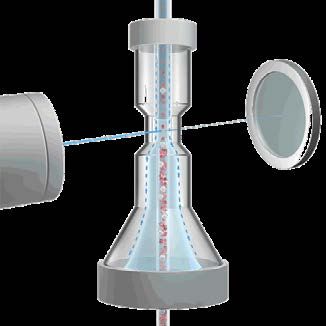
Urinary Tract Infection: How innovative technology leads to a faster diagnosis and more targeted treatment of UTI                                                      3
Urinalysis white paper | May 2020

For detecting urinary particles, two measurement channels are
available on the UF-series, the Core (CR) channel and the Surface
(SF) channel. While the SF channel detects particles that do not                                                              SSC
                                                                                             DSS                              Detector
include nucleic acid (RBC, crystals, etc.), the CR channel detects
                                                                                             Detector
nucleic acid-containing particles. Proper particle detection requires
staining of urinary particles using a diluting agent and a solution
for the fluorescence labelling of subcellular structures.
                                                                                      SFL
                                                                                      Detector
In the CR channel, the membranes of WBC (Fig. 3) and the cell
walls of bacteria are perforated by the diluent. These small per-
forations of the membranes allow the fluorescence dye to enter                                                                                           FSC
                                                                                                                                                         Detector
the cytoplasm and the nucleus and to intercalate into nucleic
acid molecules.

In the SF channel, membrane components of cellular particles                             Laser
such as RBC are stained by the fluorescence dye without affecting
                                                                                                                                      Flow Cell
the cellular integrity (Fig. 3).

The stained particles are then injected into the flow cell, where
hydrodynamic focusing ensures their separation to allow accurate
                                                                                  Fig. 5 Fluorescence flow cytometry on the UF-series. A laser beam is directed
particle counts (Fig. 4).                                                         at the flow cell, hitting all the particles passing through. Fluorescence light is
                                                                                  emitted from excited electrons of the fluorescence dyes and, depending on the
                                                                                  individual particle type, the oncoming laser light is characteristically diverted.
                                                                                  Photodetectors recognise individual particles, and based on the individual signal
        Native                    Perforation                  Detection          patterns, the signals are plotted in a scattergram.

                                                                                  Finally, the energy of a 488 nm laser beam excites electrons of
                                                                                  the fluorescence dye attached to the urinary particles, elevating
                                                                                  their energy level. Upon relaxation, photons are emitted and
                                                                                  detected by different photodetectors (Fig. 5). Depending on the
                                                                                  sub-structures of the different particles, the oncoming laser light
                                                                                  can be diverted and detected by different detectors, allowing
                                                                                  insight into the size of each cell (forward-scattered light; FSC), its
                                                                                  intracellular complexity (side-scattered light; SSC) and its nucleic
                                                                                  acid content (side-fluorescence light; SFL). Crystals are distinguished
Fig. 3 Particle-dependent reagent reaction for nucleic acid-containing cellular
particles in the CR channel (upper row) and for nucleic acid-free cellular and    from RBC by using a depolarisation filter (depolarised side-scattered
acellular urine particles (lower row)                                             light; DSS).

                                                                                  Improving screening for UTI

                                                                                  For bacteria, both quantitative and qualitative information is
                                                                                  provided in less than a minute. This includes a reliable bacteria
                                                                                  count and information on the Gram status.

                                                                                  In a representative study, the diagnostic performance of the
                                                                                  UF-series’ bacterial cell count has been identified as 0.973 (AUC).
                                                                                  Separated between male and female patients, the diagnostic
                                                                                  performance has been estimated as 0.988 for male and 0.959 for
                                                                                  female patients, respectively (Fig. 6; [11]).

Fig. 4 Hydrodynamic focusing of urine particles inside the flow cell of the
UF-series instruments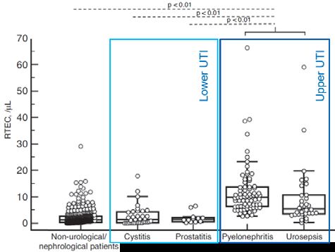
Urinary Tract Infection: How innovative technology leads to a faster diagnosis and more targeted treatment of UTI                                                   4
Urinalysis white paper | May 2020

The investigation of different cut-off values revealed a bacteria                       ‘Gram positive’           ‘Gram Pos/Neg’             ‘Gram negative’
count of ≥ 58 cells/µL as the most sensitive value for ruling out                           bacteria                  bacteria                   bacteria
urinary tract infections with a sensitivity of 99.4 % (NPV 99.7 %)
and a specificity of 78.2 % (PPV 65.4 %) [11]. However, optimal
cut-off values must be established in respect to the prevailing
patient population.

Samples suspicious of urinary tract infections are directly high-
lighted by the UTI-Info flag, based on bacteria and WBC counts
to allow targeted follow-up diagnostics.
                                                                                     Fig. 7 Detection of Gram-positive and Gram-negative bacteria by fluorescence
                                                                                     flow cytometry on the UF-5000

                                                                                     The differentiation between Gram-positive and Gram-negative
                                                                                     bacteria is based on the composition of their cell walls. Due to
                                                                                     the complexity of the Gram-positive cell wall, less fluorescence
                                                                                     dye can enter the bacterial cytoplasm, resulting in a lower side
                                                                                     fluorescence. In addition, a higher amount of laser energy is
                                                                                     available for the forward scatter and leads – in combination with
                                                                                     photons reflected from the thicker cell wall – to a higher FSC signal
                                                                                     for Gram-positive bacteria.

Fig. 6 Diagnostic accuracy of the UF-series’ bacterial count compared to
quantitative urine culture from 2,714 urine samples, including 792 positive bacte-   Gram-positive bacteria are detected with a sensitivity of 78 %
riuria samples showing a bacterial growth of ≥ 105 CFU/mL (adapted from [11]).       and a specificity of 96 %, whereas for Gram-negative bacteria,
                                                                                     both the sensitivity and specificity reach 89 %. This high degree
                                                                                     of sensitivity and specificity in pre-culture screening for urinary
                                                                                     tract infections might allow an early initiation of antibiotic UTI
Insights into the Gram status
                                                                                     therapy [12] and more targeted follow-up diagnostics.

With the BACT-Info flag, the UF-series provides additional
suspect information on the Gram dye affinity for samples                             Fungal urinary tract infections
positive for bacteriuria.
                                                                                     Fungal infections in adults are often related to immunocompromised
Based on the scattergram distribution, suspicious samples are                        individuals or other underlying conditions, such as diabetes.
highlighted with respective comments:                                                Therefore, funguria only represents around 7 % of complicated
                                                                                     urinary tract infections [3]. Fungal urinary tract infections mostly
                                                                                     manifest as lower urinary tract infections and cause classical
    	Gram Positive?
                                                                                     symptoms, whereas fungal infections of the upper urinary tract
      Based on the distribution, it can be inferred that
                                                                                     are rare, except for in immunocompromised patients, caused by
      Gram-positive bacteria are present.
                                                                                     disseminated candidiasis [13].
    	Gram Negative?
      Based on the distribution, it can be inferred that                             Along with the exclusion of bacteriuria, a recent publication also
      Gram-negative bacteria are present.                                            demonstrated a high specificity of 97.7 % (NPV 98.8 %) and a good
                                                                                     sensitivity of 89.5 % (PPV 81.0 %) for the yeast-like cell parameter
    	Gram Pos/Neg?
                                                                                     [14], allowing exclusion of fungal infections and targeted diagnostics
      Based on the distribution, it can be inferred that
                                                                                     to identify the correct treatment strategy [15].
      Gram-positive and Gram-negative bacteria are present.

    	Unclassified
      The class does not become clear from the distribution.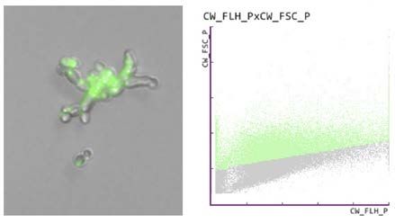
Urinary Tract Infection: How innovative technology leads to a faster diagnosis and more targeted treatment of UTI                                                   5
Urinalysis white paper | May 2020

                                                                                  Antibiotic susceptibility testing on the UF-series

                                                                                  Antibiotic susceptibility testing by agar diffusion is a mandatory
                                                                                  diagnostic procedure to identify the correct antibiotics for a
                                                                                  persisting infection to induce targeted antimicrobial therapy and
                                                                                  prevent antimicrobial resistances.

                                                                                  Briefly speaking, bacterial samples are spread on agar plates, and
                                                                                  paper disks soaked with antibiotics are placed onto the agar.
                                                                                  During incubation of the plate, the antibiotics will radially diffuse
                                                                                  and inhibit the bacterial growth, depending on their antibiotic
                                                                                  efficacy. Despite its specificity, this gold standard agar diffusion
Fig. 8 Yeast-like cells, detected by fluorescence microscopy (left) and on the
Sysmex UF-series, displayed in the respective scattergram (right)                 test has a high turn-around time of 18 – 48 hours [9].

                                                                                  A potential solution to accelerate antibiotic treatment decision
                                                                                  has been reported for conducting antibiotic susceptibility testing
Distinguishing upper and lower UTI                                                on the UF-series. Aliquots of ready-to-use microbial growth broth
                                                                                  were individually supplemented with different antibiotics and
The presence of renal tubular epithelial cells (RTEC) in urine is often           inoculated with the bacteria stemming from the patient samples.
an indicator of renal disease or tubular damage. Since RTEC line                  After incubation for up to four hours, the bacteria concentration
the entire renal tubule from the proximal to the distal segment,                  within the different cultures was determined on the UF-series.
they represent a potential diagnostic marker for renal damages                    A sensitivity of 83.3 % (PPV = 100 %) and a specificity of 100 %
when other parameters are still inconspicuous [16].                               (NPV = 91.3 %) allowed, for example, the differentiation of colistin-
                                                                                  resistant and susceptible Escherichia coli and Klebsiella pneumoniae
As a potential clinical application, the quantification of RTEC in                isolates within two hours, supported by the UF-5000 (Fig. 11; [18]).
individuals with confirmed urinary tract infection has been shown
to be a potential indicator of upper urinary tract infection (Fig. 9; [17]).      Alternative approaches combine the diagnostics for bacteriuria
                                                                                  on the UF-5000 with subsequent molecular testing for bacterial
With a diagnostic accuracy of 0.923 (AUC), the RTEC count clearly                 resistance genes [19] or mass spectrometry to identify bacteria
outperforms known markers of upper urinary tract infection, such                  and mediators of antibiotic resistance [20], allowing the installation
as α1 -microglobulin (0.735) and γ-glutamyl transferase (0.586).                  of a targeted antibiotic therapy within six hours.

The potential diagnostic value of RTEC quantification, however,
strongly depends on proper sample handling and processing, since
                                                                                                            C                                                C
their in vitro stability is impaired by storage times of two hours
                                                                                                B                                                    B
and more, as well as room temperature and acidic urinary pH [17].                                                                    37 ºC
                                                                                                        A                                                A
                                                                                                                                 18–24 h

                                                                                  Fig. 10 Schematic presentation of the antibiotic susceptibility testing by agar
                                                                                  diffusion. The different diameters of the growth inhibition zones around the
                                                                                  soaked paper disks correlate with (A) ineffective, (B) medium-effective and (C)
                                                                                  highly effective antibiotics.

                                                                                            A       B   C                        A           B   C

                                                                                                                + Antibiotics

                                                                                                                37ºC for 1–4 h

                                                                                  Fig. 11 Alternative antibiotic susceptibility testing on the UF-5000 via growth
                                                                                  monitoring of bacterial isolates in broth supplemented with different antibiotics.
Fig. 9 Renal tubular epithelial cell (RTEC) counts among non-urology/nephrology
                                                                                  The bacteria concentration correlates to (A) highly effective, (B) medium-effective
patients and patients with confirmed upper or lower urinary tract infection
                                                                                  and (C) ineffective antibiotics.
([17] modified)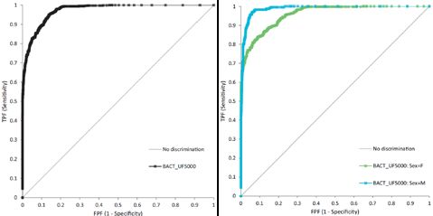
Urinary Tract Infection: How innovative technology leads to a faster diagnosis and more targeted treatment of UTI                                                      6
Urinalysis white paper | May 2020

Fighting antimicrobial resistance with
targeted diagnostics                                                                        Create awareness and understanding


                                                                                            Strengthen knowledge and scientific evidence

The adaptation of microorganisms to resist the actions of anti-
microbial agents is widely known as antimicrobial resistance, a                             Reduce infections through hygiene measures

well-recognised problem of public health of the 21st century.
                                                                                            Optimise the use of antimicrobials in human and animal health

Antimicrobial resistance, however, is a phenomenon that already
has been reported before the discovery of penicillin. After years                           Sustainable investment in new medicines, diagnostic

of extensive clinical use of the antimicrobial salvarsan for the                            tools and vaccines
treatment of syphilis, a waning effect for salvarsan had been
observed, as well as an increase in more severe clinical pictures
                                                                                       Here, laboratory diagnostics is an important factor, as it not only
of syphilis [21], indicating antimicrobial resistance.
                                                                                       aims to provide accurate information for more accurate diagnoses
                                                                                       and clinical decision support but will also contribute to allowing
Since then, irrational use of antimicrobials (e. g. inappropriate
                                                                                       a more rational use of antimicrobials.
prescriptions and self-medication), extensive use of antimicrobials
in factory farming and agriculture, but also the prolonged and
widespread use of antibiotics in therapy and prophylaxis augmented
the numbers of resistant microorganism species [22].                                      Summary and conclusion

With increasing antimicrobial resistance and slowed antimicrobial                         Considering the total amount of suspected urinary tract
drug development, antimicrobial stewardship is of utmost                                  infections that finally turn out to be negative, an optimised
importance. Without proper and immediate actions, the number                              diagnostic workflow including the Sysmex UF-series can
of deaths caused by antimicrobial resistance by 2050 will surpass                         improve the efficiency of laboratory diagnostics by ruling
those of cancer [23]. Therefore, the World Health Organisation                            out urinary tract infections within a short period (Fig. 12).
announced a global health crisis and released a global action plan                        Moreover, modern flow cytometry-based urinalysis prevents
[24] to fight antimicrobial resistance with the following actions:                        blindly prescribing unnecessary antimicrobials, and instead
                                                                                          supports a targeted and rational use of antimicrobials, thus
                                                                                          contributing to the much-needed antimicrobial stewardship.

                                                  Suspected UTI                                                           Suspected UTI
      > 1 minute

                                                     Dipstick                                                                UF-series

                                                  UTI negative?                                                            UTI negative        Rule out
                                                                                                                                                 UTI

                                                  UTI suspected                                                       UTI suspected ('UTI?')

                                                                                                                       Gram + | Gram – | Yeast
                                         Urine culture
      24 hours

                                         UTI negative
                                                                                                             Targeted urine culture
                                          UTI positive
      48 hours

                                               Antibiogram (AST)                                                        Antibiogram (AST)

                                                                                                                Targeted therapy

Fig. 12 Overview of the diagnostic workflow for the diagnosis of urinary tract infections without (left) and with automated urine particle analysis using the UF-series
(right). The UF-series allows ruling out UTI in less than a minute and reduces the unnecessary diagnostic follow-up by up to 80 % of the overall number of suspected UTI
cases. For potential UTI-positive samples (‘UTI?’) the ‘BACT Info’ flag enables more targeted diagnostics to identify the presence and type of bacterial infection.
Ruling out UTI at an early stage also helps to reduce the empirical prescription of antibiotics and supports antimicrobial stewardship.
Urinary Tract Infection: How innovative technology leads to a faster diagnosis and more targeted treatment of UTI 7
Urinalysis white paper | May 2020
References
[1] Stamm WE, Norrby SR (2001): Urinary tract infections: Disease [14] Enko D, Stelzer I, Boeckl M, Derler B, Schnedl WJ, Anderssohn P,
paranormal and challenges. J Infect Dis 183 (Suppl. 1) S1–S4. Meinitzer A and Herrmann M (2020): Comparison of the diagnostic
performance of two automated urine sediment analyzers with manual
[2] Foxman B (2014): Urinary Tract Infection Syndromes: Occurrence,
phase-contrast microscopy. Clin Chem Lab Med 58(2):268–273.
Recurrence, Bacteriology, Risk Factors and Disease Burden. Infect Dis
Clin North AM 28(1):1–13. [15] Song D, Lee HJ, Jo SY, Lee SM and Chang CL (2018): Selection of
unnecessary urine culture specimens using Sysmex UF-5000 urine flow
[3] Flores-Mireles AL, Walker JN, Caparon M and Hultgren SJ (2015):
cytometer. Ann Clin Microbiol 21(4):75–79.
Urinary tract infections: epidemiology, mechanisms of infection and
treatment options. Nat Rev Microbiol 13(5):269–284. [16] Becker GJ, Garigali G, Fogazzi GB (2016): Advance in urine microscopy.
Am J Kidney Dis 67:954–964.
[4] Lane DR and Takhar SS (2011): Diagnosis and Management of
Urinary Tract Infection and Pyelonephritis. Emerg Med Clin North [17] Oyaert M, Speeckaert M, Boelens J, Delanghe JR (2020): Renal
Am 29(3):539–552. tubular epithelial cells add value in the diagnosis of upper urinary tract
pathology. Clin Chem Lab Med 58(4):597–604.
[5] Schmiemann G, Kniehl E, Gebhardt K, Matejczyk MM,
Hummers-Pradier E (2010): The Diagnosis of Urinary Tract Infection – [18] Liste I, Cakar A, Sancak B, Hascelik G, Ozkuyumcu (2019):
A Systematic Review. Dtsch Arztebl Int 107(21):361–367. The rapid detection of colistin resistance by using a fluorescence flow
cytometry analyser. Poster 4301 presented on ‘ASM Microbe 2019’.
[6] Fischer V (2019): Ein Schritt zur schnelleren Urinanalytik.
Xtra 2:50–52. (article in German) [19] Wagner K, Mancini S, Ritter C, Böttger EC, Keller PM (2019):
Evaluation of the AID AmpC Line Probe Assay for Molecular Detection
[7] Pujades-Rodriguez M, West RM, Wilcox MH, Sandoe J (2019):
of AmpC-producing Enterobacterales. J Glob Antimicrob Resist 19:8–13.
Lower Urinary Tract Infections: Management, Outcomes and Risk
Factors for Antibiotic Re-prescription in Primary Care. E Clinical [20] Oviaño M, de la Luna Ramírez C, Pedro Barbeyto L, Bou G (2017):
Medicine 14:23–31. Rapid direct detection of carbapenemase-producing Enterobacteriaceae
in clinical urine samples by MALDI-TOF MS analysis. J Antimicrobial
[8] Wilson ML and Gaido L (2004): Laboratory diagnosis of urinary
Chemotherapy 72(5):1350–1354.
tract infections in adult patients. Clin Infect Dis 38(8):1150–1158.
[21] Silberstein S (1924): Zur Frage der salvarsanresistenten Lues. Arch
[9] Bauer AW, Kirby WM, Sherris JC, Turck M (1966): Antibiotic
Derm Syph 147:116–130. (article in German)
susceptibility testing by a standardised single disk method. Am J Path
45(4):493–496. [22] Prestinaci F, Pezzotti P, Pantosti A (2015): Antimicrobial resistance:
a global multifaceted phenomenon. Pathog Glob Health 109(7):309–318.
[10] Oyaert M and Delanghe JR (2019): Progress in automated urinalysis.
Ann Lab Med 39:15–22. [23] Review on Antimicrobial Resistance (2014): Antimicrobial Resistance:
Tackling a Crisis for the Health and Wealth of Nations.
[11] De Rosa R, Grosso S, Lorenzi S, Bruschetta G, Camporese A (2018):
[https://amr-review.org/sites/default/files/AMR%20Review%20Paper%
Evaluation of the new Sysmex UF-5000 fluorescence flow cytometry
20-%20Tackling%20a%20crisis%20for%20the%20health%20and%
analyser for ruling out bacterial urinary tract infection and for prediction
20wealth%20of%20nations_1.pdf]
of Gram-negative bacteria in urine cultures. Clinica Chimica Acta
484:171–178. [24] World Health Organization (2015): Global plan on antimicrobial
resistance. https://apps.who.int/iris/handle/10665/193736
[12] Juránkova J, Babušíková L, Protivínský J (2017): The importance
of diagnosing Gram-negative/Gram-positive bacteria in urine in the
pre-culture screening of urinary tract infections in the microbiology You can download our white papers from our website:
laboratory by fluorescence flow cytometry on the UF-4000 urine www.sysmex-europe.com/whitepapers
analyser for early initiation of targeted antibiotic therapy. Poster P2153
presented at ECCMID 2018.
[13] Lee SW (2010): An aspergilloma mistaken for a pelviureteral stone on
nonenhanced CT: a fungal bezoar causing ureteral obstruction. Korean
J Urol 51:216.
Sysmex Europe GmbH
EN.N.06/20
Bornbarch 1, 22848 Norderstedt, Germany · Phone +49 40 52726-0 · Fax +49 40 52726-100 · info@sysmex-europe.com · www.sysmex-europe.com
You will find your local Sysmex representative’s address under www.sysmex-europe.com/contactsYou can also read