Mucormycosis of the Central Nervous System in the Background of the COVID-19 Pandemic in the Absence of Antifungal Medication - An Audit of 2 Very ...
←
→
Page content transcription
If your browser does not render page correctly, please read the page content below
SVOA Neurology
                                                                                                                             ISSN: 2753-9180

                                                                                                                                 Case Report

Mucormycosis of the Central Nervous System in the
Background of the COVID-19 Pandemic in the Absence of
Antifungal Medication – An Audit of 2 Very Difficult Months in
a Tertiary Care Centre in Southern India
    Ganapathy S1*, Gangoli D2, Raykar R3 and Rao SAV4
1 Assistant   Professor, Department of Neurosurgery & Spine Surgery, St. Johns Medical College Hospital, Bangalore, India.
2 Junior   Resident, Department of Neurosurgery & Spine Surgery, St. Johns Medical College Hospital, Bangalore, India.
3   Associate Professor, Department of Neurosurgery & Spine Surgery, St. Johns Medical College Hospital, Bangalore, India.
4 Professor   and Head, Department of Neurosurgery & Spine Surgery, St. Johns Medical College Hospital, Bangalore, India.

*Corresponding Author: Dr. Sibhi Ganapathy MBBS, MS, Mch, DNB, MNAMS, FAGE, FRCS, Assistant Professor, Department of
Neurosurgery & Spine Surgery, St. Johns Medical College Hospital, Bangalore, India.

Received: September 03, 2021 Published: September 13, 2021

Abstract
Mucor of the intracranial compartment is a rare and devastating infection commonly seen in debilitated patients. The
infection commonly enters through the orbit or through the paranasal sinuses through diploic vessels or through skull
base osteomyelitis secondary to sinusitis. The onset of the COVID-19 pandemic coupled with the widespread use of
steroids reduced effective immunity of various normally immunocompetent individuals. This saw a veritable avalanche
of mucor infections in COVID affected and post COVID-19 infected patients. The presentations were different from the
commonly seen routes of entry and presented in different and unique ways. The severity was worsened due to the lack
of availability of amphotericin B required for the treatment of the infection. Many of the patients succumbed to their
ordeal despite repeated surgical debridement and abscess evacuations. We present a representation of unusual mucor
infections of the brain seen during the second COVID-19 wave in India, where an unprecedented number of infections
were seen overwhelming the health facilities of all major centres in India.
                              Keywords: Mucormycosis, COVID-19, Pandemic, Antifungal Medication

Introduction
The second wave of COVID-19 infection in India was acutely felt across the nation. Both urban and rural centres were
affected with hospitals overflowing and deaths exceeding the worst estimates possible. Patients passed away waiting for
ventilators and ICU beds while those in critical care centres wasted away without improvement. Aggressive use of ster-
oids to limit and reverse the hypersensitivity mediated immune response to the virus was advocated and taken to heart
by the harried medical community of India and steroid were even given off the counter for those who wanted it without
prescriptions from medical personnel.

As the pandemic peak abated, emergency rooms across the country began to experience a new phenomenon. Patient pre-
viously treated successfully for the COVID-19 infection began presenting with symptoms of nasal stuffiness, discharge
and eye swelling. Many also presented with seizures and features of raised intracranial tension. Careful examination
showed black coloured pus in the sputum and discharge of the patients. The skin and bony architecture of the face also
seemed involved. Imaging studies as well as microbiological assessments showed the infection to be Mucormycosis, a
rare yet deadly fungal colonisation of the paranasal sinuses and orbit. Over the next 2 months, Emergency teams, ENT
and ophthalmological surgeons were overwhelmed by the spate of cases presenting with the deadly fungus.

The problem was severely compounded by the unavailability of Amphotericin B, the supply of which was swamped by
the sudden spike in demand caused by the fungus infection. Although India is the largest manufacturer and supplier of
the drug, the sudden spike in demand exceeded all expectations and planning.

                                                                                                                                   SVOA Neurology
                                                                     148
Mucormycosis of the Central Nervous System in the Background of the COVID-19 Pandemic in the Absence of Antifungal Medication – An Audit of 2 Very Difficult Months in a Tertiary Care Centre in Southern India
By the time the drug manufacturers could ramp up production and reintroduce the medication into circulation, uncon-
trolled mucor ran amok amongst the patients presenting in weird, unusual and unique ways.
We present the novel intracranial presentations of mucor experienced by us in a tertiary care centre in Bengaluru city in
south India.
Mode of spread
It is well documented that mucor initially colonises the mucosa of the paranasal sinuses as it’s spores are transmitted by
air from the soil. The fungus then overwhelms the thin bony septae separating the sinuses from the adjoining orbit thus
leading to orbital fungal cellulitis and eventual infection of the globe. If left unchecked they can enter the intracranial
compartment as well. There have also been reports of vascular invasion of the fungal elements which lead to a wider and
more serious dissemination of the disease, especially into the brain. Lastly a rare and severe modus of spread is perineu-
ral spread, where fungal elements travel along the axons of nerves affecting passing structures along the course of the
nerve. The presentations of these entities are varied and are often confused with other commoner diseases. The different
presentations and modes of spread are shown in figure 1.
Figure 1: diagram showing the different modes of fungal spread into the intracranial cavity with the different presentations
of each subtype mentioned below it.
Case Series
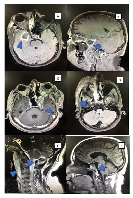
Part 1: Direct spread
The propensity of fungal cellulitis of the paranasal sinuses to spread through the diploic and lacunar vessels is well docu-
mented. The spread often is through bony divisions that separate the infected sinus or orbital lining form the intracranial
cavity. Hence common presentations of such a modus of spread are intracerebral abscesses in the frontal or temporal
lobes as shown in figure 2A & 2B. Central skull base osteomyelitis is also seen in the vicinity of the sphenoid sinus
around the cavernous venous sinuses. (figure 2C) The even rarer vault osteomyelitis was seen in absence of direct pene-
trating injury of the bone. The spread was through contiguous spread from the infected orbit as seen in Figure 2D. An-
other rare presentation was clival Mucormycosis with complete destruction of the bone and a prevertebral abscess of
mucor pus extending down all the way to the upper dorsal spine. (figures 2E & F).
Part 2: Vascular spread
Angioinvasion of the cerebral vasculature from the branches in the paranasal sinuses lead to intravascular complica-
tions. These include occlusion of vessels leading to strokes, Dissemination of the fungal elements through the blood-
stream leading to multiple abscesses far away from each other (pyemic abscesses figure 3A), and even vessel wall de-
struction leading to mycotic aneurysms (figure 3B&C) Even rarer, is the space occupying lesion presentation as seen in
figure 3D, where a vascular nidus grows into a large mass compressing the surrounding brain leading to deficits and fea-
tures of raised intracranial tension.
Part 3: Perineural spread
The most puzzling and perhaps interesting of the 3 modes of spread is the perineural infiltration of the fungus along the
axolemma and oligodendrocytes to distal structures in the brain. We saw infections leading to optic neuritis with a mass
lesion in the distal optic nerve mimicking a glioma (figure 4A), fungal trigeminal neuralgia (figure 4B) and spread along
the trigeminal nerve into the gasserian ganglion causing severe neuralgic pain (figure 4C), as well as olfactory nerve in-
fection with space occupying lesions secondary to inflammation seen. (figure 4D)
SVOA Neurology
149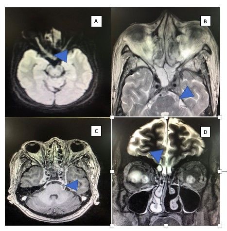
Mucormycosis of the Central Nervous System in the Background of the COVID-19 Pandemic in the Absence of Antifungal Medication – An Audit of 2 Very Difficult Months in a Tertiary Care Centre in Southern India

Figure2: (A) & (B) shows a T1 contrast MRI axial & sagittal slice of the brain showing a ring enhancing lesion with central
necrosis in the left temporal lobe suggestive of an abscess. (C) shows a T1 contrast axial MRI of the brain with enhancing
infiltrates on the right cavernous sinus and orbital apex extending posteriorly into the posterior fossa. (D) shows a T2
weighted MRI image of the brain showing significant oedema of the skin, subcutaneous tissue and bone of the temporal bone
leading to complete destruction and erosion of the bone with the pus extending into the temporal and subtemporal fossa. (E)
is sagittal MRI T2 slice of the brain showing destruction and inflammation of the clivus with pus extending below into the
prevertebral space as demonstrated clearly in (F).

                                                                                                                                                                                                          SVOA Neurology
                                                                                                          150
Mucormycosis of the Central Nervous System in the Background of the COVID-19 Pandemic in the Absence of Antifungal Medication – An Audit of 2 Very Difficult Months in a Tertiary Care Centre in Southern India
Figure 4: (A) shows an axial DWI image showing
Figure 3: (A) shows multiple ring enhancing lesions in a axial thickening and diffusion restriction along the right
T1 FLAIR sequence which also demonstrates the oedema of the optic nerve. The portion of the nerve near the chiasm
respective lesions. Other lesions were also seen in the brainstem is especially thickened almost resembling a neo-
and cerebellum. (B) a contrast T1 axial slice shows a lesion on plastic growth. (B) shows a T2 weighted axial MRI
the anterior border of the cerebellum and brainstem. An angio- slice, which shows a thickened and inflamed left tri-
gram done in (C) shows a large saccular mycotic aneurysm of geminal nerve causing severe neuralgia to the pa-
the left Superior Cerebellar Artery. (D) shows a large fungoma tient. (C) shows inflammation of the entire trigemi-
or fungal space occupying lesion which is seen far away from nal nerve from the orbit along the cavernous sinus
infected bone or sinus. The spread is thus haematological in na- upto the gasserian ganglion and beyond onto the
ture. brainstem. (D) shows a space occupying lesion seen
in close proximity to the right olfactory nerve in a
setting of severe orbital mucor inflammation. The
olfactory bulbs appear inflamed in this coronal T2
MRI brain slice.
Case Audit
Of the total of 127 Mucormycosis patients admitted in the 2 month period specified, 104 were diagnosed with rhino-
orbital Mucormycosis, of which 97 were operated. Functional endoscopic sinus surgeries (FESS) and orbital surgeries
including eventration were carried out by Otorhinolaryngologists and ophthalmologists. A total of 23 patients were re-
ferred to neurosurgery for various complaints. Most were managed conservatively especially after the focus of infection
was managed surgically as mentioned before. A total of 6 patients were operated for cerebral Mucormycosis. Surgeries
ranged from decompressive hemicraniectomies for mucor related strokes, to abscess evacuation, to skull base proce-
dures to decompress the middle or posterior cranial fossa base. The distribution of surgeries is shown in figure 5.
Figure 5: Explaining the distribution of post covid-9 Mucormycosis in St. John’s medical College Hospital, Bangalore.
SVOA Neurology
151Mucormycosis of the Central Nervous System in the Background of the COVID-19 Pandemic in the Absence of Antifungal Medication – An Audit of 2 Very Difficult Months in a Tertiary Care Centre in Southern India
Mucor progressed rapidly following the covid-19 second spike as shown in figure 6. Maximum cases were seen in May
and June mirroring the virus outbreak. The mortality of cerebral Mucormycosis was not high when amphotericin B was
available. For a period of 1 month, the drug was in short supply leading to virulent recurrent infections and progression
of the disease. Many mortalities were seen in this period mainly due to this problem.
Figure 6: Explaining the temporal relationship between the covid-19 second spike in India and the upsurge of Mucormycosis
cases.
Of the patients operated for neurosurgical mucor, the commonest presentation was skull base osteomyelitis secondary
to the infection in the paranasal sinuses and orbit. Infection of the skull base of the middle cranial fossa and rarely the
posterior fossa was seen. Osteomyelitis was also seen in the frontal bone secondary to frontal sinusitis and orbital cellu-
litis. Abscesses were also seen as an extension of the infection. Commonest locations were temporal lobe followed by
frontal lobe abscesses. These mostly were seen as an extension of the osteomyelitis. Pyemic abscesses secondary to hem-
atogenic dissemination were also seen but only rarely. Perineural abscesses which spread along the nerve sheath were
also seen as described above onto the substance of the brainstem and cerebellum. These however were small and man-
aged medically. Angioinvasion of the mucor lead to strokes and mycotic infarcts. These were seen in 1 patient with com-
plete middle cerebral artery occlusion and was managed through an emergency decompressive craniectomy. Unusual
presentations were seen in a large number of patients and have been described in detail above. They contribute to the
novelty of this report. Most unusual presentations were managed conservatively with antifungals and anticonvulsants.
All recovered well with minimal deficits. The distribution of neurosurgical presentations of mucor is summarised in fig-
ure 7.
Figure 7: Showing the different clinic-pathological presentations of cerebral Mucormycosis seen in our hospital.
Discussion
COVID-19 and mucormycosis share risk factors, such as presence of DM, which can independently contribute to mortali-
ty, but have conflicting management principles. While immune suppression with steroids may be required in moderate
to severe COVID-19, the use of steroids and the worsening glycaemic control provide an opportunity for mucor to be-
come invasive. Mucor produces keto-reductase as a virulence factor enabling them to grow in the acidic and glucose-rich
environment generated in ketoacidotic states.
SVOA Neurology
152Mucormycosis of the Central Nervous System in the Background of the COVID-19 Pandemic in the Absence of Antifungal Medication – An Audit of 2 Very Difficult Months in a Tertiary Care Centre in Southern India
Additionally, Muller et al have postulated that the human pancreas could be a possible target for the SARS-CoV-2 virus
and that the β-cell infection may result in insulin resistance. This metabolic dysregulation, in previously nondiabetic or
well-controlled diabetic COVID-19 patients, might predispose them to develop mucormycosis. Moorthy et al recently
reported the association of COVID-19 infection with uncontrolled DM and usage of corticosteroids. Similarly, Sen et al
reported a series of 6 diabetic patients with concurrent mucormycosis and COVID-19 infection. Sarkar et al reported a
series of 10 diabetic patients with ROCM postCOVID-19. All their patients had uncontrolled blood sugar values and were
treated with steroids during active COVID-19 infection. Current literature suggests that usage of systemic steroids in pa-
tients, who otherwise may have controlled diabetes, or may not be diabetics at all, can precipitate mucormycosis. Me-
konnen et al reported a case of invasive fungal rhinosinusitis with orbital involvement in a patient with COVID-19 with
uncontrolled DM and HbA1c of 14%. Mehta and Pandey reported a case of a patient with COVID-19 infection, treated
with steroids and tocilizumab, who during the course of the treatment, developed rhino-orbital mucormycosis. Waizel-
Haiat et al reported a case of rhino-orbital mucormycosis associated with ketoacidosis secondary to recent onset DM and
COVID-19 infection. Despite aggressive management the patient developed multi-organ failure and died.
In conclusion, ROCM is a known occurrence in COVID-19 affected patients. Over a third of patients can have unfavorable
final outcome. Uncontrolled DM at presentation, involvement of the orbital apex and CNS by the infection, and the usage
of steroids determined an unfavorable outcome. Involvement of the CNS was seen to be the only factor determining mor-
tality. In a similar geographic setup, as compared to previous non-COVID related cases, the coexistence of COVID-19 in
this series, did not seem to worsen the final outcome in terms of mortality. It is prudent that physicians and Neurosur-
geons, alike, involved in the care of patients with COVID-19 be aware of the outcomes of ROCM in COVID-19 patients.
Conclusion
The sheer numbers faced during the onslaught of the Mucormycosis wave which followed the second spike of covid-19
cases made diagnosis, surgery and prognostication difficult especially during the period where demand for antifungals
outstripped supply. But a combined team approach to the disease with good patient doctor counselling helped tide over
the majority of the period successfully. What it did provide was a treasure trove of experience and data which can aid
future generations in their fight against pandemics in the future. This is our humble attempt to chronicle the events and
the diversity of presentations of this period for posterity.
References
1. Chen IW, Lin CW. Rhino-orbital-cerebral mucormycosis. CMAJ. 2019;191(16):E450.
2. Munir, N., & Jones, N. (2007). Rhinocerebral mucormycosis with orbital and intracranial extension: A case report and
review of optimum management. The Journal of Laryngology & Otology, 121(2), 192-195.
3. Chakrabarti, A. and Singh, R. (2014), Mucormycosis in India: unique features. Mycoses, 57: 85-90.
4. Fouad, Yousef A et al. “Spike in Rhino-Orbital-Cerebral Mucormycosis Cases Presenting to a Tertiary Care Center
During the COVID-19 Pandemic.” Frontiers in medicine vol. 8 645270. 28 May. 2021.
5. Jiang, N., Zhao, G., Yang, S. et al. A retrospective analysis of eleven cases of invasive rhino-orbito-cerebral mu-
cormycosis presented with orbital apex syndrome initially. BMC Ophthalmol 16, 10 (2016).
6. Zafar S, Prabhu A. Rhino-orbito-cerebral mucormycosis: recovery against the odds Practical Neurology 2017;17:485-
488.
7. Gupta, Saroj et al. “Rhino-Orbital-Cerebral Mucormycosis: Battle with the Deadly Enemy.” Indian journal of otolaryn-
gology and head and neck surgery : official publication of the Association of Otolaryngologists of India vol. 72,1 (2020):
104-111. doi:10.1007/s12070-019-01774-z
8. Al Hassan, Fatimah et al. “Rhino-orbito-cerebral mucormycosis in patients with uncontrolled diabetes: A case se-
ries.” International journal of surgery case reports vol. 73 (2020): 324-327. doi:10.1016/j.ijscr.2020.07.011
9. Vaughan, C, Bartolo, A, Vallabh, N, Leong, SC. A meta-analysis of survival factors in rhino-orbital-cerebral mu-
cormycosis—has anything changed in the past 20 years? Clin Otolaryngol. 2018; 43: 1454– 1464.
10. Riley TT, Muzny CA, Swiatlo E, Legendre DP. Breaking the Mold: A Review of Mucormycosis and Current Pharmaco-
logical Treatment Options. Annals of Pharmacotherapy. 2016;50(9):747-757.
11. Zhang, Gao-Jia et al. “Fatal and Rapid Progressive Isolated Cerebral Mucormycosis Involving the Bilateral Basal Gan-
glia: A Case Report.” Frontiers in neurology vol. 11 295. 21 Apr. 2020.
12. Mehta, Salil, and Abha Pandey. “Rhino-Orbital Mucormycosis Associated With COVID-19.” Cureus vol. 12,9 e10726.
30 Sep. 2020, doi:10.7759/cureus.10726
13. Honavar, Santosh G Code Mucor, Indian Journal of Ophthalmology: June 2021 - Volume 69 - Issue 6 - p 1361-1365.
SVOA Neurology
153Mucormycosis of the Central Nervous System in the Background of the COVID-19 Pandemic in the Absence of Antifungal Medication – An Audit of 2 Very Difficult Months in a Tertiary Care Centre in Southern India
14. Dave, Tarjani Vivek M.D.*; Gopinathan Nair, Akshay M.D.†,‡; Hegde, Raghuraj M.D.§,‖; Vithalani, Nidhi M.D.*; Desai, Sa-
vari M.D.¶; Adulkar, Namrata M.D.#; Kamal, Saurabh M.D.**; Mittal, Raman M.D.††; Bradoo, Renuka A. M.D.‡ Clinical
Presentations, Management and Outcomes of Rhino-Orbital-Cerebral Mucormycosis (ROCM) Following COVID-19,
Ophthalmic Plastic and Reconstructive Surgery: July 27, 2021.
15. Mitra, Sandipta et al. “Post-COVID-19 rhino-orbito-cerebral mucormycosis: a new addition to challenges in pandem-
ic control.” European archives of oto-rhino-laryngology : official journal of the European Federation of Oto-Rhino-
Laryngological Societies (EUFOS) : affiliated with the German Society for Oto-Rhino-Laryngology - Head and Neck Sur-
gery, 1–6. 26 Jul. 2021,
16. Al-Tawfiq, Jaffar A et al. “COVID-19 and mucormycosis superinfection: the perfect storm.” Infection, 1–21. 24 Jul.
2021
17. Revannavar, Shweta Mallikarjun et al. “COVID-19 triggering mucormycosis in a susceptible patient: a new phenome-
non in the developing world?.” BMJ case reports vol. 14,4 e241663. 27 Apr. 2021.
18. Eswaran, Sudhagar et al. “Acute Fulminant Mucormycosis Triggered by Covid 19 Infection in a Young Pa-
tient.” Indian journal of otolaryngology and head and neck surgery : official publication of the Association of Otolaryn-
gologists of India, 1-5. 8 Jul. 2021.
19. Buil, Jochem B et al. “Case series of four secondary mucormycosis infections in COVID-19 patients, the Netherlands,
December 2020 to May 2021.” Euro surveillance : bulletin Europeen sur les maladies transmissibles = European com-
municable disease bulletin vol. 26,23 (2021): 2100510.
Citation: Ganapathy S, Gangoli D, Raykar R, Rao SAV. “Mucormycosis of the Central Nervous System in the Background
of the COVID-19 Pandemic in the Absence of Antifungal Medication – An Audit of 2 Very Difficult Months in a Tertiary
Care Centre in Southern India”. SVOA Neurology 2:5 (2021) Pages 148-154.
Copyright: © 2021 All rights reserved by Ganapathy S., et al. This is an open access article distributed under the
Creative Commons Attribution License, which permits unrestricted use, distribution, and reproduction in any medium,
provided the original work is properly cited.
SVOA Neurology
154You can also read