MVMM-REGNET: A NEW IMAGE REGISTRATION FRAMEWORK BASED ON MULTIVARIATE MIXTURE MODEL AND NEURAL NETWORK ESTIMATION
←
→
Page content transcription
If your browser does not render page correctly, please read the page content below
MvMM-RegNet: A new image registration
                                         framework based on multivariate mixture model
                                                and neural network estimation

                                                                                                       ?
                                                                   Xinzhe Luo and Xiahai Zhuang

                                                       School of Data Science, Fudan University, Shanghai, China

                                                 Abstract. Current deep-learning-based registration algorithms often ex-
                                                 ploit intensity-based similarity measures as the loss function, where dense
arXiv:2006.15573v2 [cs.CV] 14 Jul 2020

                                                 correspondence between a pair of moving and fixed images is optimized
                                                 through backpropagation during training. However, intensity-based met-
                                                 rics can be misleading when the assumption of intensity class corre-
                                                 spondence is violated, especially in cross-modality or contrast-enhanced
                                                 images. Moreover, existing learning-based registration methods are pre-
                                                 dominantly applicable to pairwise registration and are rarely extended to
                                                 groupwise registration or simultaneous registration with multiple images.
                                                 In this paper, we propose a new image registration framework based on
                                                 multivariate mixture model (MvMM) and neural network estimation. A
                                                 generative model consolidating both appearance and anatomical infor-
                                                 mation is established to derive a novel loss function capable of imple-
                                                 menting groupwise registration. We highlight the versatility of the pro-
                                                 posed framework for various applications on multimodal cardiac images,
                                                 including single-atlas-based segmentation (SAS) via pairwise registration
                                                 and multi-atlas segmentation (MAS) unified by groupwise registration.
                                                 We evaluated performance on two publicly available datasets, i.e. MM-
                                                 WHS-2017 and MS-CMRSeg-2019. The results show that the proposed
                                                 framework achieved an average Dice score of 0.871 ± 0.025 for whole-
                                                 heart segmentation on MR images and 0.783 ± 0.082 for myocardium
                                                 segmentation on LGE MR images1 .

                                         1     Introduction

                                             The purpose of image registration is to align images into a common co-
                                         ordinate space, where further medical image analysis can be conducted, in-
                                         cluding image-guided intervention, image fusion for treatment decision, and
                                         atlas-based segmentation [14]. In the last few decades, intensity-based regis-
                                         tration has received considerable scholarly attention. Commonly used similarity
                                         measures comprise intensity difference and correlation-based methods for intra-
                                         modality registration, and information-theoretic metrics for inter-modality reg-
                                         istration [10, 14, 18, 22, 23].
                                         ?
                                             Corresponding author: zxh@fudan.edu.cn.
                                         1
                                             Code is available from https://zmiclab.github.io/projects.html.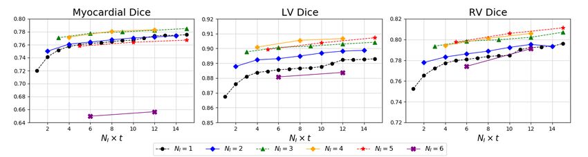
2       Xinzhe Luo et al.

    Recently, deep learning (DL) techniques have formulated registration as a
parameterized mapping function, which not only made registration in one shot
possible but achieved state-of-the-art accuracies [4, 8, 11, 24]. de Vos et al. [24]
computed dense correspondence between two images by optimizing normalized
cross-correlation between intensity pairs. While intensity-based similarity mea-
sures are widely used for intra-modality registration, there are circumstances
when no robust metric, solely based on image appearance, can be applied. Hu et
al. [11] therefore resorted to weak labels from corresponding anatomical struc-
tures and landmarks to predict voxel-level correspondence. Balakrishnan et al. [4]
proposed leveraging both intensity- and segmentation-based metrics as loss func-
tions for network optimization. More recently, Dalca et al. [8] developed a prob-
abilistic generative model and derived a framework that could incorporate both
of the intensity images and anatomical surfaces.
    Meanwhile, in the literature several studies have suggested coupling regis-
tration with segmentation, in which image registration and tissue classification
are performed simultaneously within the same model [2, 6, 20, 25]. However, the
search for the optimal solution of these methods usually entails computationally
expensive iterations and may suffer from problems of parameter tuning and local
optimum. A recent study attempted to leverage registration to perform Bayesian
segmentation on brain MRI with an unsupervised deep learning framework [9].
Nevertheless, it can still be difficult to apply unsupervised intensity-based ap-
proaches to inter-modality registration or to datasets with poor imaging quality
and obscure intensity class correspondence. Besides, previous DL-integrated reg-
istration methods have mainly focused on pairwise registration and are rarely
extended to groupwise registration or simultaneous registration with multiple
images.
    In this paper, we consider the scenario in which multiple images from various
modalities need to be co-registered simultaneously onto a common coordinate
space, which is set onto a reference subject or can be implicitly assumed during
groupwise registration. To this end, we propose a probabilistic image registra-
tion framework based on both multivariate mixture model (MvMM) and neural
network estimation, referred to as MvMM-RegNet. The model incorporates both
types of information from the appearance and anatomy associated with each im-
age subject, and explicitly models the correlation between them. A neural net-
work is then employed to estimate likelihood and achieve efficient optimization
of registration parameters. Besides, the framework provides posterior estimation
for MAS on novel test images.
    The main contribution of this work is four-fold. First, we extend the con-
ventional MvMM for image registration with multiple subjects. Second, a DL-
integrated groupwise registration framework is proposed, with a novel loss func-
tion derived from the probabilistic graphical model. Third, by modelling the
relationship between appearance and anatomical information, our model out-
performs previous ones in terms of segmentation accuracy on cardiac medical
images. Finally, we investigate two applications of the proposed framework on
cardiac image segmentation, i.e. SAS via pairwise registration and MAS unified
MvMM-RegNet 3
(a) Groupwise registration (b) Graphical model
Fig. 1. (a) Groupwise registration framework, (b) Graphical representation of the pro-
posed generative model, where random variables are in circles, deterministic parameters
are in boxes, observed variables are shaded and plates indicate replication.
by groupwise registration, and achieve state-of-the-art results on two publicly
available datasets.
2 Methods
Groupwise registration aims to align every subject in a population to a com-
mon coordinate space Ω [5, 7], referred to as the common space [25]. Assume
we have NI moving subjects I = {Ii }N i=1 , of which each is defined on spatial
I
domain Ωi . For each subject Ii , we can observe its appearance Ai from medical
imaging as well as labels of anatomical structures Si in various cases for image
registration tasks. Thus, we can formulate Ii = (Ai , Si ) as a pair of appearance
and anatomical observations for each subject.
Associated with the moving subjects is a set of spatial transforms φ that
map points from the common space to counterparts in each subject space:
φ = {φi : yi = φi (x), i = 1, . . . , NI }, (1)
where x ∈ Ω, yi ∈ Ωi . The framework is demonstrated in Fig. 1(a).
2.1 Multivariate mixture model
The proposed method builds on a generative model of the appearance and
anatomical information over a population of subjects. The likelihood function is
computed as a similarity measure to drive the groupwise registration process.
For spatial coordinates in the common space, an exemplar atlas can be de-
termined a priori, providing anatomical statistics of the population regardless of
their corresponding appearances through medical imaging. For notational con-
venience, we denote tissue types using label values kx , where k ∈ K, K is the set
of labels, with its prior distribution defined as πkx = p(kx ). Assuming
Q indepen-
dence of each location, the likelihood can be written as L(φ|I) = x∈Ω p(Ix |φ).
Moreover, by summing over all states of the hidden variable kx , we have
Y X
L(φ|I) = πkx p(Ix |kx , φ). (2)
x∈Ω k∈K4 Xinzhe Luo et al.
Given the common-space anatomical structures, the multivariate mixture
model assumes conditional independence of the moving subjects, namely
NI
Y NI
Y
p(Ix |kx , φ) = p(Ii,x |kx , φi ) = p(Ii,yi |kx ), (3)
i=1 i=1
where Ii,yi denotes a patch of observations centred at yi = φi (x). Given anatom-
ical structures of each subject, one can further assume its appearance is condi-
tional independent of the groupwise anatomy, i.e. p(Ai,yi |Si,yi , kx ) = p(Ai,yi |Si,yi ).
Hence, we can further factorize the conditional probability into
p(Ii,yi |kx ) = p(Ai,yi |Si,yi ) p(Si,yi |kx ). (4)
Accordingly, the log-likelihood is given by
( NI
)
X X Y
l(φ|I) = log πkx p(Ai,yi |Si,yi ) p(Si,yi |kx ) . (5)
x∈Ω k∈K i=1
In practice, we optimize the negative log-likelihood as a dissimilarity measure
to obtain the desired spatial transforms φ̂. The graphical representation of the
proposed model is shown in Fig. 1(b).
2.2 The conditional parameterization
In this section, we specify in detail the conditional probability distributions
(CPDs) for a joint distribution that factorizes according to the Bayesian network
structure represented in Fig. 1(b).
Spatial prior. One way to define the common-space spatial prior is to average
over a cohort of subjects [2], and the resulting probabilistic atlas is used as a
reference. To avoid bias from a fixed reference and consider the population as a
whole, we simply
P adopt a flat prior over the common space, i.e. πkx = ck , ∀x ∈ Ω
satisfying k∈K ck = 1, where ck is the weight to balance each tissue class.
Label consistency. Spatial alignment of a group of subjects can be measured
by their label consistency, defined as the joint distribution of the anatomical
information p(Si,yi , kx ), where Si,yi = {Si,yi }N
i=1 . Each CPD p(Si,yi |kx ) gives the
I
likelihood of the anatomical structure around a subject location being labelled
as kx , conditioned on the transform that maps from the common space to each
subject space. We model it efficiently by a local Gaussian weighting:
X
p(Si,yi |kx ) = wz · δ(Si (z) = kx ), (6)
z∈Nyi
where δ(·) is the Kronecker delta function, Nyi defines a neighbourhood around yi
of radius rs and wz specifies the weight for each voxel within the neighbourhood.
This formulation is equivalent to applying Gaussian filtering using an isotropic
standard deviation σs to the segmentation mask [11], where we set rs = 3 σs .MvMM-RegNet 5
Fig. 2. Visualization of different appearance models computed from a coronal view of a
whole heart MR image subject at background areas, where ”Mask”, ”MOG”, ”NCC”
and ”ECC” denote appearance model using ROI mask, mixture of Gaussians, nor-
malized cross correlation and entropy cross correlation, respectively. For comparison,
values are normalized to intervals between 0 and 1.
Appearance model. Finally, we seek to specify the term p(Ai,yi |Si,yi ). A com-
mon approach adopted by many tissue classification algorithms [2, 9, 13, 16, 25]
is to model this CPD as a mixture of Gaussians (MOG), where intensities of
the same tissue type should be clustered and voxel locations are assumed inde-
pendent. Nevertheless, we hypothesize that using such an appearance model can
mislead the image registration when the assumption of intensity class correspon-
dence is violated, due to poor imaging quality, particularly in cross-modality or
contrast enhanced images [19]. Let ∇(·) and k · k be the voxel-wise gradient and
Euclidean-norm operators, respectively. A vanilla means is to use a mask around
the ROI boundaries:
(
1 if ∃ z ∈ Nyi s.t. ∇kSi (z)k > 0
p(Ai,yi |Si,yi ) = (7)
0 otherwise,
which ignores the appearance information. However, we argue that a reasonable
CPD design should reflect fidelities of medical imaging and serve as a voxel-wise
weighting factor for likelihood estimation. Thus, we formalize a CPD that 1)
is defined with individual subjects, 2) is zero on voxels distant to the ROIs, 3)
has increasing values at regions where appearance and anatomy have consistent
rate of change. Therefore, we speculate that voxels with concordant gradient
norms between appearance and anatomy are more contributory to determining
the spatial correspondence. Based on these principles, one can estimate the CPD
as a Gibbs distribution computed from an energy function or negative similarity
measure between gradient-norm maps of appearance and anatomy, i.e.
1
p(Ai,yi |Si,yi ) = exp {−E(k∇Ai,yi k, k∇Si,yi k)} , (8)
Z
where Z is the normalization factor and E(·) can be the negative normalized
cross-correlation (NCC) [3] or negative entropy correlation coefficient (ECC) [17].
Fig. 2 visualises the different appearance models.6 Xinzhe Luo et al.
2.3 Neural network estimation
We formulate a neural network gθ (·) parameterized by θ that takes as input
a group of NI images to predict the deformation fields, based on a 3D UNet-
style architecture designed for image registration [11]. To discourage non-smooth
displacement, we resort to bending energy as a deformation regularization term
and incorporate it into the loss function [11, 24]. Hence, the final loss function
for network optimization becomes
Loss(θ; I) = −l(φθ |I) + λ · R(φθ ), (9)
where R(·) denotes the deformation regularization term and λ is a regularization
coefficient.
2.4 Applications
In this section, we present two applications from the proposed MvMM-
RegNet framework, which are validated in our experiments.
Pairwise MvMM-RegNet for SAS. Pairwise registration can be considered
as a specialization of groupwise registration where the number of subjects equals
two and one of the spatial coordinate transforms is the identity mapping. We
will demonstrate the registration capacity of our model by performing pairwise
registration on a real clinical dataset, referred to as pMvMM-RegNet.
Groupwise MvMM-RegNet for MAS. During multi-atlas segmentation
(MAS), multiple expert-annotated images with segmented labels, called atlases,
are co-registered to a target space, where the warped atlas labels are combined
by label fusion [12]. Delightfully, our model provides a unified framework for
this procedure through groupwise registration, denoted as gMvMM-RegNet. By
setting the common space onto the target as the reference space, we can derive
the following segmentation formula:
ŜT (x) = argmax p(kx |Ix , φ)
k∈K
(10)
( NI
)
Y
= argmax πkx p(Ii,x |kx , φi ) .
k∈K i=1
In practice, the MAS result with NI × t atlases can be generated from t times of
groupwise registration over NI subjects followed by label fusion using Eq. (10).
3 Experiments and Results
In this section, we investigate two applications of the proposed framework
described in Section 2.4. In both of the two experiments, the neural networksMvMM-RegNet 7
Table 1. Average substruture Dice and Hausdorff distance (HD) of MR-to-MR and
CT-to-MR inter-subject registration, with * indicating statistically significant improve-
ment given by a Wilcoxon signed-rank test (p < 0.001).
MR-to-MR CT-to-MR
Methods
Dice HD (mm) Dice HD (mm)
WeaklyReg 0.834 ± 0.031 17.45 ± 2.482 0.842 ± 0.033 17.99 ± 2.681
Baseline-MOG 0.832 ± 0.027 19.65 ± 2.792 0.851 ± 0.028* 19.03 ± 2.564
Baseline-Mask 0.840 ± 0.028* 16.91 ± 2.374* 0.851 ± 0.032 17.51 ± 2.687*
Baseline-ECC 0.844 ± 0.026* 16.69 ± 2.355* 0.850 ± 0.032 17.70 ± 2.659
Baseline-NCC 0.847 ± 0.028* 16.83 ± 2.422 0.850 ± 0.032 17.78 ± 2.721
MVF-MvMM Dice=0.871 ± 0.025 HD (mm)=17.21 ± 4.408
were trained on a NVIDIAr RTXTM 2080 Ti GPU with the spatial transformer
module adapted from open-source code in VoxelMorph [4], implemented in Ten-
sorFlow [1]. The Adam optimizer was adopted [15], with a cyclical learning rate
bouncing between 1e-5 and 1e-4 to accelerate convergence and avoid shallow
local optima [21].
3.1 pMvMM-RegNet for SAS on whole heart MRI
Materials and baseline. This experiment was performed on the MM-WHS
challenge dataset, which provides 120 multi-modality whole-heart images from
multiple sites, including 60 cardiac CT and 60 cardiac MRI [26, 27], of which 20
subjects from each of the modalities were selected as training data. Intra- (MR-
to-MR) and inter-modality (CT-to-MR) but inter-subject registration tasks were
explored on this dataset, resulting in 800 propagated labels in total for 40 test
MR subjects.
An optimal weighting of bending energy could lead to a low registration error,
when maintaining the global smoothness of the deformations. To be balanced,
we set λ = 0.5 as the default regularization strategy2 . We analysed different
variants of the appearance model described in Section 2.2, i.e. ”MOG”, ”Mask”,
”NCC” and ”ECC”, and compared with a reimplementation of [11], known as
”WeaklyReg”, which exploited the Dice similarity metric for weakly-supervised
registration. In addition, with the propagated labels obtained from pairwise reg-
istrations, we evaluated the performance of MAS by applying a simple majority
vote to the results, denoted as ”MVF-MvMM”.
Results and discussion. Table 1 presents the Dice statistics of both intra-
and inter-modality registration tasks on the MM-WHS dataset. With increas-
ingly plausible modelling of the relationship between appearance and anatomy,
we have observed better registration accuracy especially for MR images, in-
dicating efficacy of the proposed framework. Fusing labels by majority vote
2
See Fig. 1 in the supplementary material for an empirical result.8 Xinzhe Luo et al.
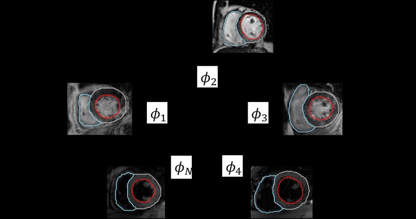
Fig. 3. Dice scores of MAS results using NI × t atlases, where NI denotes the number
of subjects used in each groupwise registration and t counts the number of groupwise
registrations performed before label fusion.
(”MVF-MvMM”) can produce a better segmentation accuracy, reaching an av-
erage Dice score of 0.871 ± 0.0253 , comparable to the inter-observer variability
of 0.878 ± 0.014 reported in [26].
3.2 gMvMM-RegNet for MAS on LGE CMR
Materials and baseline. In this experiment, we explored MAS with the appli-
cation of Eq. (10) on MS-CMRSeg challenge dataset [25]. The dataset consists of
45 patients scanned using three CMR sequences, i.e. the LGE, T2 and bFFSP,
from which 20 patients were chosen in random for training, 5 for validation
and 20 for testing. We implemented inter-subject and inter-modality groupwise
registration and evaluated the MAS results on LGE CMR images.
A 2D version of the network architecture described in Section 2.3 was devised
to jointly predict the deformation fields for NI atlases by optimizing Eq. (9). The
MAS result was generated by t times of groupwise registration over NI randomly
sampled subjects followed by label fusion using Eq. (10).
Results and discussion. The comparison between SAS and MAS highlights
that more accurate and realistic segmentation is generated by groupwise regis-
tration than pairwise registration, especially for apical and basal slices4 . Fig. 3
further reports the mean Dice scores for each cardiac substructure obtained from
MAS using t times of groupwise registration with NI subjects. With a fixed total
number of atlases, label fusion on 2D slices resulting from groupwise registration
outperforms those from conventional pairwise registration, reaching the average
myocardium Dice score of 0.783 ± 0.082. However, we also observe decline in
accuracy when having a large number of subjects (NI ≥ 5) to be groupwise reg-
istered. This discrepancy could be attributed to the lack of network parameters
compromising the predicted deformations.
3
See Fig.2 in the supplementary material for evaluation statistics on all cardiac sub-
structure.
4
See Fig. 3 in the supplementary material for visualization of the segmentation results.MvMM-RegNet 9
4 Conclusion
In this work, we propose a probabilistic image registration framework based
on multivariate mixture model and neural network estimation, coupling group-
wise registration and multi-atlas segmentation in a unified fashion. We have
evaluated two applications of the proposed model, i.e. SAS via pairwise registra-
tion and MAS unified by groupwise registration, on two publicly available cardiac
image datasets and compared with state-of-the-art methods. The proposed ap-
pearance model along with MvMM has shown its efficacy in realizing registration
on cardiac medical images characterizing inferior intensity class correspondence.
Our method has also proved its superiority over conventional pairwise registra-
tion algorithms in terms of segmentation accuracy, highlighting the advantage
of groupwise registration as a subroutine to MAS.
Acknowledgement
This work was supported by the National Natural Science Foundation of
China (grant no. 61971142).
References
1. Abadi, M., Agarwal, A., Barham, P., Brevdo, E., Chen, Z., Citro, C., Corrado,
G.S., Davis, A., Dean, J., Devin, M., Ghemawat, S., Goodfellow, I.J., Harp, A.,
Irving, G., Isard, M., Jia, Y., Józefowicz, R., Kaiser, L., Kudlur, M., Levenberg,
J., Mané, D., Monga, R., Moore, S., Murray, D.G., Olah, C., Schuster, M., Shlens,
J., Steiner, B., Sutskever, I., Talwar, K., Tucker, P.A., Vanhoucke, V., Vasudevan,
V., Viégas, F.B., Vinyals, O., Warden, P., Wattenberg, M., Wicke, M., Yu, Y.,
Zheng, X.: Tensorflow: Large-scale machine learning on heterogeneous distributed
systems. ArXiv abs/1603.04467 (2015)
2. Ashburner, J., Friston, K.J.: Unified segmentation. NeuroImage 26, 839–851 (2005)
3. Avants, B.B., Epstein, C.L., Grossman, M., Gee, J.C.: Symmetric diffeomorphic
image registration with cross-correlation: Evaluating automated labeling of elderly
and neurodegenerative brain. Medical image analysis 12 1, 26–41 (2008)
4. Balakrishnan, G., Zhao, A., Sabuncu, M.R., Guttag, J.V., Dalca, A.V.: Voxel-
morph: A learning framework for deformable medical image registration. IEEE
Transactions on Medical Imaging 38, 1788–1800 (2019)
5. Balci, S.K., Golland, P., Shenton, M.E., Wells, W.M.: Free-form b-spline deforma-
tion model for groupwise registration. Medical image computing and computer-
assisted intervention : MICCAI ... International Conference on Medical Image
Computing and Computer-Assisted Intervention 10 WS, 23–30 (2007)
6. Bhatia, K.K., Aljabar, P., Boardman, J.P., Srinivasan, L., Murgasova, M., Coun-
sell, S.J., Rutherford, M.A., Hajnal, J.V., Edwards, A.D., Rueckert, D.: Groupwise
combined segmentation and registration for atlas construction. Medical image com-
puting and computer-assisted intervention : MICCAI ... International Conference
on Medical Image Computing and Computer-Assisted Intervention 10 Pt 1, 532–
40 (2007)10 Xinzhe Luo et al.
7. Bhatia, K.K., Hajnal, J.V., Hammers, A., Rueckert, D.: Similarity metrics for
groupwise non-rigid registration. Medical image computing and computer-assisted
intervention : MICCAI ... International Conference on Medical Image Computing
and Computer-Assisted Intervention 10 Pt 2, 544–52 (2007)
8. Dalca, A.V., Balakrishnan, G., Guttag, J.V., Sabuncu, M.R.: Unsupervised learn-
ing of probabilistic diffeomorphic registration for images and surfaces. Medical
image analysis 57, 226–236 (2019)
9. Dalca, A.V., Yu, E.M., Golland, P., Fischl, B., Sabuncu, M.R., Iglesias, J.E.: Un-
supervised deep learning for bayesian brain mri segmentation. In: MICCAI (2019)
10. Hill, D.L.G., Batchelor, P.G., Holden, M., Hawkes, D.J.: Medical image registra-
tion. Physics in medicine and biology 46 3, R1–45 (2001)
11. Hu, Y., Modat, M., Gibson, E., Li, W., Ghavami, N., Bonmati, E., Wang, G.,
Bandula, S., Moore, C.M., Emberton, M., Ourselin, S., Noble, J.A., Barratt, D.C.,
Vercauteren, T.: Weakly-supervised convolutional neural networks for multimodal
image registration. Medical Image Analysis 49, 1–13 (2018)
12. Iglesias, J.E., Sabuncu, M.R.: Multi-atlas segmentation of biomedical images: A
survey. Medical image analysis 24 1, 205–219 (2014)
13. Iglesias, J.E., Sabuncu, M.R., Leemput, K.V.: A unified framework for cross-
modality multi-atlas segmentation of brain mri. Medical Image Analysis 17, 1181–
1191 (2013)
14. Khalil, A., Ng, S.C., Liew, Y.M., Lai, K.W.: An overview on image registration
techniques for cardiac diagnosis and treatment. Cardiology research and practice
(2018)
15. Kingma, D.P., Ba, J.: Adam: A method for stochastic optimization. CoRR
abs/1412.6980 (2014)
16. Leemput, K.V., Maes, F., Vandermeulen, D., Suetens, P.: Automated model-based
tissue classification of mr images of the brain. IEEE Transactions on Medical Imag-
ing 18, 897–908 (1999)
17. Maes, F., Collignon, A., Vandermeulen, D., Marchal, G., Suetens, P.: Multimodal-
ity image registration by maximization of mutual information. IEEE Transactions
on Medical Imaging 16, 187–198 (1997)
18. Mäkelä, T., Clarysse, P., Sipilä, O., Pauna, N., Pham, Q.C., Katila, T., Magnin,
I.E.: A review of cardiac image registration methods. IEEE Transactions on Med-
ical Imaging 21, 1011–1021 (2002)
19. Pluim, J.P.W., Maintz, J.B.A., Viergever, M.A.: Mutual-information-based regis-
tration of medical images: a survey. IEEE Transactions on Medical Imaging 22,
986–1004 (2003)
20. Pohl, K.M., Fisher, J.W., Grimson, W.E.L., Kikinis, R., Wells, W.M.: A bayesian
model for joint segmentation and registration. NeuroImage 31, 228–239 (2006)
21. Smith, L.N.: Cyclical learning rates for training neural networks. 2017 IEEE Winter
Conference on Applications of Computer Vision (WACV) pp. 464–472 (2015)
22. Sotiras, A., Davatzikos, C., Paragios, N.: Deformable medical image registration:
A survey. IEEE Transactions on Medical Imaging 32, 1153–1190 (2013)
23. Viergever, M.A., Maintz, J.B.A., Klein, S., Murphy, K., Staring, M., Pluim, J.P.W.:
A survey of medical image registration - under review. Medical image analysis 33,
140–144 (2016)
24. de Vos, B.D., Berendsen, F.F., Viergever, M.A., Sokooti, H., Staring, M., Išgum,
I.: A deep learning framework for unsupervised affine and deformable image regis-
tration. Medical Image Analysis 52, 128143 (2018)MvMM-RegNet 11
25. Zhuang, X.: Multivariate mixture model for myocardial segmentation combining
multi-source images. IEEE Transactions on Pattern Analysis and Machine Intelli-
gence 41, 2933–2946 (2019)
26. Zhuang, X., Li, L., Payer, C., Štern, D., Urschler, M., Heinrich, M.P., Oster, J.,
Wang, C., Smedby, Ö., Bian, C., Yang, X., Heng, P.A., Mortazi, A., Bagci, U.,
Yang, G., Sun, C., Galisot, G., Ramel, J.Y., Brouard, T., Tong, Q., Si, W., Liao,
X., Zeng, G., Shi, Z., Zheng, G., Wang, C., MacGillivray, T., Newby, D.E., Rhode,
K.S., Ourselin, S., Mohiaddin, R., Keegan, J., Firmin, D.N., Yang, G.: Evaluation
of algorithms for multi-modality whole heart segmentation: An open-access grand
challenge. In: Medical Image Analysis (2019)
27. Zhuang, X., Shen, J.: Multi-scale patch and multi-modality atlases for whole heart
segmentation of mri. Medical image analysis 31, 77–87 (2016)You can also read