Unexpected intracranial location of a Cephenemyia stimulator larva in a roe deer, Capreolus capreolus, revealed by computed tomography - SECEM
←
→
Page content transcription
If your browser does not render page correctly, please read the page content below
Galemys, 33: xx-xx, 2021
                                                                                                    ISSN 1137-8700
                                                                                                  e-ISSN 2254-8408
                                                                                      DOI: 10.7325/Galemys.2021.A2

Unexpected intracranial location of a Cephenemyia stimulator larva
in a roe deer, Capreolus capreolus, revealed by computed tomography
Inesperada ubicación intracraneal de una larva de Cephenemyia stimulator en un corzo,
Capreolus capreolus, detectada mediante tomografía computerizada

Luis E. Fidalgo1, Ana M. López-Beceiro1, Carlos Martínez-Carrasco2, Noelia
Caparrós-Fontarosa3, Antonio Sánchez3, Mónica Vila1, Daniel Barreiro1, Mathieu
Sarasa4 & Jesús M. Pérez5,6 *
1. Departamento de Ciencias Clínicas Veterinarias, Universidad de Santiago de Compostela, Avda. Carballo Calero
   s.n., 22741 Lugo, Spain.
2. Departamento de Sanidad Animal, Campus de Excelencia Internacional Regional “Campus Mare Nostrum”,
   Universidad de Murcia, Campus Espinardo, 30100 Murcia, Spain.
3. Departamento de Biología Experimental, Universidad de Jaén, Campus Las Lagunillas s.n., 23071 Jaén, Spain.
4. BEOPS, 1 Esplanade Compans Caffarelli, 31000 Toulouse, France.
5. Departamento de Biología Animal, Biología Vegetal y Ecología, Universidad de Jaén, Campus Las Lagunillas s.n.,
   23071 Jaén, Spain.
6. Wildlife Ecology & Health group (WE&H).
*Corresponding author: jperez@ujaen.es

             Abstract
             In this study we describe the finding of a Cephenemyia stimulator larva in the brain of a roe deer (Capreolus
             capreolus) after performing a computed tomography (CT) scan of its head. Despite this anatomical
             location of oestrid larvae could be relatively frequent in other genera, such as Oestrus, to our knowledge,
             this is the first reported case involving the genus Cephenemyia. Concretely, a second-instar C. stimulator
             larva was found in the basis of the cranium. The location of a macroscopic hemorrhagic lesion involving
             the brain parenchyma peripheral to the location of the larva suggests that tissue colonization occurred
             before the animal was hunted. Since no detectable alterations or damage to the cranial bones were
             observed, we suggest a possible larval migration route drilling the skull bones. Finally, we propose the
             use of the term “neuromyiasis” to be referred to the invasion of the central nervous system by dipteran
             larvae, particularly oestrids.
             Keywords: Cephenemyia stimulator, cerebral myiasis, computed tomography, neuromyiasis, roe deer

             Resumen
             En este estudio describimos el hallazgo de una larva de Cephenemyia stimulator en el cerebro de un
             corzo (Capreolus capreolus) tras realizar una tomografía computerizada (TC) de su cabeza. Aunque esta
             localización anatómica de larvas de Oéstridos puede ser relativamente frecuente en otros géneros, como
             Oestrus, que sepamos, este es el primer caso que involucra al género Cephenemyia. Concretamente, una
             larva de segundo estadio de C. stimulator se encontró en la base del cráneo. La localización de una
             lesión macroscópica hemorrágica que afectaba al parénquima cerebral periférico a la ubicación de la
             larva sugiere que la colonización del tejido se produjo antes de que el animal fuese abatido. Dado que
             no se detectaron alteraciones o daños en los huesos craneales, sugerimos una posible ruta de migración
             larvaria a través de perforaciones de los huesos del cráneo. Finalmente, proponemos el uso del término
             “neuromiasis” para referirnos a la invasión del sistema nervioso central del hospedador por larvas de
             dípteros, particularmente de Oéstridos.
             Palabras clave: Cephenemyia stimulator, corzo, miasis cerebral, neuromiasis, tomografía computerizada

                                                                                                                        1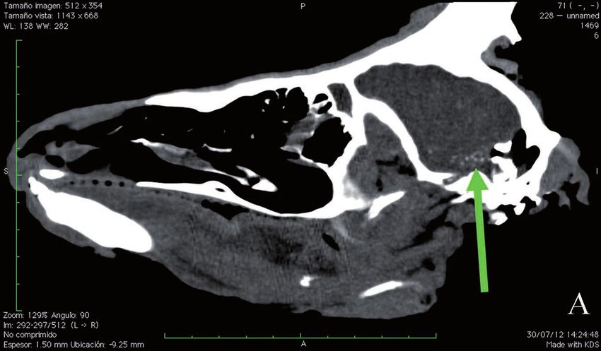
Galemys 33, 2021

Introduction                                              old child after detecting the hematoma produced
                                                          by the larva by means of computed tomography
    Cephenemyia stimulator (Clark, 1815) causes           (Kalelioglu et al. 1989).
naso-pharyngeal myiasis in roe deer, Capreolus                In Europe, Cephenemyia stimulator was described
capreolus (Linnaeus, 1758), throughout the                in the early XIXth century and its veterinary
Palaearctic (Colwell et al. 2006). This oestrid species   importance is known since the first half of the XXth
has unusually been found infesting red deer, Cervus       (Ullrich 1938, Zumpt 1965, Dudzinski 1970). It
elaphus Linnaeus, 1758 (Király & Egri 2004) and           has been reported from Austria (Kutzer 2000),
an uncommon infection by the moose throat bot             the Czech Republic (Salaba et al. 2013), Estonia
fly, C. ulrichii, in a roe deer has also been reported    (Jögisalu 2010), Fennoscandia (Norway, Sweden,
in Finland (Nilssen et al. 2008). Cephenemyia             Finland and Denmark) (Stéen et al. 1998), France
stimulator larvae usually develop closely each other      (Maes & Bullard 2001), Germany (Nickel et al.
within a single or various pouches in the naso-           1986), Hungary (Sugár 1974), Italy (Rivosecchi et
olfactive area and, with certain frequency, in the        al. 1978) and Poland (Drozdz 1961).
oesophagous and respiratory organs of the host,               The first cite of C. stimulator in Spain comes
such as trachea and lungs (bronchioles) (Dudzinski        from Ciudad Real and is recent (Notario &
1970, Bernard & Biesemans 1975). C. stimulator            Castresana 2001). In northwestern Spain, this
larvae have also been found in the eustachian tube,       parasite was found by the first time in 2005 (Arias
in the arytenoid cavity and near the hypophysis           et al. 2016). In 2011 and 2012 it was reported
of a parasitized roe deer (Ullrich 1938) and might        parasitizing roe deer in Cataluña (de la Fuente
occasionally be swallowed and pass through the            2014) and Extremadura (Calero-Bernal & Habela
digestive tract of the host (Blickle 1956).               2013), respectively. This oestrid has also been
    Larval migratory routes of first-instar Hypoderma     collected from roe deers from Cantabria and País
spp. larvae include mainly connective tissues and         Vasco (Arias et al. 2014). In Galicia, Asturias
nerves (Colwell 2006). An intracranial myiasis            and León, seropositive animals were detected
in a horse caused by a first-instar Hypoderma             since 2007 and the seroprevalence for the period
bovis (Linnaeus 1758) larva was associated with           2007-2014 reached 38% (Arias et al. 2016). In
incoordination of gait, circling to the left, head tilt   an epidemiological survey on cephenemyiosis
to the right, partial paralysis of the face, impaired     in roe deer in Galicia, based on direct diagnosis,
vision and, after necropsy, with haemorrhage              prevalence was slightly lower: 31% (López-Beceiro
and oedema in the brain tissue close to the larva         et al. 2015). Within this context, the computed
(Hadlow et al. 1977).                                     tomography (CT), as a no-invasive technique,
    Aberrant migration of Cuterebra larvae into the       was adapted to detect Cephenemyia larvae within
central nervous system of cats and dogs were also         intact (not necropsied) roe deer heads (Fidalgo et
described (Cook et al. 1985, Sartin et al. 1986, Glass    al. 2015).
et al. 1998). Histopathological findings included             This work describes a case with an unusual
presence of parasitic track lesions, superficial          location of a Cephenemyia stimulator larva in the
laminar cerebrocortical necrosis, cerebral infarction,    brain of a roe deer, which was first detected with
subependymal rarefaction and astrogliosis and             the aid of the CT.
subpial astrogliosis. These features were related to
the feline ischemic encephalopathy (Glass et al.
1998).                                                    Materials and methods
    With regards to human hosts, several fatal
cases of cerebral myiasis caused by the warble fly,          Material used in this study was the head of an
Dermatobia hominis (Linnaeus Jr. In Pallas, 1781),        adult male roe deer, Capreolus capreolus, (4 yr-old),
were described (Dunn 1934, Rossi & Zucoloto               which was selectively hunted (trophy) in 27th June
1973). Such cases involved young patients (children       2012 in Santa Colomba de Somoza Council (León
aging less than 2 years) and Dermatobia larvae, which     province, northwestern Spain). This animal was
commonly cause cutaneous myiasis in humans,               repeatedly observed during the two previous months.
entered the cerebral cavity through the bregmatic         When shot, the animal was in good condition and
fontanelle. One case of intracerebral myasis due to       showed no signs of abnormal behaviour and/or
a Hypoderma bovis larva was diagnosed in an 8 yr-         locomotion. The head was removed, introduced in

2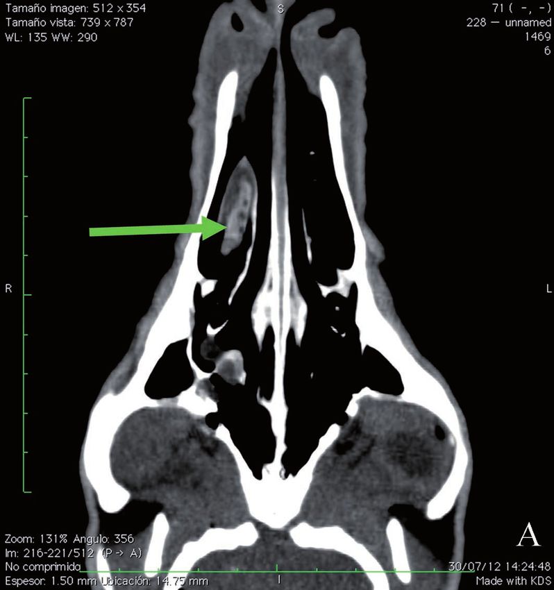
Finding of a Cephenemyia larva in the brain of a roe deer                                     L. E. Fidalgo et al.

a plastic bag and kept at 4ºC in a refrigerator until       instar larvae in the encephalum, concretely at
CT analysis.                                                the level of the middle fossa (Fig. 3). Brain
   Twenty hours after being harvested, the head             parenchyma surrounding this larva presented
of this roe deer was analyzed with an ECLOS                 clear macroscopic hemorrhagic lesions.
16™system (Hitachi Medical Systems, Inc., Tokyo,
                                                               The morphology and size of collected
Japan) with acquisition parameters of 120 kVp, 150
                                                            larvae fitted the descriptions of Cephenemyia
mA and 1 s per rotation. Scans of the deer head
were obtained in sections of 1.25 mm and 0.63               stimulator given by Zumpt (1965) and Bernard
mm in thickness, using soft tissue and bone filters,        & Biesemans (1975). Sequences obtained from
respectively (Fidalgo et al. 2015).                         positive clones (GenBank accession numbers:
   After CT analysis, the head was cut as described         MG763915 and MG763916) were practically
by Fidalgo et al. (2015) and all anatomical parts were      identical each other and reached a 99.4 % of
carefully examined looking for oestrid larvae and           identity with regards to the COI sequence for
posible associated lesions. Oestrid larvae found were       C. stimulator available at the GenBank.
individually fixed in absolute ethanol. Their mor-             The fact of finding C. stimulator larvae of
phological identification was carried out following         different instars simultaneously in the same
descriptions by Zumpt (1965) and Bernard &
                                                            host could be explained by the production of
Biesemans (1975). Larval DNA was extracted by
                                                            several larval generations per year (Dudzinski
means of the HotSHOT technique (Truett et al.
2000). Two specific primers were used to amplify a          1970), by the ability of first-instar larvae to
689 bp fragment of the cytochrome c oxidase subunit         become hypobiotic and overwinter into the host
I (COI) (UEA7: ´-TACAGTTGGAATAGACGTT-                       head cavities and/or also by an asynchronous
GATAC-3´; UEA10: -TCCAATGCACTAATCT-                         development of each larva (Colwell et al.
GCCATATTA-3´) previously described by Zhang                 2006). In our case, only one larva was found
& Hewitt (1997). Amplification products were                associated with macroscopic hemorrhagic
resolved in 1% agarose gels stained with ethidium           lesions of the surrounding brain tissue, but
bromide, purified with the QIAamp Gel Extration             no clinical nervous signs in the animal before
Kit (Qiagen) and cloned in JM109 bacteria using             being shot and, as could be expected (Rossi
PGEMT-easy vector (Promega). Finally, two positive
                                                            & Zucoloto 1973). This ectopic location
clones were sequenced in both directions.
                                                            without apparent signs of disease could be
                                                            explained by a very recent migration of the
Results and Discussion                                      larva or even by a post-mortem migration.
                                                            Dudzinski (1970) suggested that, if necropsy
   The CT revealed the presence of ovoid                    is delayed with regards to the host death, then
structures in nasal and ethmoidal turbinates                larvae can move within the head cavities and
of the right side (Fig. 1A, B). They were well              reach a “random” distribution. In our case, the
delimitated from the surrounding tissues                    short time elapsed between sample collection
and previous results indicated that they were               and CT analysis, and the fact that during this
probably oestrid larvae. CT also detected                   period it was maintained at 4 ºC, make larval
ovoid structures of soft-tissue and well                    movement very unlikely. Histologic variation
delimitated ovoid structures in the right side              of lesions caused by Hypoderma lineatum
of the cranium basis, at the level of the middle            suggests that larval migration in the horse brain
fossa, with small radio-transparent foci (Fig.              can last several days (Olander 1967). In fact, we
2A, B). No osteolysis was observed in the                   found a “normal” location of the 8 third-instar
nasal turbinates or in the nasal septum, nor                larvae and an absence of larvae in the proximal
mucous thickening or associated rhinitis. No                section of the trachea and esophagous. On
macroscopic alterations of the cranium bones                the other hand, the presence of surrounding
nor the encephalic tissue were detected. After              hemorrhagic lesions is associated with active
performing necropsy, eight third-instar larvae              blood circulation through the vascular system
were found in the nasal passages and a second-              (Brooks 2016).

                                                                                                                3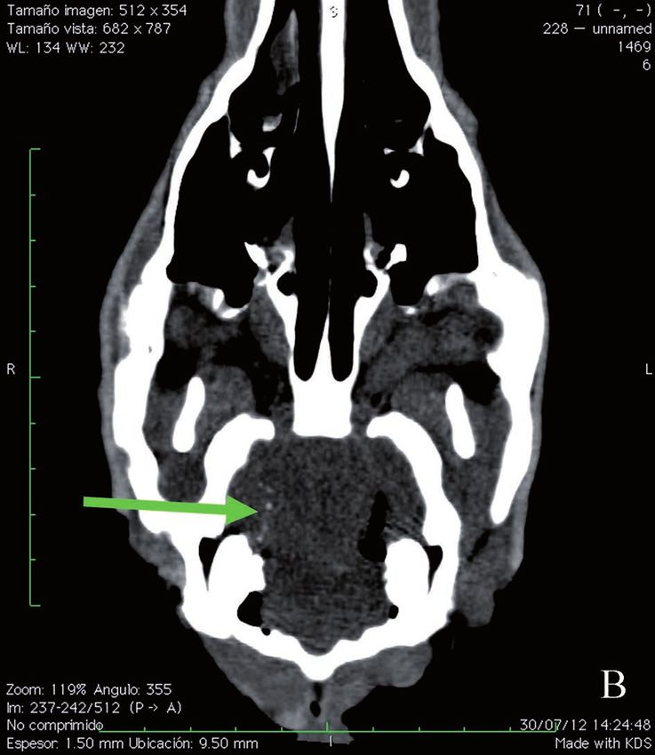
Galemys 33, 2021

    Moreover, the ability of the larva to reach the        medium-long term. Anyway, this hypothesis implies
cerebral cavity without causing damage to cranial          certain ability of the larva to perforate soft tissues.
bones is noteworthy, in particular taking into                When possible, the methods commonly used
account that it was a second-instar larva, with a          to diagnose cephenemyiosis in roe deer and other
relatively large size (> 4 mm in diameter). Possible       hosts, would benefit from being complemented
larval migration routes include the labyrinthus            with observational studies of the host behaviour in
ethmoidalis, through the meatus ethmoidalis to             order to address the frequency of the cases like that
reach the cribosum plate of the ethmoides, which           described here (which was the only one of 75 scans
delimitates the nasal and cranial cavities each other.     made). Finally, we want to emphasize that the brain
This is a very thin plate containing numerous              is not usually sampled in necropsies of ungulate
orifices. After passing this plate, the larva could have   heads for collecting oestrid larvae. Therefore, the
followed the sub-dural via to reach its final location.    use of computerized tomography can become very
If so, and even in the case of one or several nervous      useful for determining the real frequency of this
connections become damaged, few or none host               “aberrant” larval location in this and other hosts,
behavioural signs would be expected both at short or       even when still alive.

                                                                      Figure 1. A) Sagittal computed tomography
                                                                         scan of the roe deer head. Arrow points a
                                                                         well-delimitated ovoid structure (a third-
                                                                           instar oestrid larva). B) Axial computed
                                                                       tomography. Arrow points well-delimitated
                                                                      ovoid structures (third-instar oestrid larvae).

4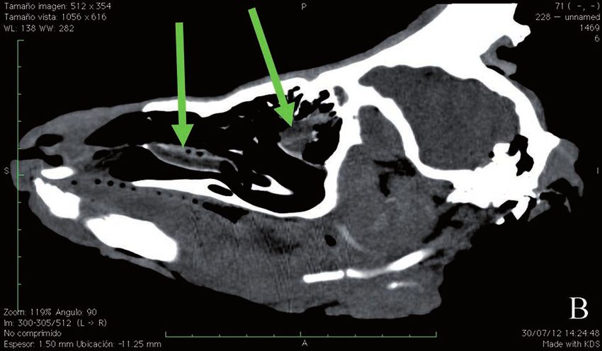
Finding of a Cephenemyia larva in the brain of a roe deer L. E. Fidalgo et al.
Figure 2. A) Axial computed tomography scan
of the roe deer head. Arrow points soft-tissue
and well-delimitated ovoid structures in the
cranium basis, at the level of the middle fossa,
with small radio-transparent foci. In this location,
a second-instar Cephenemyia stimulator larvae
was found after necropsy. B) Sagittal computed
tomography scan. Arrow points soft-tissue and
well-delimitated ovoid structures in the right side
of the cranium basis, with small radio-transparent
foci. In this location, a second-instar Cephenemyia
stimulator larvae was found after necropsy
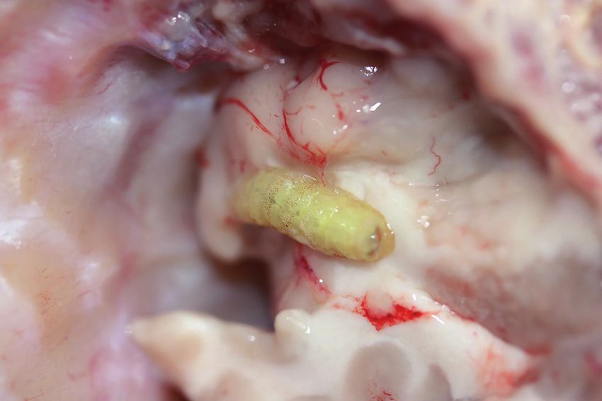
Figure 3. Location of a
second-instar Cephenemyia
stimulator larva in the host
brain of the roe deer after
necropsy. The parenchyma
of the brain peripheral to
the location of the larva
appeared embedded in
hemorrhagic fluid.
5Galemys 33, 2021
In conclusion, despite this is the first reported The Oestrid Flies: Biology, Host-parasite Relationships,
case in which a Cephenemyia stimulator larva has Impact and Management. CABI Publishing,
been found within the host brain, the occurrence Wallingford.
and implications of the invasion of the cranial cavity Colwell D.D., Hall M.J.R. & Scholl P.J. 2006. A synopsis
of the biology, hosts, distribution, disease significance
by larvae of Oestridae species underline the need for
and management of the genera. Pp. 220-305. In: D.D.
a reappraisal of our understanding of host-myiasis Colwell, M.J.R. Hall & P.J. Scholl (eds). The Oestrid
interactions. For cases involving the invasion of Flies: Biology, Host-parasite Relationships, Impact and
the central nervous system by dipteran larvae, we Management. CABI Publishing, Wallingford.
propose the use of the term “neuromyiasis”. Cook J.R. Jr., Levesque D.C. & Nuehring L.P. 1985.
Intracranial cuterebral myiasis causing acute
Acknowledgements lateralizing meningoencephalitis in two cats. Journal
of the American Animal Hospital Association, 21:
This study was funded by the Spanish Federation of
279-284.
Hunters (FEC), the Foundation for the Study and
de la Fuente A.M. 2014. Análisis morfológico y molecular
Defence of the Nature and Game (FEDENCA), and
de especies de la subfamilia Oestrinae (Diptera,
the National Federation of Hunters (France) (Project:
Oestridae). Master Thesis, University of Jaén, Spain,
FNC-PSN-PR5-2013). The research activities of NC-
69 pp.
F, AS and JMP are partially supported by the Junta de
Drozdz J. 1961. Cephenomyinae (Diptera: Oestridae)
Andalucía, Plan Andaluz de Investigación (BIO-220 and
of cervids in Poland. Wiadomosci Parazytologiczne, 8:
RNM-118 groups). The procedures used in this study
381-382.
were carried out in compliance with Spanish legislation
Dudzinski W. 1970. Studies on Cephenemyia stimulator
on animal experimentation and welfare.
(Clark) (Diptera, Oestridae), the parasite of European
roe deer, Capreolus capreolus (L.). I. Biology. Acta
References Parasitologica Polonica, 18: 555-572.
Dunn L.H. 1934. Prevalence and importance of the
Arias M.S., Pajares G., Díez-Baños N., Pérez-Creo tropical warble fly Dermatobia hominis Linn. in
A., Prieto A., Díez-Baños P. & Morrondo P. 2016. Panama. Journal of Parasitology, 20: 219-226.
Cephenemyiosis, an emergent myiasis in roe deer Fidalgo L.E., López-Beceiro A.M., Vila-Pastor M.,
(Capreolus capreolus) from northwestern Spain. Martínez-Carrasco C., Barreiro-Vázquez J.D. & Pérez
Parasitology Research, 115: 4605-4610. DOI: J.M. 2015. Use of computed tomography as a non-
10.1007/s00436-016-5251-7 invasive method for diagnosing cephenemyiosis in
Arias M.S., Pajares G., Paz-Silva A., Díez-Baños N., roe deer (Capreolus capreolus). Medical and Veterinary
Suárez J.L., Díez-Baños P., Sánchez-Andrade R. & Entomology, 29: 110-113. DOI: 10.1111/mve.12087
Morrondo P. 2014. Antigen characterization from Glass E.N., Cornetta A.M., de Lahunta A., Center S.A.
second instars of oestrid bot flies for the detection of & Kent M. 1998. Clinical and clinicopathologic
anti-Cephenemyia stimulator antibodies by ELISA in features in 11 cats with Cuterebra larvae myiasis in the
roe deer (Capreolus capreolus). Medical and Veterinary central nervous system. Journal of Veterinary Internal
Entomology, 28 Suppl. 1: 83-89. DOI: 10.1111/ Medicine, 12: 365-368. DOI: 10.1111/j.1939-
mve.12080 1676.1998.tb02136.x
Bernard J. & Biesemans W. 1975. A propos des oestrides Hadlow W.J., Ward J.K. & Krinsky W.L. 1977.
parasites du cheuvreuil en Belgique. Bulletin Annuelle Intracranial myiasis by Hypoderma bovis (Linnaeus)
de la Societé Royale Belge d’Entomologie, 111: 71-95. in a horse. Cornell Veterinary, 67: 272-281.
Blickle R.L. 1956. Notes on the life history of Jögisalu I. 2010. Roe deer nose botfly (Cephenemyia
Cephenomyia phobifer Clark (Diptera). Entomological stimulator Clark, 1815) (Diptera: Oestridae) larvae
News, 67: 13-14. and helminths impact on the European roe deer
Brooks J.W. 2016. Postmorten changes in animal (Capreolus capreolus Linnaeus, 1758). Master Thesis.
carcasses and estimation of the postmortem University of Tartu, Estonia, 48 pp.
interval. Veterinary Pathology, 53: 929-940. DOI: Kalelioglu M., Aktürk G., Aktürk F. et al. 1989.
10.1177/0300985816629720 Intracerebral myiasis from Hypoderma bovis larva in
Calero-Bernal R. & Habela M.A. 2013. First report of a child. Case report. Journal of Neurosurgery, 71: 929-
Cephenemyia stimulator (Diptera, Oestridae) parasitizing 931. DOI: 10.3171/jns.1989.71.6.0929
roe deer (Capreolus capreolus) in Extremadura (Spain). Király I. & Egri B. 2004. Naso-pharyngeal bot infestation
Galemys, Spanish Journal of Mammalogy, 25: 29-34. of the roe deer population of Tolna county. Magyar
DOI: 10.7325/Galemys.2013.A03 Allatorvosok Lapja, 126: 433-438.
Colwell D.D. 2006. Life cycle strategies. Pp. 67-77. Kutzer E. 2000. The treatment of oestrinosis and
In: D.D. Colwell, M.J.R. Hall & P.J. Scholl (eds). hypodermosis in red deer (Cervus elaphus hippelaphus)
6Finding of a Cephenemyia larva in the brain of a roe deer L. E. Fidalgo et al.
and roe deer (Capreolus c. capreolus) by means of stimulator and Hypoderma diana infection of roe
ivermectin (Ivomec (R)). Berliner und Munchener deer in the Czech Republic over an 8-year period.
Tierarztliche Wochenschrift, 113: 149-151. Parasitology Research, 112: 1661-1666. DOI:
López-Beceiro A.M., Martínez-Carrasco C., Pérez J.M., 10.1007/s00436-013-3322-6
Colins B. & Fidalgo, L.E. 2015. Prevalencia de Sartin E.A., Hendrix C.M., Dillehay D.L. & Nicholls
Cephenemyia stimulator en Galicia. 33èmes Rencontres B. 1986. Cerebral cuterebrosis in a dog. Journal of
du GEEFSM, Balme, Italy. the American Veterinary Medicine Association, 189:
Maes S. & Boulard C. 2001. Deer myiasis in France. 1338-1339.
Pp. 181-186. En: M. Good et al. (eds.). Mange and Stéen M., Faber W.E. & Oksanen A. 1998. Disease and
myiasis of livestock. COST Action 833. genetical investigations of Fennoscandian Cervids - a
Nickel E.A., Danner G. & Stubbe I. 1986. review. Alces, 34: 287-310.
Morphologische und metrische untersuchungen Sugár L. 1974. The occurence of nasal throat bot
an larven I von Cephenemyia stimulator (Diptera, flies (Oestridae) in wild ruminants in Hungary.
Oestridae). Angewandte Parasitologie, 27: 187-192. Parasitologia Hungarica, 7: 181-189.
Nilssen A.C., Isomursu M. & Oksanen A. 2008. Truett G.E., Heeger P., Mynatt R.L., Truett A.A.,
The moose throat bot fly Cephenemyia ulrichii Walker, J.A. & Warman M.L. 2000. Preparation of
larvae (Diptera: Oestridae) found developing in PCR-quality mouse genomic DNA with hot sodium
roe deer (Capreolus capreolus) for the first time. hydroxide and tris (HotSHOT). BioTechniques: 29,
Acta Veterinaria Scandinavica, 50: 14. DOI: 52-54. DOI: 10.2144/00291bm09
10.1186/1751-0147-50-14 Ullrich H. 1938. Zur biologie der Rachenbremsen unseres
Notario A. & Castresana L. 2001. Contribution to the einheimischer Wildes, Genus Cephenomyia Latreille
knowledge of Cephenemyia stimulator Clark, 1815 und Genus Pharyngomyia Schiner. Verhandlungen VII
(Diptera, Oestridae) in Spain. Folia Venatoria, 30-31: Kongress Entomologie, III: 2149-2162.
325-326. Zhang D.X. & Hewitt G.M. 1997. Assessment of
Olander H.J. 1967. The migration of Hypoderma the universality and utility of a set of conserved
lineatum in the brain of a horse. A case report and mitochondrial COI primers in insects. Insect
review. Veterinary Pathology, 4: 477-483. Molecular Biology, 6: 143-150. DOI: 10.1111/j.1365-
Rivosecchi L., Zanin E., Cavallini C. & De Paoli 2583.1997.tb00082.x
C. 1978. Infestation of roe deer by Cephenemyia Zumpt F. 1965. Myiasis in man and animals in the Old
stimulator (Clark) (Diptera, Oestridae) in Trent World. Butterworths, London. 267 pp.
province. Parassitologia, 20: 143-152.
Rossi M.A. & Zucoloto S. 1973. Fatal cerebral myiasis
caused by the tropical warble fly, Dermatobia hominis.
American Journal of Tropical Medicine and Hygiene, Submitted: 5 November 2020
22: 267-269. DOI: 10.4269/ajtmh.1973.22.267 Accepted: 14 February 2021
Salaba O., Vadlejch J., Petrtyl M., Valek P., Kudrnacova
M., Jankovska I., Bartak M. et al. 2013. Cephenemyia Associate editor was Francisco Ruiz Fons
7You can also read