The wide spectrum of ultrasound diagnosis of holoprosencephaly
←
→
Page content transcription
If your browser does not render page correctly, please read the page content below
Original papers                                                                                Med Ultrason 2019, Vol. 21, no. 2, 163-169
                                                                                               DOI: 10.11152/mu-1614

The wide spectrum of ultrasound diagnosis of holoprosencephaly
Cringu Antoniu Ionescu1, Simona Vladareanu2, Stefania Tudorache3, Liana Ples4,
Catalin Gabriel Herghelegiu5, Adrian Neacsu4, Dan Navolan6, Ioana Dragan5, Daniela-Nuti
Oprescu5

1Carol Davila University of Medicine and Pharmacy, Department of Obstetrics Gynecology Sf Pantelimon Emer-
gency Hospital, Bucharest, 2Carol Davila University of Medicine and Pharmacy, Department Obstetrics, Gynecology,
and Neonatology, Elias Emergency Clinical Hospital, Bucharest, 3University of Medicine and Pharmacy Craiova,
Department of Obstetrics Gynecology, Filantropia Hospital, Craiova, 4Carol Davila University of Medicine and
Pharmacy, Department of Obstetrics Gynecology, Sf Ioan Emergency Hospital, Bucharest, 5Carol Davila University
of Medicine and Pharmacy, Department of Obstetrics and Gynecology, INSMC Polizu Hospital, Bucharest, Romania,
6Victor Babeș University of Medicine and Pharmacy, Department Obstetrics Gynecology Timisoara

         Abstract
       Aim: Holoprosencephaly (HPE) is the most common brain malformation. A wide spectrum of anatomical variants are
   characterized by a lack of midline separation of the cerebral hemispheres. The aim of this study was to assess the ultrasound
   diagnostic criteria for HPE. Material and method: A database of 175 fetuses with central nervous system anomalies identified
   by ultrasound was collected retrospectively from 2006 to 2016 in this multicenter, retrospective, observational study. Among
   them 18 cases (10.2%) with HPE were identified. Results: The prevalence of HPE was 2.5:10.000 with the sex distribution
   male:female of 1:1.6. Six cases were alobar subtype, 3 were semilobar, 7 were lobar and 2 were middle interhemispheric vari-
   ant. In the second trimester, we consider that the abnormal fusion of the lateral ventricles and the absence of the cavum septum
   pellucidum are the most important landmarks for HPE. Facial abnormalities varied considerably. Conclusion: This study il-
   lustrates the heterogeneity of HPE with different cerebral and facial appearances.
       Keywords: holoprosencephaly; ultrasound; cavum septum pellucidum

    Introduction                                                     tion includes a wide spectrum of anatomical variants that
                                                                     are characterized by lack of midline separation of the
   Holoprosencephaly (HPE) represents a complex mal-                 cerebral hemispheres (telencephalon) and diencephalic
formation of the forebrain, determined by an absence or              structures, extending from the anterior to the posterior
incomplete cleavage of the prosencephalon (forebrain)                depending on the severity.
during the 4th week of embryogenesis [1,2]. The condi-                   This malformation has been suggested to be the result
                                                                     of a defect in the ventral induction and patterning of the
                                                                     rostral neural tube by the precordal mesenchyma. Since
Received 08.10.2018 Accepted 16.01.2019                              ventral induction is related to facial development, many
Med Ultrason
2019, Vol. 21, No 2, 163-169
                                                                     cases of HPE have craniofacial abnormalities, leading to
Corresponding author: Cringu Antoniu Ionescu                         a so-called “holoprosencephaly sequence” [1]. The most
                       “Carol Davila” University of                  common facial anomalies include anophthalmia, cyclo-
                       Medicine and Pharmacy,                        pia, ethmocephaly, synophthalmia, cebocephaly, probos-
                       Department of Obstetrics Gynecology,
                       Dionisie Lupu str, no 37,
                                                                     cis, median cleft lip and palate and hypotelorism present
                       020031, Bucharest, Romania                    in up to 80% of cases [3,4]. This brain-face correlation
                       email: antoniuginec@yahoo.com                 was also observed by William DeMyer, who studied a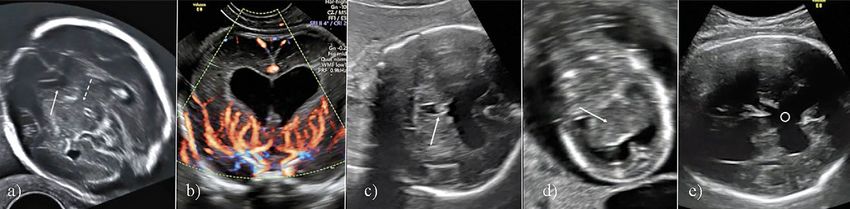
164   Cringu Antoniu Ionescu et al                                   The wide spectrum of ultrasound diagnosis of holoprosencephaly

      group of patients with HPE and in the end concluded that         used. Postnatal confirmation was made using transfonta-
      “The face predicts the brain” [5].                               nellar ultrasound in all cases, magnetic resonance imag-
          Although HPE is a rare malformation, it is nonethe-          ing (MRI) in 6 cases or by necropsy. Also, data about
      less the most common malformation of the brain and face          clinical evaluation of the newborns that survived was
      in humans, with a prevalence of around 1–1.34 in 10.000          collected. Genetic counseling and testing (amniocentesis
      births and, if aborted embryos are included in the esti-         or biopsy of the chorionic villi) was offered to all cases,
      mate, prevalence is much higher at around 1 in 200–250           but only in 10 cases the parents opted for these investiga-
      [4,6–8]. It seems that the lower the gestational age is, the     tions.
      higher the prevalence and this can be explained by the               Statistical analysis
      high intrauterine lethality of fetuses with HPE, probably            A descriptive analysis was performed and continu-
      due to the associated genetic and structural defects [9].        ous data are expressed as means and percentages. For
          The exact cause of HPE is difficult to be identified         statistical analysis and revealing the patterns in our data,
      as this pathology seems to have a multifactorial etiology        we provide univariate and bivariate distributions for our
      including teratogen exposure, genetic abnormalities and          analysed variables in terms of descriptive statistics, us-
      syndromic association [10]. Maternal diabetes is consid-         ing Pivot Tables in Excel 2016, a product of Microsoft
      ered the most important risk factor, potentially increasing      Office 365.
      the risk by 200-fold. Other risk factors include alcohol,
      cigarette smoking, salicylates, retinoic acid, and cyto-            Results
      megalovirus infection [11].
          HPE is considered by some a “continuum of fore-                  The prevalence of HPE was 2.5:10.000 with the sex
      brain malformations with no clear-cut distinction among          distribution male:female of 1:1.6. The mean fetal age
      the different subcategories” thus an accurate diagnosis of       at diagnosis of the 18 cases with HPE was 23 weeks+5
      the exact HPE subtype is often difficult to be established       days (range, 12–37 weeks). Six cases (33.3%) were diag-
      [12]. Since the prognosis of the newborn depends on the          nosed with alobar HPE, 3 (16.6%) with semilobar HPE,
      HPE subtype, we evaluated the outcome of these fetuses           7 (38.8%) with lobar HPE and 2 (11.1%) with middle
      diagnosed with HPE in the second and third trimester.            interhemispheric (MIH) variant HPE. The details about
          For the classification of the HPE cases we chose the         these cases are presented in Table I and examples of HPE
      one proposed by DeMyer et al [5], which consists of three        cases in figure 1 and 2. The mean age of mothers was
      subtypes of HPE: alobar, semilobar, and lobar, depending         27.5 years (range, 20–34 years) and no relevant medical
      on the degree of cleavage of the hemispheres. We also            history or exposure to teratogens was found in any case.
      included a more recent subtype – middle interhemispher-              The ultrasound findings found in the study group are
      ic (MIH) variant, described by Barkovich et al [13].             presented in Table II. Only 7 babies were born alive be-
                                                                       tween 36-38 weeks and from these only 2 survived until
         Materials and methods                                         one year old. In 7 cases the parents decided to terminate
                                                                       the pregnancy and there were 4 cases of intrauterine
          Patients selections                                          death.
          From a total of 71.160 births over a 10-year period
      (2006-2016) in four tertiary university hospitals in Ro-            Discussions
      mania (“Elias” Hospital, Bucharest; INSMC “Polizu”
      Hospital, Bucharest; “Sf. Pantelimon” Hospital, Bucha-               Our study confirm the variety of prenatal ultrasound
      rest; “Filantropia” Hospital, Craiova) we identified 175         findings that can be encountered in HPE and the impor-
      anomalies of the central nervous system. From these, 18          tance of ultrasonography in assessing the subtypes of this
      cases of HPE (with or without facial anomalies) with rel-        pathology, especially in relation with the prognostic of
      evant cerebral anomalies for each subtype were included.         the foetuses. The lobar subtype was the most frequent-
      Data were collected retrospectively from medical files           ly encountered, while the alobar subtype had the worst
      and approval from the local Ethics Committee was ob-             prognosis.
      tained. We excluded cases with septo-optical dysplasia,              The alobar subtype is the most severe expression of
      ventriculomegaly associated anomalies, isolated absent           HPE and is characterized by a complete failure of cleav-
      cavum septum pellucidum or other central nervous sys-            age of the prosencephalon, resulting in a single midline
      tem anomalies.                                                   forebrain with a single forebrain monoventricle [10,11].
          For prenatal ultrasonography GE Voluson 730 Pro,             In all of our 6 cases of alobar HPE the cerebral hemi-
      Voluson 730 Expert, and E8 ultrasound machines were              spheres were completely fused into a holosphere and the
Med Ultrason 2019; 21(2): 163-169 165
interhemispheric fissure was completely absent, so the roof of the ventricle, the brain was shaped from the sagit-
resulting brain was smaller than normal. Also a dorsal tal view like a ball where the cortex encircles the mono-
cyst was observed in 2 cases, these being the pathogno- ventricle in 4 cases or a cup where the monoventricle was
monic characteristics for this subtype of HPE [12]. Three not completely encircled in 2 cases. We did not encoun-
cases were diagnosed as early as the first trimester, due ter the literature reported shape of a pancake where the
to the absence of the “butterfly sign” formed normally by cortex is flattened at the base of the skull [10,12]. Since
the two choroid plexuses [14]. Depending on the degree in all our cases there was a complete fusion of the two
to which the cerebrum surrounds the dorsal membranous hemispheres, no midline structures were present, includ-
Table I. Clinical, ultrasound and genetic characteristics of the cases with holoprosencephaly
Patient HPE Corpus Face Karyotype Age at diag- Outcome
No Type callosum nostic (weeks)
1 Alobar Absent Synophthalmia absent nose proboscis NP 18 TOP
2 Alobar Absent Cyclopia proboscis NP 22 ID
3 Alobar - Hypotelorism Triploidy 12 TOP
4 Alobar - Hypotelorism NP 13 TOP
5 Alobar Absent Hypotelorism Mosaicism at 38 Birth 38w
cebocephaly chromosome 18
6 Alobar Absent Proboscis T13 14 TOP
7 Semilobar Absent Hypotelorism T13 27 ID
8 Semilobar Absent Hypotelorism T13 37 Birth 37w
9 Semilobar Absent Hypotelorism NP 24 TOP
10 Lobar Hypoplastic Hypotelorism NP 25 Birth 36 w
11 Lobar Absent Normal NP 22 TOP
12 Lobar Hypoplastic Normal T13 27 ID
13 Lobar Absent Normal NP 23 ID
14 Lobar Hypoplastic Normal Normal 28 Birth at 37w
15 Lobar Hypoplastic Hypotelorism Normal 32 Birth at 38w
16 Lobar - Hypotelorism NP 14 TOP
17 MIH Hypoplastic Normal Normal 26 Birth at 39w
18 MIH Hypoplastic Normal Normal 27 Birth at 38w
T13 – trisomy 13; No – number of patients; MIH – middle interhemispheric variant of holoprosencephaly; NP – not performed, TOP – ter-
mination of pregnancy; ID – intrauterine death
Table II. Brain ultrasound characteristics encountered for each subtype of HPE
Alobar Semilobar Lobar MIH
(n=6) (n=3) (n=7) (n=2)
Cortical hemispheres complete anterior half basal frontal posterior frontal and
fusion parietal
Interhemispheric fissure absent present posteriorly only hypoplastic anteriorly present in the anterior
and falx cerebri and present posteriorly and posterior poles
Corpus callosum absent absent or thin and hypo- thin and hypoplastic absent or thin and
plastic hypoplastic
Cavum septum pelu- absent absent absent or absent or
cidum dysplastic dysplastic
Lateral ventricles monoventricle fusion of the anterior half hypoplastic and partially fused at their middle
of the lateral ventricles fused frontal horns portion (body)
Third ventricle absent absent visible visible
Dorsal cyst present (n=2) absent absent absent
Deep grey nuclei often completely fussed incompletely separated partially fused normal
Doppler findings - - “rete of vessels” -
branching from the
internal carotids (n=1)
Head circumference macrocephaly (n=1) microcephaly (n=1) no no
n – number of cases166 Cringu Antoniu Ionescu et al The wide spectrum of ultrasound diagnosis of holoprosencephaly
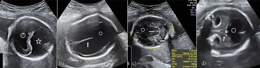
Fig 1. a) 18 weeks old fetus with alobar HPE, the cerebral hemispheres are completely fused with a single midline ventricle in the
middle (circle). In the occipital region a large dorsal cyst is present (star), pushing anteriorly the dorsal part of the prosencephalon;
b) 27 weeks old fetus with semilobar HPE: severely dilated occipital horns of the lateral ventricles, the rest of the body and frontal
horns are fused forming a single “midline ventricle” (circle). The interhemispheric fissure (arrow); c) 23 weeks fetus with semilo-
bar HPE. The cerebral hemispheres are fused in the anterior half (circle) and the BPD and HC are smaller than the 5th percentile;
d) 24 weeks old fetus with semilobar HPE: lack of cleavage of the anterior half of the hemispheres, with fused ventricles and absent
midline structures (circle), also the deep grey nuclei appear to be incompletely separated (arrow).
Fig 2. a) 27 weeks fetus with lobar HPE: hypoplastic anterior interhemispheric fissure, fused rudimentary frontal horns, absent
cavum septum pellucidum (arrow) and partially fused deep grey nuclei (dotted arrow); b) fetus of 22 weeks with lobar HPE: fused
lateral ventricles, the interhemispheric fissure is present but falx cerebri is hypoplastic, abnormal vasculature with a “rete of vessels”
branching from the internal carotids; c) 28 weeks fetus with lobar HPE: fussed lateral ventricles, hypoplastic falx cerebri partially
separating the cerebral and fussed fornices (arrow); d)fetus of 14 weeks old with hypoplastic interhemispheric fissure and falx cer-
ebri, partially fused lateral ventricles and thalami that appear to be at least partially fused (arrow); e) 38 weeks fetus of middle inter-
hemispheric HPE with both the anterior and posterior poles of the hemispheres well separated, normally defined anterior ventricular
horns but with completely fused bodies of the lateral ventricles (circle).
ing the falx cerebri, interhemispheric fissure, cavum sep- middle cerebral arteries being replaced by a “rete of ves-
tum pellucidum, or corpus callosum as in other published sels” arising from the internal carotid and basilar arteries,
cases [4]. In one case where necropsy was performed, we could not identify these features in any of our cases
it was clearly visible that the basal ganglia, hypothala- [4,12,15].
mus and thalamus nuclei were fussed in the midline so The semilobar HPE is an intermediate form in which
no third ventricle was visible, a finding that is frequently the anterior halves of the hemispheres fail to separate,
described in literature [10,15]. In all of our cases the but there is a degree of separation of the posterior hemi-
brainstem and the cerebellum were grossly normal, but spheres [1,15] with a hypoplastic posterior interhemi-
we could not be sure if there was only a single cerebral spheric fissure and falx cerebri [4,10]. All of these fea-
peduncle. We suspected this finding because there was tures were present in our 3 cases of semilobar and also
only one cerebral holosphere but we could not character- the non-cleaved lobes were smaller than normal, result-
ize the corticospinal tracts (hypoplastic or absent) com- ing in microcephaly. Another characteristic feature of
paring with other studies [4,12]. semilobar HPE that we found was that the frontal horns
Despite the fact that in most/many cases of alobar of the lateral ventricles were fused forming a single
HPE presented in literature, there is an abnormal devel- “midline ventricle”, but the posterior horns and trigones
opment of the anterior vasculature, with the anterior and were present.Med Ultrason 2019; 21(2): 163-169 167
A dorsal cyst may be present, when the thalami are Although HPE can be successfully diagnosed in the
fused and may lead to macrocephaly [12]; also the hip- first trimester [14,24] less than a quarter of our cases
pocampus is usually incompletely formed appearing nor- were diagnosed before 14 weeks, with a mean age of 23
mal only in the temporal lobes [16]. In our cases only the weeks+5 days. One explanation for this is that an im-
deep grey nuclei appear to be incompletely separated and portant percentage (50%) of our patients missed the first
no dorsal cyst was identified. trimester scan (9 fetuses). Alobar and semilobar types
Case reports described that in the most severe cases of HPE are easy to diagnose in the first trimester with
the vasculature is abnormal (similar to alobar HPE); most ultrasound, with reports of cases detected as early as 10
of the time an unpaired anterior cerebral artery displaced weeks[10]. In our series of cases, 50% (n = 3) of alobar
anteriorly being identified creating the “snake under the HPE cases were diagnosed in the first trimester, while the
skull sign” [10,15]. We did not identify this sign in our rest of the patients missed the first trimester ultrasound.
series of cases. The lobar HPE is the least severe form The characteristic finding indicating HPE in the first tri-
and is characterized by a near complete cleavage of the mester, when evaluating the fetal head, is the presence of
hemispheres, with the interhemispheric fissure present a midline “monoventricle” and the absence of the typical
along the entire midline and fused only the most ventral echogenic “butterfly” sign corresponding to the choroid
aspects of the frontal neocortex [12,15]. These features plexuses. Although, there is no consensus concerning the
were present in all our cases of lobar HPE. Furthermore, mode of delivery, in all our cases over 35 weeks, the deliv-
the falx cerebri was present anteriorly although it was ery was through cesarean section, but the reason was not
hypoplastic due to the partial fusion of the frontal lobes, fetal macrocephaly in all cases, but because of “defensive
a characteristic finding [16]. The cavum septum pelluci- medicine” leading to an increase in the incidence of CS
dum was absent in all cases, as was the most anterior part in our country [25,26]. As for milder forms of HPE, the
of the corpus callosum (usually the rostrum and genu), correct diagnosis becomes harder and harder to be estab-
but most of the posterior body and the splenium were lished as the spectrum of anatomical variants gets closer
present. Characteristic for the lobar type, the third ven- and closer to normal brain anatomy [27]. For these milder
tricle was normal and the dorsal cyst was absent [12,15]. forms identified at the second trimester ultrasound scan,
Another characteristic sign of lobar HPE, described by the most valuable clue in our opinion is the absence of the
Pilu et al [17], is the intraventricular fusion of the for- cavum septum pellucidum. This structure is easily identi-
nices, that appears on ultrasound as a hyperechogenic fied as it is visualized in the “standard” section for fetal
structure, a sign that was also identifiable in our series head biometry and is on the “checklist” of most scanning
of cases. In one case we identified abnormal vasculature, protocols. Most importantly, its absence is a hallmark of
forming the characteristic “rete of vessels” that is present all forms of HPE. All cases we reviewed had anterior fu-
in HPE, sometimes seen arising from the internal carotid sion of the lateral ventricles with a degree of hypoplasia
and basilar arteries or a single azygous anterior cerebral of the frontal ventricular horns, depending on the sever-
artery present [15]. ity of the case. The rest of the typical brain anomalies
In the “middle interhemispheric” variant of HPE or in HPE are agenesis/hypoplasia of the corpus callosum,
syntelencephaly, first described by Barkovich et al, a fusion of the deep grey nuclei, and absence of the third
degree of middle interhemispheric fusion was encoun- ventricle Vascular abnormalities can vary depending on
tered [13]. Though it was considered at first a subtype of the severity of the malformation and are usually difficult
semilobar HPE, close analysis of the few cases reported to document by ultrasound (we identified only one case),
[18–21] led to the conclusion that the MIH is a different requiring experienced sonographers and, in some cases,
and distinct clinic-neuro-radiologic form of HPE, and it 3D power Doppler ultrasound or MRI evaluation [28].
is classified as a new 4th type of HPE, alongside the 3 The outcome in cases with HPE is generally poor,
“classic” types described by DeMyer [2,22]. In both of with high rates of mortality, however, some children
our cases of middle interhemisheric variant, we observed survive for many years. Higher mortality will correlate
an abnormal midline continuity in the posterior frontal with the severity of brain malformations and facial mal-
and anterior parietal regions of the cerebral hemispheres formations, the presence of genetic abnormalities and the
with fusion of the bodies of the lateral ventricles, but presence of other congenital malformations [29–31]. We
with normal interhemispheric separation of the anterior also observed this trend in our case series, with intrauter-
frontal lobes and occipital region as described in litera- ine mortality reaching 37% (4 cases out of 11 in which
ture [12,15]. The cavum septum pellucidum was absent pregnancy was not terminated). The grim prognosis was
or dysplastic and also the callosal body was absent or at confirmed by the low 1 year survival rate below 30%
least partially absent, as described in literature [15,23]. (2 cases of 7 born between 36-38 weeks).168 Cringu Antoniu Ionescu et al The wide spectrum of ultrasound diagnosis of holoprosencephaly
The management of HPE cases is challenging. There 6. Bulakbasi N, Cancuri O, Kocaoğlu M. The middle inter-
are several clinical manifestations commonly observed hemispheric variant of holoprosencephaly: magnetic reso-
in children with HPE that survive after birth, including nance and diffusion tensor imaging findings. Br J Radiol
2016;89:20160115.
the developmental delay. The degree of delay is variable,
7. Matsunaga E, Shiota K. Holoprosencephaly in human
correlating with the severity of the brain malformation,
embryos: epidemiologic studies of 150 cases. Teratology
but tends to be severe. Seizures are common, and may 1977;16:261-272.
be difficult to control. Approximately half of the children 8. Orioli IM, Castilla EE. Epidemiology of holoprosenceph-
with HPE in a cohort study had at least one seizure [29]. aly: Prevalence and risk factors. Am J Med Genet Part C
One limitation of our study is that it is retrospective Semin Med Genet 2010;154C:13-21.
and with a small number of cases (18 cases). Although 9. Kagan KO, Staboulidou I, Syngelaki A, Cruz J, Nicolaides
the spectrum of anomalies found was very wide, we were KH. The 11–13-week scan: diagnosis and outcome of holo-
able to identify the great majority of structural defects prosencephaly, exomphalos and megacystis. Ultrasound
reported as characteristic for each type of HPE in litera- Obstet Gynecol 2010;36:10-14.
ture. Furthermore, even with these limitations, the key 10. Winter TC, Kennedy AM, Woodward PJ. Holoprosenceph-
aly: a survey of the entity, with embryology and fetal imag-
marker for ultrasound diagnosis of HPE is the degree
ing. Radiographics 2015;35:275-290.
of fusion of the lateral ventricles and the absence of the 11. Raam MS, Solomon BD, Muenke M. Holoprosencephaly: a
cavum septum pellucidum in the second trimester. Also, guide to diagnosis and clinical management. Indian Pediatr
we could not compare ultrasound and MRI according to 2011;48:457-466.
gestational age, because a MRI was performed in a few 12. Marcorelles P, Laquerriere A. Neuropathology of holo-
cases only. Another limit is linked to the fact that ultra- prosencephaly. Am J Med Genet C Semin Med Genet
sound evaluation of the fetal brain requires appropriate 2010;154C:109-119.
technical skills to obtain correct diagnostic images and is 13. Barkovich AJ, Quint DJ. Middle interhemispheric fusion:
highly dependent on fetal position, so different expertise an unusual variant of holoprosencephaly. AJNR Am J Neu-
in individual centers may affect the accuracy of the spe- roradiol 1993;14:431-440.
14. Sepulveda W, Dezerega V, Be C. First-trimester sonograph-
cific diagnosis.
ic diagnosis of holoprosencephaly: value of the “butterfly”
Conclusions sign. J Ultrasound Med 2004;23:761-765.
15. Hahn JS, Barnes PD. Neuroimaging advances in holoprosen-
This study confirmed the heterogeneity of ultrasound cephaly: Refining the spectrum of the midline malformation.
findings in HPE. Although alobar and semilobar HPE Am J Med Genet C Semin Med Genet 2010;154C:120-132.
can be recognized by ultrasound prenatally during the 16. Simon EM, Hevner R, Pinter JD, et al. Assessment of the
deep gray nuclei in holoprosencephaly. AJNR Am J Neuro-
first and early second trimester, a clear differentiation
radiol 2000;21:1955-1961.
between the subtypes of this pathology is sometimes dif- 17. Pilu G, Ambrosetto P, Sandri F, et al. Intraventricular fused
ficult and a complete diagnostic may be available only fornices: a specific sign of fetal lobar holoprosencephaly.
after birth. Ultrasound Obstet Gynecol 1994;4:65-67.
18. Fujimoto S, Togari H, Banno T, Wada Y. Syntelencephaly
Conflict of interest: none associated with connected transhemispheric cleft of focal
Reference cortical dysplasia. Pediatr Neurol 1999;20:387-389.
19. Sener RN. Holoprosencephaly manifesting with fusion of
1. Golden JA. Towards a greater understanding of the patho- the gyri cinguli. J Neuroradiol 1998;25:52-54.
genesis of holoprosencephaly. Brain Dev 1999;21:513-521. 20. Robin NH, Ko LM, Heeger S, Muise KL, Judge N, Bangert
2. Lewis AJ, Simon EM, Barkovich AJ, et al. Middle inter- BA. Syntelencephaly in an infant of a diabetic mother. Am
hemispheric variant of holoprosencephaly: a distinct clini- J Med Genet 1996;66:433-437.
coneuroradiologic subtype. Neurology 2002;59:1860-1865. 21. Atalar MH, Icagasioglu D, Sener RN. Middle interhemi-
3. Pucciarelli V, Bertoli S, Codari M, Veggiotti P, Battezzati A, spheric variant of holoprosencephaly associated with bilater-
Sforza C. Facial Evaluation in Holoprosencephaly. J Crani- al perisylvian polymicrogyria. Pediatr Int 2008;50:241-244.
ofac Surg 2017;28:e22-e28. 22. Simon EM, Barkovich AJ. Holoprosencephaly: new con-
4. Volpe P, Campobasso G, De Robertis V, Rembouskos G. cepts. Magn Reson Imaging Clin N Am 2001;9:149-164.
Disorders of prosencephalic development. Prenat Diagn 23. Simon EM, Hevner RF, Pinter JD, et al. The middle in-
2009;29:340-354. terhemispheric variant of holoprosencephaly. AJNR Am J
5. DeMyer W, Zeman W, Palmer CG. The face predicts the Neuroradiol 2002;23:151-156.
brain: diagnostic significance of median facial anoma- 24. Tongsong T, Wanapirak C, Chanprapaph P, Siriangkul S.
lies for Holoprosencephaly (arhinencephaly). Pediatrics First trimester sonographic diagnosis of holoprosencepha-
1964;34:256-263. ly. Int J Gynaecol Obstet 1999;66:165-169.Med Ultrason 2019; 21(2): 163-169 169
25. Ionescu CA, Dimitriu M, Poenaru E, et al. Defensive cae- A case report and literature review. Medicine (Baltimore)
sarean section: A reality and a recommended health care 2017;96:e7483.
improvement for Romanian obstetrics. J Eval Clin Pract 29. Levey EB, Stashinko E, Clegg NJ, Delgado MR. Man-
2019;25:111-116. agement of children with holoprosencephaly. Am J
26. Mocuta D, Popovici LR, Dumitriu AS, Burlibasa L, Iones- Med Genet C Semin Med Genet 2010;154C:183-
cu C, Sfeatcu R. Life Quality - Condition of social welfare. 190.
Metal Int 2009;14:62-64. 30. Pleș L, Sima RM, Moisei C, Moga MA, Dracea L. Abnor-
27. Mercier S, Dubourg C, Belleguic M, et al. Genetic coun- mal ultrasound appearance of the amniotic membranes –
seling and “molecular” prenatal diagnosis of holopros- diagnostic and significance: a pictorial essay. Med Ultrason
encephaly (HPE). Am J Med Genet C Semin Med Genet 2017;19:211-215.
2010;154C:191-196. 31. Ionescu CA, Calin D, Navolan D, et al. Alobar holopros-
28. Herghelegiu D, Ionescu CA, Pacu I, Bohiltea R, Herghel- encephaly associated with a rare chromosomal abnormal-
egiu C, Vladareanu S. Antenatal diagnosis and prognostic ity: Case report and literature review. Medicine (Baltimore)
factors of aneurysmal malformation of the vein of Galen: 2018; 97:e11521.You can also read