Catastrophic Antiphospholid Syndrome - An Unusual Case Report - Indian Journal of ...
←
→
Page content transcription
If your browser does not render page correctly, please read the page content below
CASE REPORT

Catastrophic Antiphospholid Syndrome – An Unusual Case
Report
Sneha Madkaiker

 A b s t r ac t
 Antiphospholipid syndrome (APLS) is characterised by venous or arterial thrombosis and/or adverse pregnancy outcome in the presence of
 persistent laboratory evidence of antiphospholipid antibodies. Catastrophic Antiphospholipid Syndrome (CAPS) is a severe and rare form of
 antiphospholipid syndrome characterised by multiple site thrombosis involving small, medium and large blood vessels occurring over a short
 period of time (usually 1 week) causing multiorgan failure.
 We present an unusual case of left upper limb acute arterial thrombosis with purpura fulminans like skin lesions precipitated by swine flu (H1N1)
 infection with adult respiratory distress syndrome subsequently developing acute renal failure, retinal infarcts, multiple acute cerebral infarcts,
 cardiac valvular vegetations and hemolytic anemia with recurrent bleeding episodes. A positive lupus anticoagulant confirmed the diagnosis
 of CAPS. In spite of early initiation of triple therapy (anticoagulation, high dose steroids, plasmapheresis) our patient did not survive. This rare
 case of probable CAPS is presented with an aim to study the clinical manifestations, laboratory findings, efficacy of therapy and prognosis in
 the medical ICU.
 Keywords: Catastrophic antiphospholipid syndrome, Cerebral infarcts, Lupus anticoagulant, Purpura, Renal failure
 Indian Journal of Critical Care Medicine (2019): 10.5005/jp-journals-10071-23180

Introduction                                                                        Critical Care Medicine, Lilavati Hospital and Research Centre, Mumbai,
Antiphospholipid syndrome (APLS) also called Hughes syndrome                        Maharashtra, India
is a heterogenous autoimmune disorder of hypercoagulation,                          Corresponding Author: Sneha Madkaiker, Critical Care Medicine,
manifested mostly as arterial and venous thrombosis, recurrent                      Lilavati Hospital and Research Centre, Mumbai, Maharashtra, India,
fetal loss, thrombocytopenia and by the presence of aPL antibodies                  Phone: 02226300832, 0961959198, e-mail: drsnehajk@gmail.com
such as anticardiolipin antibodies and lupus anticoagulant.1,2                      How to cite this article: Madkaiker S. Catastrophic Antiphospholid
Catastrophic antiphospholipid syndrome (CAPS) also known as                         Syndrome – An Unusual Case Report. Indian J Crit Care Med
Asherson’s syndrome is a rare and fatal form of APLS resulting in                   2019;23(6):276–280.
multiorgan falure. Although CAPS develops in 1% of the patients                     Source of support: Nil
with APLS, it is associated with a mortality as high as 50%. 3-5                    Conflict of interest: None
APLS occurs either as a primary or a secondary condition in the
setting of an underlying autoimmune disease usually systemic
lupus erythromatosis (SLE).1,4,6 Antiphospholipid antibodies such
as lupus anticoagulant and antibodies against cardiolipin and β2
glycoprotein are serological hallmarks of CAPS.4,6,7
     Since it is associated with high morbidity and mortality5, early
diagnosis and an aggressive therapy seems to be the best treatment
option.6,7
     Here we present a case of probable CAPS presenting as an
acute arterial thrombosis with skin involvement and rapidly
progressing to acute renal failure, acute ischemic cerebral infarcts,
retinal infarcts, cardiac valvular vegetations and hemorrhagic
manifestations precipitated by a viral infection.

Case
A 45-year-old female, hyperthyroid since 10 years, presented to
us with fever and chills since 7 days, cough and throat pain since
4 days and progressive breathlessness since 2 days. She was treated
at a local hospital as viral interstitial pneumonitis with acute                    Fig. 1: Xray chest on presentation 1 showing bilateral infiltrates
respiratory distress syndrome (ARDS) and shifted to our hospital
in view of worsening hypoxia. On examination, she was afebrile,                     on both sides, with bilateral diffuse crepts while other systemic
warm, pulse 110 beats per minute, blood pressure (BP) 140/90                        examination was unremarkable.
mm Hg, SpO2 75% on 15L O2, with visible respiratory distress. She                       ECG was normal, X-ray chest showed bilateral infiltrates (Fig. 1),
was conscious, alert and neurologically intact. Air entry was equal                 Arterial blood gas with pH7.42/ pCO230.9/ pO261.7/ HCO320/ Sat

© The Author(s). 2019 Open Access This article is distributed under the terms of the Creative Commons Attribution 4.0 International License (https://creativecommons.
org/licenses/by-nc/4.0/), which permits unrestricted use, distribution, and non-commercial reproduction in any medium, provided you give appropriate credit to
the original author(s) and the source, provide a link to the Creative Commons license, and indicate if changes were made. The Creative Commons Public Domain
Dedication waiver (http://creativecommons.org/publicdomain/zero/1.0/) applies to the data made available in this article, unless otherwise stated.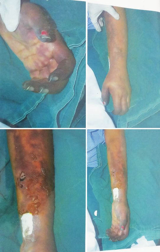
Catastrophic Antiphospholid Syndrome – An Unusual Case Report
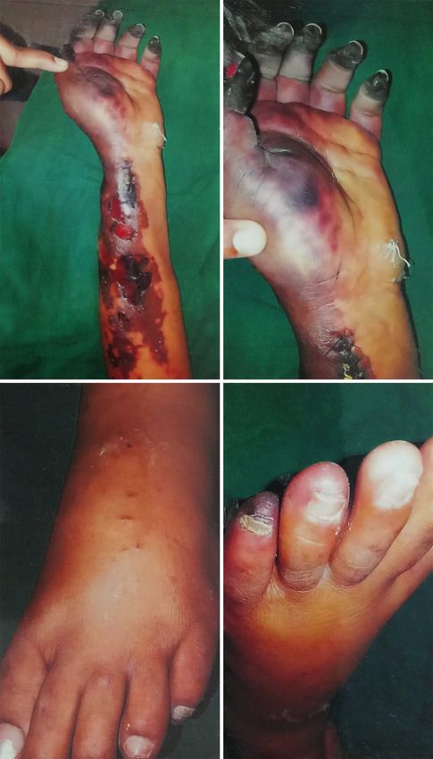
Fig. 2: Digital ischemia with forearm and hand skin changes
88.9% on 60% FiO2 noninvasive ventilation. She was initiated on fingers showed features of severe gangrene with blistering and
intravenous meropenem, tab clarithromycin, tab oseltamivir and extensive ecchymotic changes involving the whole forearm and
supportive treatment. In view of persistent hypoxia patient was hand. Due to irreversible vascular thrombosis, it was decided to go
intubated and ventilated during which she required a low dose of conservative and hence injection enoxaparin 40 mg subcutaneous
noradrenaline. Left radial artery was cannulated for BP monitoring. twice a day was continued.
Initial lab test showed a hemoglobin 8 g/dL, total counts 5560/cu Over the next few days, although the PaO2/FiO2 ratio improved,
mm, platelet 1.73 lakh/cu mm with normal coagulation profile, PEEP was reduced and noradrenaline tapered off, patient became
normal renal and liver functions and procalcitonin of 0.5U. febrile and drowsy. Dose of meropenem increased to 2 gm
Transthoracic 2Decho was normal with an ejection fraction of 50%, 8 hourly and linezolid added pending cultures. Her throat swab
no regional wall abnormalities and normal valves. was positive for swine flu (HINI) virus, thyroid stimulating hormone
On day two evening, there was no back flow in the arterial line (TSH) ≤ 0.002 U/dL. Neomercazole was reintroduced. Subsequently
and hence repositioned to left dorsalis pedis artery. Next morning, patient developed hypertension which required nitroglycerine
the patient developed deep cyanotic changes in all the finger tips infusion. Serum creatinine increased to 2.65 mg/dL. Urine routine
of left upper limb with patchy ecchymotic skin lesions involving the showed significant proteinuria and hematuria. Urine culture
whole forearm with no radial pulsations (Fig. 2). Urgent left upper grew Enterococcus feacium which was covered with appropriate
limb arterial doppler showed an echogenic thrombus in left radial antibiotics. Blood cultures were negative. Fundoscopy revealed
artery from mid forearm to wrist. Urgent left radial embolectomy retinal infarcts raising suspicion of vasculitis. Patient developed
was performed. Post procedure radial pulsations were well felt. pervaginal bleeding although platelet count and coagulation was
But on the next day, radial pulsations were lost again and her left normal. Ultrasound pelvis revealed multiple uterine fibroids.
Indian Journal of Critical Care Medicine, Volume 23 Issue 6 (June 2019) 277
Catastrophic Antiphospholid Syndrome – An Unusual Case Report

Fig. 3: CT brain showing large MCA infarct with multiple small infarcts        Fig. 5: CT brain showing intracerebral bleed with ventricular extention
at other sites
                                                                                   Patient continued to have a small persistant pervaginal
                                                                               bleed but this time associated with progressively worsensing
                                                                               thrombocytopenia. On workup her peripheral smear showed a few
                                                                               schistocytes with a direct coomb test positive. Her serum ADAMTS-
                                                                               13 was sent and a diagnosis of thrombotic thrombocyptopenic
                                                                               purpura (TTP) was contemplated and plasmapheresis was
                                                                               decided. Subsequently her lupus anticoagulant was positive,
                                                                               protein C and protein S were low and factor V Leiden was also
                                                                               positive. Plasmapheresis and hemodialysis were initiated in
                                                                               view of catastrophic antiphospholipid syndrome and worsening
                                                                               acute kidney injury with anuria. Since there was no improvement
                                                                               seen in the neurological status and with worsening metabolic
                                                                               acidosis, recurrence of fever, anuria, intermittent PV bleeding,
                                                                               worsening thrombocytopenia, repeated negative blood, urine
                                                                               and endotracheal cultures and coagulopathy despite five sessions
                                                                               of plasmapheresis, high dose steroids were initiated and only
                                                                               clopidogrel was continued while anticoagulation was already
Fig. 4: 2D echo short axis view showing ball like vegetations on mitral        stopped.
and aortic valve                                                                   After two weeks of ventilation , tracheostomy was planned but
                                                                               she developed a sudden massive drop in hemoglobin to 6.6 g/dL
    She continued to have fever and obtunded sensorium with                    on the same day. On neurological examination she was comatosed
stable hemodynamics and a better gas exchange but with severe                  with fixed and dialated pupils. Urgent CT brain showed a large
dry gangrenous changes of the left upper limb. We concluded that               intraparenchymal bleed in left occipital region with perilesional
the source could be the gangreneous upper limb with secondary                  edema with extension in both lateral, 3rd and 4th ventricles and
infection so antibiotics were escalated and planned for amputation             bilateral extensive subarachanoid hemorrhage (Fig. 5).
once patient was stable. Her serum creatinine increased to 4.6 mg/                 Prognosis was explained to the relatives. In view of irreversible
dL although the urine output was maintained. CT brain done as a                neurological damage they took a decision of withdrawal of care.
part of the work up revealed a large acute right MCA infarct with              Patient sustained cardiac arrest after two days.
minimal midline shift and few small infarcts in the left parietal, left
temporal, B/L occipital lobes, and right cerebellar hemisphere with
no evidence of hemorrhage (Fig. 3). Dual antiplatelets were started.           Discussion
Antinuclear antibodies, anti double stranded DNA antibodies,                   Antiphospholipid antibody syndrome is a heterogeneous
Antineutrophil cytoplasmic antibodies were negative and C3,                    multisystem autoimmune disorder of hypercoagulation1. It is
C4 was normal. A repeat 2 Decho revealed nodular echogenic                     regarded as primary or secondary if there are other associated
shadows on anterior mitral leaflet and on right coronary cusps with            connective tissue disorders like SLE.1,3,6 It is predominantly seen
preserved left ventricular function which was later confirmed on               in females , with a female to male ratio of 3.5:1.1,6 Catastrophic
transesophageal echocardiography (Fig. 4). So an acute left radial             antiphospholipid syndrome, first described by Asherson R. in 1992,
artery thrombosis with gangrene of left upper limb, probably                   is a fatal variant of APLS occurring in 1% of APLS patients.3-6
embolic or thrombotic phenomenon with vegetations on mitral and                     Putative pathogenetic mechanisms for APLS can be arbitraily
aortic valves probably mirantic, multiple non watershed cerebral               divided into four interrelated groups which include cellular
infarcts, in H1N1 positive patient suggested a vasculitic process              activation, inhibition of anticoagulation, inhibition of fibrinolysis
with gangrene triggered by H1N1 viral infection.                               and complement activation.7 The development of thrombi and

278      Indian Journal of Critical Care Medicine, Volume 23 Issue 6 (June 2019)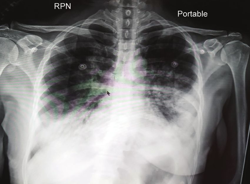
Catastrophic Antiphospholid Syndrome – An Unusual Case Report

the high risk of recurrent thrombotic events has been linked to the             a pathological hallmark of CAPS could not be demonstrated on
high titres of lupus anticoagulant and anticardiolipin antibodies1.             histology in our patient due to overall poor condition.6,7
     Our patient was a 45-year-old female, with no underlying SLE                   Laboratory findings in CAPS include thrombocytopenia,
or other autoimmune disorders, suggesting possibility of a primary              hemolytic anemia which is often accompanied by small number
CAPS. But subsequently she was positive for factor V Leiden with                of schistocytes in about 14% and disseminated intravascular
deficient protein C and protein S so an underlying prothrombotic                coagulation.1,3,4,7,14 These are also the hematological manifestations
state could not be ruled out. As per the CAPS registry created in               of CAPS. The autoantibodies to diagnose APLS are anti β 2
the year 2000 of 280 patients, CAPS is commonly found in females                glycoprotein-1, anticardiolipin antibody and lupus anticoagulant.1,6,7
(72%) with a mean age of 37 ± 14 years with majority suffering                  The 2006 revised classification criteria for APLS updated the time
from primary APLS (40%). 6,7 Interestingly CAPS was the first                   frame for the presence of elevated titres of aPL antibodies from 6
manifestation of APLS in 46% of patients, as in our case.4 Concurrent           weeks to 12 weeks.1,6,11 In our patient we demonstrated haemolytic
autoimmune disease like SLE is seen in 40%, SLE like syndrome in                anaemia with peripheral schistocytes, thrombhocytopenia and
5% and other autoimmune pathologies in 9%.7 Triggering factors                  lupus anticoagulant which could not be repeated due to early
leading to CAPS are infection(22%), surgery(10%), withdrawal                    death of our patient.
of anticoagulation(8%), medication(7%), obstetric complication,
neoplasm, and lupus flare.4,6-9 In our case H1N1 viral infection was            Updated Antiphospholipid Syndrome
a precipitating factor for the thrombotic microangiopathic state.               Classification Criteria10-12
     The diagnosis of APLS is based on the clinical features                    Clinical Criteria
(depending on the organ involved by thrombosis ) and the                        • Vascular thrombosis:
laboratory criteria.10 -12 The major organs involved during                     	≥1 clinical episodes of arterial, venous, or small vessel
catastrophic episodes were renal (71%), lungs (64%), brain (62%),                 thrombosis, in any tissue or organ
heart (51%), and skin (50%), liver and Gastrointestinal tract as per            • Pregnancy morbidity:
CAPS registry.4,6,7                                                               – ≥1 unexplained deaths of a morphologically normal fetus
     Our patient developed left upper limb ischemic skin changes                      at or beyond the 10th week of gestation, or
in all digits which was due to acute arterial thrombosis and                      – ≥1 premature births of a morphologically normal neonate
progressed to digital gangrene and purpura fulminans like skin                        before the 34th week of gestation because of: eclampsia,
lesions. Cutaneous involvement in CAPS includes livedo reticularis,                   severe preeclampsia, or recognized features of placental
acrocyanosis, purpura, ecchymosis, splinter hemorrhages and                           insufficiency, or
necrosis resulting in ulcerations.7,13 Renal involvement is defined               – ≥3 unexplained consecutive spontaneous abortions
by ≥50% increase in creatinine concentration, proteinuria of >                        before the 10th week of gestation, with maternal anatomic
0.5gms/day, severe hypertension (BP >180/100) or combination of                       or hormonal abnormalities and paternal and maternal
these.8 Renal infarction is rare.5,7 In our case there was a sudden                   chromosomal causes excluded.
increase in serum creatinine with proteinuria and hypertension.
Lung involvement includes ARDS, pulmonary embolism and                          Laboratory Criteria
rarely alveolar hemorrhages.1,7 Common CNS presentations being                  •  Lupus anticoagulant present in plasma, on ≥2 occasions at
hypertensive encephalopathy, ischemic encephalopathy, stroke,                      least 12 weeks apart
seizure, cortical venous thrombosis and altered mental status.6,7               • Anticardiolipin antibody of IgG and/or IgM isotype, in medium
Our patient also developed an acute worsening of sensorium due                     or high titer (>40 GPL or MPL, or > the 99th percentile), on ≥2
to a large MCA and other territorial multiple infarcts including                   occasions, at least 12 weeks apart.
cerebellum progressing terminally to intracerebral bleed. Cardiac               • Anti-β2-glycoprotein-I antibody of IgG and/or IgM isotype, in
involvement presents as coronary thrombosis causing unstable                       medium or high titer (> the 99th percentile), on ≥2 occasions,
angina and myocardial infarction, valvular regurgitant lesions, and                at least 12 weeks apart.
valvular sterile vegetations.1,6,7 In our patient we also observed                 Definite APS is present if at least one of the clinical criteria and
fluffy ball like vegetations involving the anterior mitral leaflet              one of the laboratory criteria are met.
and the aortic cusps with no regional wall motion abnormality.
Ocular involvement is rare with only 5.8% of the cases reporting                Preliminary Classification Criteria for Catastrophic
retinal infarcts and choroidal thrombosis.4,7 A rare case of bilateral          Antiphospholipid Syndrome10-12
central retinal artery occlusion with profound vision loss has been             •   Evidence of involvement of three or more organs, systems
reported.7 Retinal infarcts was the first hint for an underlying                    and/or tissues
vasculitic process in our patient. Hematological involvement                    •   Development of manifestations simultaneously or in less than
forms the most important part of the clinical spectrum of CAPS                      a week
with thrombotic and hemorrhagic manifestations occurring                        •   Confirmation by histopathology of small-vessel occlusion*
concurrently most of the time and was also seen in our patient.7,14             •   Laboratory confirmation of the presence of antiphospholipid
Thrombotic microangiopathy and small vessel occlusive disease6,7,                   antibodies†

*
 Vasculitis may coexist, but significant thrombosis must be present as well.
†
 “Positive aPL” twice 12 weeks apart (of note, the original Sapporo APS classification criteria required two positive aPL tests 6 weeks apart, which has
been changed to 12 weeks as part of the updated Sapporo APS classification criteria.

                                                                           Indian Journal of Critical Care Medicine, Volume 23 Issue 6 (June 2019)     279
Catastrophic Antiphospholid Syndrome – An Unusual Case Report
Definite Catastrophic Antiphospholipid Syndrome References
All four criteria present 1. Zahid H, Hassan S, Gul S, Rizwan K, Khan SU, Maghazi MA. Co-existing
bilateral pulmonary embolism and intra-cardiac mass: A case of
Probable Catastrophic Antiphospholipid Syndrome catastrophic antiphospholipid syndrome-like disease. Cureus. 2018;
• All four criteria, except only two organs, systems, and/or tissues 10(10): e3438. doi: 10.7759/cureus.3438.
involved 2. Uthman I, Noureldine MHA, Ruiz-Irastorza G, Khamashta M. Manage
• All four criteria, except for the absence of laboratory ment of antiphospholipid syndrome. Ann Rheum Dis. 2019;78:155–
161.
confirmation of antiphospholipid antibodies
3. Gu JY, Lu C, Shi H, Yang CD. Case series and clinical analysis of 14 cases
• Criteria 1, 2, and 4 of catastrophic antiphospholipid syndrome. Beijing Da Xue Xue Bao
• Criteria 1, 3, and 4, with the development of a third event Yi Xue Ban. 2018;50(6):1033–1038.
more than 1 week but within 1 month of presentation, despite 4. Saraf SS, Patel YP, Desai A, Desai UR. Catastrophic antiphospholipid
anticoagulation syndrome presenting as bilateral central retinal artery occlusions. Case
Histologically CAPS is characterised by acute thrombotic Rep Ophthalmol Med. 2015; 2015: 206906. doi: 10.1155/2015/206906.
microangiopathy.6,7 Hence CAPS should be differentiated from 5. Prasad N, Bhadauria D, Agarwal N Gupta A, Gupta P, Jain M et al.
other thrombotic microangiopathies such as hemolytic uremic Catastrophic antiphospholipid antibody syndrome in a child with
syndrome, thrombotic thrombocytopenic purpura (T TP), thrombotic microangiopathy. Indian J Nephrol. 2012;22 : 310–313.
doi: 10.4103/0971-4065.101266
disseminated intravascular coagulopathy, heparin induced
6. Strakhan M, Hurtado-Sbordoni M, Galeas N, Bakirhan K, Alexis K, Elrafei
thrombocytopenic thrombosis and severe sepsis.4,8 Sepsis and TTP T. 36year old female with catastrophic antiphospholipid syndrome
were the differential diagnosis in our patient. However, it should treated with eculizumab: acase report and review of literature. Case
be noted that her ADAMTS 13 activity was negative and her lupus Rep Hematol. 2014; 2014: 704371. doi: 10.1155/2014/704371
anticoagulant was positive with persistently negative cultures. 7. Nayer A, Ortega LM. Catastrophic antiphospholipid syndrome:
CAPS is life threatening systemic disease and is associated a clinical review. J Nephropathol. 2014; 3(1):9–17. doi: 10.12860/
with high morbidity and mortality.1,3,6,7 Therefore aggressive jnp.2014.03
multidisciplinary collaborative treatment strategy is indicated. 8. Mendoza-Pinto C, García-Carrasco M, Cervera R. Role of infectious
diseases in the antiphospholipid syndrome (including its catastrophic
A combination of anticoagulation and antiplatelets, high dose
variant). Curr Rheumatol Rep. 2018; 20:62.
steroids, IVIg or plasma exchange, cyclophosphamide and the 9. Cruz-Tapias P, Blank M, Anaya JM Shoenfeld Y. Infections and vaccines
latest Eculizumab has been found to be effective. 3,6,7,11,12 Our in the etiology of antiphospholipid syndrome. Curr Opin Rheumatol.
patient did receive the triple therapy in the form of therapeutic 2012;24(4): 389–393. doi: 10.1097/BOR.0b013e32835448b8
enoxaparin, dual antipletelets, plasmapheresis and high dose 10. Asherson R, Cervera R, de Groot., Erkan D, Boffa M, Piette J, et al
methylprednisolone, but the disease progression was so rapid and Catastrophic Antiphospholipid syndrome : international consensus
simultaneous that she ultimately succumbed to massive intracranial statement on classification criteria and treatment guidelines Lupus
hemorrhage within three weeks of admission. But the recent 2003;12(7):530–534. doi: 10.1191/0961203303lu394oa
11. Aguiar CL, Erkan D. Catastrophic antiphospholipid syndrome: how
studies have shown a significant decline in the mortality rate in
to diagnose a rare but highly fatal disease. Ther Adv Musculoskeletal
CAPS from a high of 53–33% which is attributed to early diagnosis Dis, 2013;5(6):305–314. doi: 10.1177/1759720X13502919
and the rapid initiation of the aforementioned treatment strategy 12. Cervera R, Rodríguez-Pintó I, Espinosa G. The diagnosis and clinical
in combination.7,15 management of the catastrophic antiphospholipid syndrome: A
comprehensive review. J Autoimmun. 2018;92: 1–11. doi: 10.1016/j.
C o n c lu s i o n jaut.2018.05.007
13. PluB M, Zeisberg M, Muller GA, Vasko R, Korsten P. Therapeutic
CAPS patient suffer from a life threatening acute multiple organ response to glucocorticoids, anticoagulation and plasma exchange
thrombosis, usually accompanied by microthrombosis and in a patient with primary antiphospholipid syndrome presenting
hematoloical manifestations with high titre of antiphospholipid with purpura fulminans. Lupus. 2018;27(13):2170–2073. doi:
antibodies. In our patient the occurrence of so many manifestations 10.1177/0961203318804884.
simultaneously over a short span confirmed the diagnosis of 14. Vieregge GB, Harrington TJ, Andrews DM, Carpintero MF, Green DF,
Nayer A. Catastrophic antiphospholipid syndrome with severe acute
catastrophic APLA syndrome. Without early diagnosis and
thrombotic microangiopathy and hemorrhagic complications. Case
treatment, prognosis is poor. It is important to anticipate CAPS and Rep Med. 2013;2013:915309. doi: 10.1155/2013/915309
CAPS like disease in young patients with multiorgan involvement 15. Costedoat-Chalumeau N, Coutte L, Le Guern V, Morel N, Leroux
and overlapping other thrombotic microangiopathies and known G, Paule R et al. 2016 review on catastrophic antiphospholipid
triggering factors in order to halt the mortality and improve syndrome. Presse Med. 2016; 45(12 Pt 1):1084–1092. doi: 10.1016/j.
prognosis. lpm.2016.07.023
280 Indian Journal of Critical Care Medicine, Volume 23 Issue 6 (June 2019)
You can also read