Practical guidelines on the management and prevention of the diabetic foot
←
→
Page content transcription
If your browser does not render page correctly, please read the page content below
DIABETES/METABOLISM RESEARCH AND REVIEWS GUIDELINES
Diabetes Metab Res Rev 2008; 24(Suppl 1): S181–S187.
Published online in Wiley InterScience (www.interscience.wiley.com) DOI: 10.1002/dmrr.848
Practical guidelines on the management and
prevention of the diabetic foot
Based upon the International Consensus on the Diabetic Foot (2007)
Prepared by the International Working Group on the Diabetic Foot
J. Apelqvist1 Introduction
K. Bakker2
Foot complications are among the most serious and costly complications of
W. H. van Houtum3 *
diabetes mellitus. Amputation of all or part of a lower extremity is usually
N. C. Schaper4 preceded by a foot ulcer. A strategy which includes prevention, patient
on behalf of the International and staff education, multi-disciplinary treatment of foot ulcers, and close
Working Group on the monitoring can reduce amputation rates by 49–85%. Therefore, several
Diabetic Foot (IWGDF) countries and organizations, such as the World Health Organization and
Editorial Board the International Diabetes Federation, have set goals to reduce the rate of
amputations by up to 50%.
1
Department of Endocrinology, The basic principles of prevention and treatment described in these
University Hospital of Malmö, S-205 guidelines are based on the International Consensus on the Diabetic Foot.
02 Malmö, Sweden Depending on local circumstances, these principles have to be translated
2
International Working Group on the for local use, taking into account regional differences in socio-economics,
Diabetic Foot (IWGDF), accessibility to healthcare, and cultural factors. These practical guidelines are
Heemsteedsedreef 90, 2102 KN aimed at healthcare workers involved in the care of people with diabetes.
Heemstede, The Netherlands For more details and information on treatment by specialists in foot care, the
3
Department of Internal Medicine, reader is referred to the International Consensus document.
Spaarne Hospital Hoofddorp,
Hoofddorp, The Netherlands
4
Department of Internal Medicine,
Pathophysiology
Division of Endocrinology. University
Although the spectrum of foot lesions varies in different regions of the world,
Hospital Maastricht, PO Box 5800,
6202 AZ Maastricht, The Netherlands
the pathways to ulceration are probably identical in most patients. Diabetic
foot lesions frequently result from two or more risk factors occurring together.
*Correspondence to: W. H. van In the majority of patients, diabetic peripheral neuropathy plays a central
Houtum, Department of Internal role: up to 50% of people with type 2 diabetes have neuropathy and at-risk
Medicine, Spaarne Hospital feet.
Hoofddorp, Spaarnepoort 1, 2134 Neuropathy leads to an insensitive and sometimes deformed foot, often
TM, Hoofddorp, The Netherlands. with an abnormal walking pattern. In people with neuropathy, minor
E-mail: houtum@tiscali.nl trauma – caused for example by ill-fitting shoes, walking barefoot, or an acute
injury – can precipitate a chronic ulcer. Loss of sensation, foot deformities,
and limited joint mobility can result in abnormal biomechanical loading of
the foot. Thickened skin (callus) forms as a result. This leads to a further
increase of the abnormal loading and, often, subcutaneous haemorrhage.
Whatever the primary cause, the patient continues walking on the
insensitive foot, impairing subsequent healing (Figure 1). Peripheral vascular
disease, usually in conjunction with minor trauma, may result in a painful,
purely ischaemic foot ulcer.
However, in patients with both neuropathy and ischaemia (neuro-ischaemic
Received: 9 October 2007
ulcer), symptoms may be absent, despite severe peripheral ischaemia. Micro-
Accepted: 30 January 2008
angiopathy should not be accepted as a primary cause of an ulcer.
Copyright 2008 John Wiley & Sons, Ltd.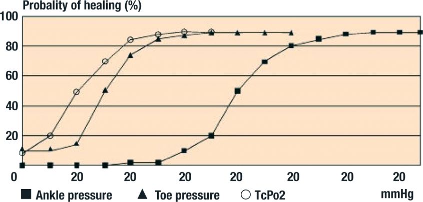
S182 Guidelines
Figure 1. Illustration of ulcer due to repetitive stress
Cornerstones of foot management Education of patients, family, and
healthcare providers
There are five key elements which underpin foot
management: Education, presented in a structured and organized
1. Regular inspection and examination of the foot at manner, plays an important role in the prevention of
risk. foot problems. The aim is to enhance motivation and
2. Identification of the foot at risk. skills. People with diabetes should learn how to recognize
3. Education of patient, family, and healthcare providers. potential foot problems and be aware of the steps they
4. Appropriate footwear. need to take in response. The educator must demonstrate
5. Treatment of non-ulcerative pathology. the skills, such as how to cut nails appropriately.
Education should be provided in several sessions over
time, and preferably using a mixture of methods. It is
Regular inspection and examination essential to evaluate whether the person with diabetes
has understood the message, is motivated to act, and has
All people with diabetes should be examined at least once sufficient self-care skills. An example of instructions for the
a year for potential foot problems (Table 1). Patients with high-risk patient and family is given below. Furthermore,
demonstrated risk factor(s) should be examined more physicians and other healthcare professionals should
often – every 1–6 months. Absence of symptoms does not receive periodic education to improve care for high-risk
mean that the feet are healthy; a patient might have individuals.
neuropathy, peripheral vascular disease, or even an ulcer
without any complaints. The feet should be examined
with the patient lying down and standing up, and the Table 1. Easy to use foot screening assessment sheet for clinical
shoes and socks should also be inspected. examination
The foot is at risk if any of the below are present
Deformity or bony prominences Yes/No
Identification of the at-risk foot Skin not intact(ulcer) Yes/No
Neuropathy
Following examination of the foot, each patient can - Monofilament undetectable Yes/No
- Tuning fork undetectable Yes/No
be assigned to a risk category, which should guide - Cotton wool undetectable Yes/No
subsequent management (Figure 2). Abnormal pressure, callus Yes/No
Loss of joint mobility Yes/No
Foot pulses
Progression of risk categories: - Tibial posterior artery absent Yes/No
- Dorsal pedal artery absent Yes/No
• Sensory neuropathy and/or foot deformities or bony Discoloration on dependency Yes/No
prominences and/or signs of peripheral ischaemia Any others
and/or previous ulcer or amputation - previous ulcer Yes/No
- amputation Yes/No
• Sensory neuropathy Inappropriate footwear Yes/No
• Non-sensory neuropathy
Copyright 2008 John Wiley & Sons, Ltd. Diabetes Metab Res Rev 2008; 24(Suppl 1): S181–S187.
DOI: 10.1002/dmrr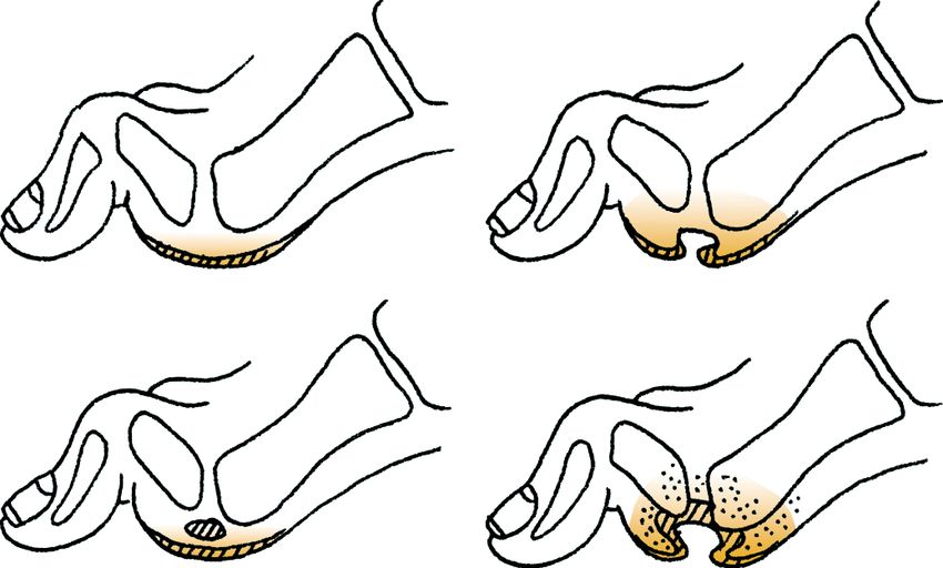
Guidelines S183
History and examination
History Previous ulcer/amputation, previous foot education,
social isolation, poor access to healthcare, walking
barefoot
Neuropathy Symptoms, such as tingling or pain in the lower limb,
especially at night
Vascular status Claudication, rest pain, pedal pulses
Skin Colour, temperature, oedema
Bone/joint Deformities (e.g. claw toes, hammer toes) or bony
prominences
Footwear/socks Assessment of both inside and outside
Sensory loss
Sensory loss due to diabetic polyneuropathy can be assessed using the
following techniques:
Pressure perception Semmes–Weinstein monofilaments (10 g, see
addendum) The risk of future ulceration can be
determined with a 10-g monofilament
Vibration perception 128-Hz tuning fork (hallux, see addendum)
Discrimination Pin prick (dorsum of foot, without penetrating
the skin)
Tactile sensation Cotton wool (dorsum of foot)
Reflexes Achilles tendon reflexes • Avoidance of barefoot walking indoors or outdoors and
of wearing of shoes without socks
• Chemical agents or plasters to remove corns and
calluses - should not be used
• Daily inspection and palpation of the inside of the shoes
• Do not wear tight shoes or shoes with rough edges and
uneven seams
• Use of lubricating oils or creams for dry skin - but not
between the toes
• Daily change of socks
• Wearing of stocking with seams inside out or preferably
without any seams
• Never wear tight or knee-high socks
• Cutting nails straight across (see Figure 3)
• Corns and calluses - should be cut by a healthcare
provider
• Patient awareness of the need to ensure that feet are
examined regularly by a healthcare provider
• Notifying the healthcare provider at once if a blister,
cut, scratch or sore has developed
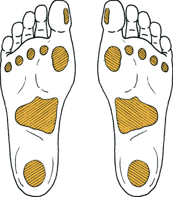
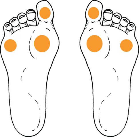
Figure 2. Areas at risk
Items which should be covered when instructing the
high-risk patient
• Daily feet inspection, including areas between the toes
• The need for another person with skills to inspect feet,
should the people with diabetes be unable to do so
(If vision is impaired, people with diabetes should not
attempt their own foot care)
• Regular washing of feet with careful drying, especially
between the toes
• Water temperature – always below 37 ◦ C
• Do not use a heater or a hot-water bottle to warm your
feet Figure 3. How to cut nails
Copyright 2008 John Wiley & Sons, Ltd. Diabetes Metab Res Rev 2008; 24(Suppl 1): S181–S187.
DOI: 10.1002/dmrr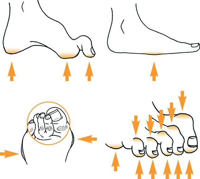
S184 Guidelines
Appropriate footwear Foot ulcers
Inappropriate footwear is a major cause of ulceration.
A standardized and consistent strategy for evaluating
Appropriate footwear should be used both indoors
wounds is essential, and will guide further therapy. The
and outdoors, and should be adapted to the altered
following items must be addressed:
biomechanics and deformities – essential for prevention.
Patients without loss of protective sensation can select
off-the-shelf footwear by themselves. In patients with Cause. Ill-fitting shoes are the most frequent cause of
neuropathy and/or ischaemia, extra care must be taken ulceration, even in patients with ‘pure’ ischaemic ulcers.
when fitting footwear, particularly when foot deformities Therefore, shoes should be examined meticulously in all
are also present. Shoes should not be too tight or too loose patients.
(Figure 4). The inside of the shoes must be 1–2 cm longer
than the feet. The internal width should be equal to the Type. Most ulcers can be classified as neuropathic,
width of the foot at the site of the metatarsal phalangeal ischaemic, or neuro-ischaemic. This will guide further
joints, and the height should allow enough room for the therapy. Assessment of the vascular tree is essential in the
toes. The fit must be evaluated with the patient in standing management of a foot ulcer.
position, preferably at the end of the day. If the fit is too If one or more pedal pulses are absent or if an ulcer does
tight due to deformities or if there are signs of abnormal not improve despite optimal treatment, more extensive
loading of the foot (e.g. hyperaemia, callus, ulceration), vascular evaluation should be performed. As a first step,
patients should be referred for special footwear (advice the ankle brachial pressure can be measured. An ankle
and/or construction), including insoles and orthoses. brachial pressure index (ABPI) below 0.9 is a sign of
peripheral arterial disease. However, ankle pressure might
be falsely elevated due to calcification of the arteries.
Treatment of non-ulcerative pathology Preferably, other tests such as measurements of toe
pressure or transcutaneous pressure of oxygen (TcPo2)
In a high-risk patient callus, and nail and skin pathology
should be used. Figure 5 gives an estimate of the chance
should be treated regularly, preferably by a trained foot
of healing using the tests. If a major amputation is being
care specialist. If possible, foot deformities should be
contemplated, the option of revascularization should be
treated non-surgically (e.g. with an orthosis).
considered first.
Site and depth. Neuropathic ulcers frequently occur on
the plantar surface of the foot or in areas overlying a
bony deformity. Ischaemic and neuro-ischaemic ulcers
are more common on the tips of the toes or the lateral
border of the foot.
The depth of an ulcer can be difficult to determine,
due to the presence of overlying callus or necrosis. There-
fore, neuropathic ulcers with callus and necrosis should be
debrided as soon as possible. This debridement should not
be performed in ischaemic or neuro-ischaemic ulcers with-
out signs of infection. In neuropathic ulcers, debridement
Figure 4. Internal width of the shoe can usually be performed without (general) anaesthesia.
Figure 5. A schematic estimate of the probability of healing of foot ulcers and minor amputations in relation to ankle blood
pressure, toe blood pressure, and transcutaneous oxygen pressure (TcPo2) based on selected reports
Copyright 2008 John Wiley & Sons, Ltd. Diabetes Metab Res Rev 2008; 24(Suppl 1): S181–S187.
DOI: 10.1002/dmrr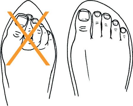
Guidelines                                                                                                                    S185

Signs of infection. Infection in a diabetic foot presents a       ◦ debridement with removal of all necrotic tissue and
direct threat to the affected limb, and should be treated           oral antibiotics targeted at Staphylococcus aureus
promptly and actively. Signs and/or symptoms of infection           and streptococci
such as fever, pain, or increased white blood count/ESR         • Deep (limb-threatening) infection
are often absent. However, if present, substantial tissue         ◦ surgical drainage as soon as possible (emergency
damage or even development of an abscess is likely.                 referral) with removal of necrotic or poorly vascular-
   The risk of osteomyelitis should be determined. After            ized tissue, including infected bone
initial debridement, if it is possible to touch the bone with     ◦ revascularization if necessary
a sterile probe, it is likely that the underlying bone is         ◦ broad-spectrum antibiotics intravenously, aimed
infected.                                                           at Gram-positive and negative micro-organisms,
   A superficial infection is usually caused by Gram-               including anaerobes
positive bacteria. In cases of (possible) deep infections,
Gram stains and cultures from the deepest tissue involved
are advised – not superficial swabs. These infections are       Metabolic control and treatment of comorbidity
usually polymicrobial, involving anaerobes and Gram-            • Optimal diabetes control, if necessary with insulin
positive/negative bacteria.                                       (blood glucose
S186 Guidelines
auditing. Local variations in resources and staffing will Addendum
often determine the way care is provided. Ideally, a foot
care programme should provide the following:
Sensory foot examination
• Education of patients, carers, and healthcare staff in
hospitals, primary healthcare, and the community
Neuropathy can be detected using the 10-g (5.07
• A system to detect all people who are at risk, with
Semmes–Weinstein) monofilament, tuning fork (128 Hz),
annual foot examination of all known patients
and/or cotton wisp.
• Measures to reduce risk, such as podiatry and
appropriate footwear
• Prompt and effective treatment Semmes–Weinstein monofilament
• Auditing of all aspects of the service to ensure that local
• Sensory examination should be carried out in a quiet
practice meets accepted standards of care
and relaxed setting. First apply the monofilament on
• An overall structure which is designed to meet the
the patient’s hands (or elbow or forehead) so that he or
needs of patients requiring chronic care, rather than
she knows what to expect.
simply responding to acute problems when they occur.
• The patient must not be able to see whether or where
the examiner applies the filament. The three sites to be
In all countries, at least three levels of foot care tested on both feet are indicated in Figure 6.
management are needed. • Apply the monofilament perpendicular to the skin
surface (Figure 7(a)).
• Apply sufficient force to cause the filament to bend or
Level 1 – General practitioner, podiatrist, and diabetic
buckle (Figure 7(b)).
nurse.
Level 2 – Diabetologist, surgeon (general and/or vascular
and/or orthopaedic), podiatrist, and diabetic nurse.
Level 3 – Specialized foot centre with multiple disciplines
specialized in diabetic foot care.
Setting up a multi-disciplinary foot care team has been
found to be accompanied by a drop in the number of
amputations. If it is not possible to create a full team
from the outset, this should be built up step by step,
introducing the various disciplines at different stages.
This team must work in both primary and secondary care
settings.
Ideally, a foot care team would consist of a
diabetologist, surgeon, podiatrist, orthotist, educator,
and plaster technician, in close collaboration with an
orthopaedic, podiatric, and/or vascular surgeon and
dermatologist. Figure 6. Sites to be tested with monofilaments
(a) (b)
Figure 7. (a) and (b) Application of the monofilaments
Copyright 2008 John Wiley & Sons, Ltd. Diabetes Metab Res Rev 2008; 24(Suppl 1): S181–S187.
DOI: 10.1002/dmrrGuidelines S187
Protective sensation is absent with two out of three
incorrect answers; the patient is then considered to be
at risk of ulceration.
• Encourage the patient during testing by giving a positive
feedback.
• The healthcare provider should be aware of the possible
loss of buckling force of the monofilament if used for
too long a period of time.
Tuning fork
• The sensory examination should be carried out in a
quiet and relaxed setting. First, apply the tuning fork
on the patient’s wrists (or elbow or clavicle) so that
he/she knows what to expect.
• The patient must not be able to see whether or where
the examiner applies the tuning fork. The tuning fork is
applied on a bony part on the dorsal side of the distal
phalanx of the first toe.
• The tuning fork should be applied perpendicularly with
Figure 8. How to use a tuning fork
constant pressure (Figure 8).
• Repeat this application twice, but alternate this with at
• The total duration of the approach – skin contact and least one ‘mock’ application in which the tuning fork is
removal of the filament – should be approximately 2 s. not vibrating.
• Apply the filament along the perimeter of, not on, an • The test is positive if the patient incorrectly answers
ulcer site, callus, scar, or necrotic tissue. at least two out of three applications, (’at risk for
• Do not allow the filament to slide across the skin or ulceration’) and negative with two out of three correct
make repetitive contact at the test site. answers. (A test is positive if it identifies the risk).
• Press the filament to the skin and ask the patient • If the patient is unable to sense the vibrations on the
whether he/she feels the pressure applied (‘yes’/‘no’) big toe, the test is repeated more proximally (malleolus,
and next, where he/she feels the pressure (‘left tibial tuberosity).
foot’/’right foot’). • Encourage the patient during testing by giving a positive
• Repeat this application twice at the same site, but feedback.
alternate this with at least one ‘mock’ application in
which no filament is applied (totally three questions
per site).
Conflict of interest
• Protective sensation is present at each site if the The authors have no conflicts of interest.
patient correctly answers two out of three applications.
Copyright 2008 John Wiley & Sons, Ltd. Diabetes Metab Res Rev 2008; 24(Suppl 1): S181–S187.
DOI: 10.1002/dmrrYou can also read