Rheumatic Heart Disease with Multiple Systemic Emboli: A Rare Occurrence in a Single Subject - Cureus
←
→
Page content transcription
If your browser does not render page correctly, please read the page content below
Open Access Case
Report DOI: 10.7759/cureus.2964
Rheumatic Heart Disease with Multiple
Systemic Emboli: A Rare Occurrence in a
Single Subject
Ravi R. Pradhan 1 , Ashish Jha 2 , Gaurav Nepal 1 , Manju Sharma 3
1. Internal Medicine, Tribhuvan University Institute of Medicine, Kathmandu, NPL 2. Medicine, Institute
of Medicine, Tribhuvan University Teaching Hospital, Kathmandu, NPL 3. Cardiology, Manmohan
Cardiothoracic Vascular and Transplant Center, Kathmandu, NPL
Corresponding author: Ravi R. Pradhan , drravipradhan@iom.edu.np
Disclosures can be found in Additional Information at the end of the article
Abstract
Valvular heart disease is one of the more common diseases in low- and middle-income
countries, when associated with atrial fibrillation (AF), carries a risk of multisystemic
embolizations. We report a case of 37-year-old man with multiple systemic emboli consisting
of acute ischemic stroke, acute myocardial infarction, and acute limb ischemia. This is a rare
occurrence in a single subject. The patient had a background of rheumatic heart disease (RHD)
involving severe mitral stenosis (MS) with AF, who was not compliant with his medications. A
computed tomography (CT) scan of the head showed right-sided ischemic stroke involving
more than one-third of the middle cerebral artery territory. An electrocardiogram (ECG)
showed AF and ST-segment elevation in V4 to V6. Cardiac enzymes were elevated. A
transthoracic echocardiogram demonstrated hypokinetic left ventricular anterolateral wall,
severe MS, and a left atrial clot. An arterial Doppler of the right lower limb showed an occluding
thrombus of the right common femoral artery and right popliteal artery with no flow in color
Doppler. Patient adherence to medications in cases of RHD prevents devastating outcomes.
Categories: Cardiac/Thoracic/Vascular Surgery, Cardiology, Internal Medicine
Keywords: rheumatic heart disease, mitral stenosis, acute ischemic stroke, acute myocardial infarction,
acute limb ischemia
Introduction
Acute rheumatic fever (ARF) results from the body’s autoimmune response to a throat infection
caused by Streptococcus pyogenes, a group A beta-hemolytic Streptococcus. Rheumatic heart
disease (RHD) refers to the long-term cardiac damage caused by either a single severe episode
Received 06/29/2018 or multiple recurrent episodes of ARF. RHD involving mitral valve causes inflammation and
Review began 07/05/2018
fibrosis with disruption of the atrial architecture. There is increased left atrial pressure that
Review ended 07/10/2018
contributes to left atrial dilatation and increased wall stress predisposing to the development of
Published 07/11/2018
atrial fibrillation leading to left atrial clot and subsequent systemic embolism.
© Copyright 2018
Pradhan et al. This is an open access
article distributed under the terms of RHD ranks among the important non-communicable diseases in low- and middle-income
the Creative Commons Attribution countries, and nearly eradicated in high-income countries. It is a sentinel of social inequality
License CC-BY 3.0., which permits and a physical manifestation of poverty, and thus continues to be a substantial health care
unrestricted use, distribution, and
challenge in less privileged regions of the world [1]. Nevertheless, it is a cause of significant
reproduction in any medium, provided
the original author and source are
morbidity and mortality.
credited.
This case demonstrates a rare and atypical presentation of RHD with severe mitral stenosis
How to cite this article
Pradhan R R, Jha A, Nepal G, et al. (July 11, 2018) Rheumatic Heart Disease with Multiple Systemic
Emboli: A Rare Occurrence in a Single Subject. Cureus 10(7): e2964. DOI 10.7759/cureus.2964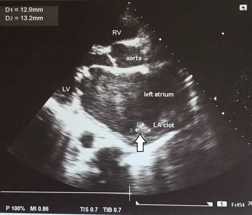
(MS) and atrial fibrillation (AF) leading to multiple systemic emboli, that presented with acute
                                   ischemic stroke, acute myocardial infarction, and acute limb ischemia. Early recognition and
                                   aggressive management led to a satisfactory outcome in this case.

                                   Case Presentation
                                   A 37-year-old man from the Himalayan region of Nepal presented with swelling of the right leg
                                   for ten days and sudden onset weakness of the left half of the body for two days. The swelling of
                                   the right leg had an insidious onset and was gradually progressive. The pain disabled the
                                   patient to move his right leg. Meanwhile, the patient developed weakness of the left half of the
                                   body, predominantly in the upper left limb. The patient also developed slurring of speech and
                                   deviation of the face towards the right side. He denied any history of chest pain, diaphoresis,
                                   shortness of breath, loin pain, nausea or vomiting. He had decreased urine output and red
                                   colored urine and was a non-smoker and non-alcoholic. There was no history of hypertension,
                                   chronic kidney disease, and diabetes mellitus in this patient.

                                   On detail inquiry, the patient gave a history of recurrent throat infection during childhood,
                                   however, he was not medically managed then. Three years back, when he visited a cardiac
                                   centre with complaints of shortness of breath and palpitations, a diagnosis of RHD with severe
                                   MS and AF was made. On reviewing the past record of the patient, a significantly elevated
                                   serum antistreptolysin O (ASO) titer was seen. He was planned for per-cutaneous trans-mitral
                                   commissurotomy (PTMC) by his physician. However, he lost the follow-up and was non-
                                   compliant to his medications.

                                   On physical examination, his blood pressure measured 110/70 mm Hg; pulse rate was
                                   irregularly irregular at 77 beats per minute; respiratory rate was 16 breaths per minute, and
                                   body temperature measured 37.1 o C orally. The patient was alert, conscious, and cooperative.
                                   There were no appreciable pallor, icterus, clubbing, splinter hemorrhages, rashes and cyanosis.
                                   On cardiovascular systemic examination, the first heart sound (S1) was variable in intensity and
                                   second heart sound (S2) was normal. There was a low pitched rough rumbling mid-diastolic
                                   murmur at the apex heard best when the patient was lying on the left side with breath held in
                                   expiration using the bell of the stethoscope. There was a high pitched blowing pansystolic
                                   murmur at the left lower sternal border. Examination of the abdomen was unremarkable. On
                                   neurological examination, the patient had a left-sided central facial nerve palsy, muscle
                                   strengths in the left upper and lower limbs were 2/5 and 3/5 respectively, and there was an
                                   ipsilateral Babinski sign. A thorough eye examination along with fundoscopy was
                                   unremarkable. Local examination of the right leg revealed swelling below the level of the knee
                                   and blackish discoloration of toes and lower third of the leg on inspection. Vesicles could be
                                   seen over the lateral aspects of the leg and dorsum of the foot. The limb was cold to touch and
                                   tender on palpation. Dorsalis pedis and posterior tibial pulses were not palpable (Figure 1).

2018 Pradhan et al. Cureus 10(7): e2964. DOI 10.7759/cureus.2964                                                               2 of 11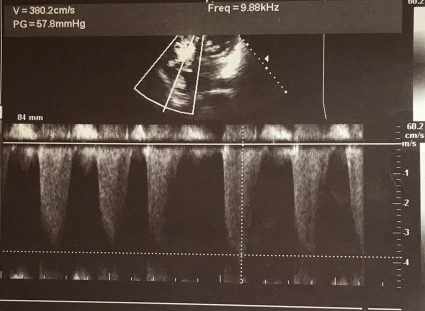
FIGURE 1: Right leg with swelling below the level of knee and
blackish discoloration of toes and lower third of the leg
Vesicles could be seen over the lateral aspects of the leg and dorsum of the foot. The limb was cold
to touch and tender on palpation. Dorsalis pedis and posterior tibial pulses were not palpable.
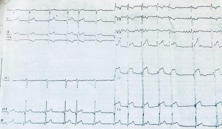
On investigation, electrocardiogram (ECG) showed ST segment elevation of more than two
millimetre (mm) in chest leads V4, V5 and V6, suggesting ST elevation myocardial infarction
(STEMI) and AF with normal ventricular rate (Figure 2). Transthoracic echocardiography
showed thickened, calcified mitral valve leaflets with mitral valve area (MVA) of 1.01 cm 2 on
planometry, mitral valve pressure half-time (PHT) of 344 millisecond, and mean pressure
gradient of 9 mmHg (features suggestive of rheumatic severe mitral stenosis), left atrial
enlargement and a left atrial clot measuring 12.8 mm x 13.2 mm, severe tricuspid regurgitation
(TR) (pressure gradient of 57.8 mm Hg), hypokinesia of antero-lateral wall of the left ventricle
with left ventricle ejection fraction of 35% to 40% (Figures 3-5).
2018 Pradhan et al. Cureus 10(7): e2964. DOI 10.7759/cureus.2964 3 of 11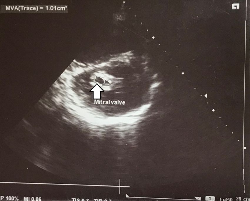
FIGURE 2: Electrocardiogram showing atrial fibrillation with
                                     normal ventricular rate and ST-elevation from V4 to V6

                                     FIGURE 3: Transthoracic echocardiography on short axis view
                                     showing thickened, calcified mitral valve leaflets with MVA of
                                     1.01 cm2 on planometry
                                     MVA: mitral valve area

2018 Pradhan et al. Cureus 10(7): e2964. DOI 10.7759/cureus.2964                                      4 of 11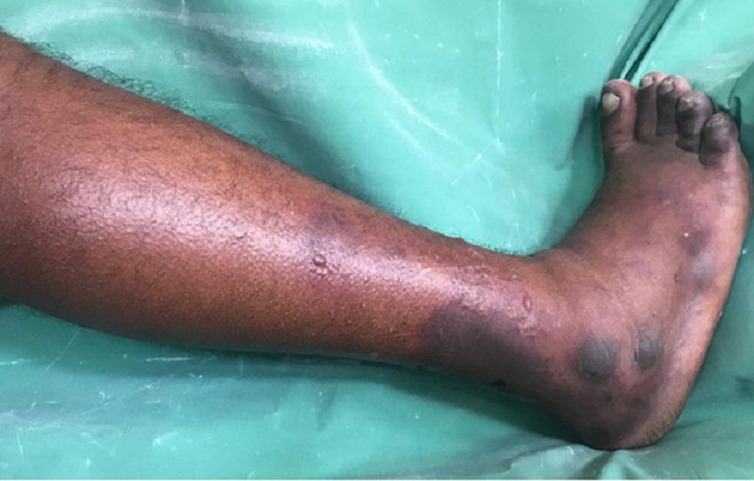
FIGURE 4: Transthoracic echocardiography on parasternal
                                     long axis view showing left atrial enlargement, left atrial clot
                                     measuring 12.8 mm x 13.2 mm (arrow), and thickened and
                                     calcified mitral valve
                                     LV: left ventricle; LA: left atrium; RV: right ventricle; D: diameter.

2018 Pradhan et al. Cureus 10(7): e2964. DOI 10.7759/cureus.2964                                              5 of 11
FIGURE 5: Transthoracic echocardiogram in four-chamber view
with continuous wave (CW) mode showing severe tricuspid
regurgitation with a pressure gradient of 57.8 mmHg
V: velocity; PG: pressure gradient.
Chest radiography revealed cardiomegaly with cardiothoracic ratio 70%, straightening of left
heart border, double right heart border, cephalization (upturned moustache sign or Antler
sign), and widened carinal angle (more than 90 o ) typical of left atrial enlargement. The cardiac
border was displaced laterally and downwards implying left ventricular enlargement (Figure 6).
2018 Pradhan et al. Cureus 10(7): e2964. DOI 10.7759/cureus.2964 6 of 11FIGURE 6: Chest radiograph (posteroanterior view) showing
cardiomegaly with cardiothoracic ratio 70%, straightening of
left heart border (yellow line), double right heart border (white
arrow), cephalization (white circle), and widened carinal angle
typical of left atrial enlargement
The cardiac border was displaced laterally and downwards implying left ventricular enlargement.
A computed tomography (CT) scan of the head showed right-sided ischemic stroke involving
more than one-third of the middle cerebral artery territory (Figure 7).
2018 Pradhan et al. Cureus 10(7): e2964. DOI 10.7759/cureus.2964 7 of 11FIGURE 7: A computed tomography (CT) scan of the head
showed right-sided ischemic stroke (white arrow) involving
more than one-third of the middle cerebral artery territory
Arterial doppler of the right leg showed nearly occluding echogenic content in right femoral
artery with minimal triphasic flow and occluding echogenic content in right popliteal artery
with no flow in color and spectral Doppler study. No flow was noted in the right tibioperoneal
trunk, anterior tibial artery, posterior tibial artery, and dorsalis pedis artery. Venous Doppler of
the right leg demonstrated deep vein thrombosis (DVT) involving right femoral and popliteal
vein. Abdominal ultrasonography was normal.
2018 Pradhan et al. Cureus 10(7): e2964. DOI 10.7759/cureus.2964 8 of 11At workup, the patient had total creatine phosphokinase (CPK) 10000 unit per liter (U/L)
(normal= 10 to 120 U/L), creatine phosphokinase-MB (CPK-MB) 3000 U/L (normal= 0-25 U/L),
troponin I 40.6 nanogram per mililiter (ng/ml) (positive for > 0.12 ng/ml), total white cell count
18,210 per microliter (uL) (normal= 4000-11000 per uL), hemoglobin 15.4 gram per deciliter
(g/dl), creatinine 3.28 milligram per deciliter (mg/dl) (normal= 0.6 to 1.1 mg/dl), sodium 122
milliequivalent per liter (mEq/L) (normal= 135 to 145 mEq/L), potassium 6.2 mEq/L (normal=
3.5 to 4.5 mEq/L), total billirubin 1.4 mg/dl (normal is < 1 mg/dl), alanine trasaminase (ALT) 516
U/L (normal= 30 to 65 U/L), aspartate trasaminase (AST) 2030 U/L (normal= 0 to 45 U/L),
alkaline phosphatase 117 U/L (normal= 40 to 140 U/L), total protein 6.8 g/dl (normal= 6.4 to 8.2
g/dl), albumin 3.2 g/dl (normal= 3.8 to 4.9 g/dl), and prothrombin time (PT) was 13.3 second
with international normalized ratio (INR) 1.1 (control= 12 second). Serum ASO titer was not
done as there was no active rheumatic activity.
A provisional diagnosis of RHD with severe MS and TR with AF leading to multiple systemic
embolizations was formulated. Anticoagulation was started with low molecular weight heparin
(enoxaparin 60 mg subcutaneous twice daily). The patient received aspirin 75 mg and digoxin
0.125 mg once daily. Hyperkalaemia was managed medically with intravenous calcium
gluconate one gram over 10 minutes, 50 ml of 50% dextrose with regular insulin of 10 U and
salbutamol nebulization. Subsequently, the patient underwent above knee amputation of the
right lower limb. His liver and kidney function tests were monitored until they were normalized.
He was discharged on warfarin 6 mg, aspirin 75 mg, spirinolactone 25 mg, enalapril 2.5 mg,
digoxin 0.125 mg once daily and frusemide 20 mg twice daily. The patient was adviced for
regular follow-up for monitoring his clinical status and adherence to medications. He paid two
visits (one week apart) to the hospital after discharge and his clinical conditions were
improving.
Discussion
Of all native valvular diseases, rheumatic mitral valve disease carries the highest risk of
systemic embolization. The incidence of AF in patients with RHD is 43.61% [2]. The presence of
AF increases the risk of systemic embolization and mitral stenosis carries a higher risk of
embolization compared with mitral regurgitation [2]. After the first episode of embolization,
recurrent embolization occurs in 30%-65% of patients; with more than half of recurrences
occurring during the first one year [3]. Similarly, in our case, the cardioembolic phenomenon
associated with RHD was the prime cause of the patient's illness.
The non-valvular atrial fibrillation (NVAF) increases the risk for ischemic stroke by five-fold,
the risk is even greater among patients with mitral stenosis, increasing up to 17-fold [4]. Our
patient had severe MS with AF and was at increased risk of cardioembolic stroke. Stroke in
patients with AF is generally more severe and the outcome is markedly poorer than in patients
with sinus rhythm. Studies in patients with both rheumatic mitral stenosis and atrial fibrillation
have shown that warfarin is effective in preventing cerebral embolism [5]. Mitral valve repair
and mitral valve replacement are also considered in the prevention of cerebral embolism for
patients with hemodynamically significant rheumatic mitral stenosis or insufficiency [3].
When the afflicted patients are young, the tragic consequences for family, friends, and
occupation are particularly catastrophic and unexpected as with our case. In a developing
country like Nepal, where there is no facility for regular monitoring of PT/INR, anticoagulation
with warfarin may not be feasible. However, the evidence regarding the use of newer oral
anticoagulants like rivaroxaban and dabigatran in rheumatic valvular AF is yet to be proven. In
this case, we discharged the patient with warfarin as an anticoagulant with provision for
regular monitoring of PT/INR risking the adherence of the patient with it.
RHD with mitral stenosis presenting for the first time as acute STEMI is rare. In a prospective
2018 Pradhan et al. Cureus 10(7): e2964. DOI 10.7759/cureus.2964 9 of 11study (n=376) conducted by Jose VJ et al. [6], the overall prevalence of coronary artery disease in
a group of patients with rheumatic heart disease undergoing valve surgery was 12.2%. In our
case, the patient was diagnosed with STEMI based on ECG changes, cardiac biomarkers, and
echocardiography, however, he didn’t give a history of chest pain. Primary percutaneous
coronary intervention (PPCI) was not performed in this patient as there was no evidence of
ongoing myocardial ischemia and no features suggestive of heart failure or cardiogenic shock.
Acute peripheral arterial occlusion is characterized by "the six Ps": pain of sudden onset, pallor,
pulselessness, paresthesias, paresis, and prostration with the symptoms of shock. Femoral
bifurcation, the superficial femoral artery, and the popliteal artery are the most common sites
for lodgment of embolus [7] and diseased heart is the main source of arterial emboli [8]. Our
case is in accordance with the above-mentioned studies. Surprisingly, our patient also suffered
from DVT in the right limb, which is an unusual finding. This might have arisen due to a
restriction of movement in the right limb caused by ischemic pain.
Apart from cerebral, coronary and peripheral arterial embolisms, such patients are also
vulnerable for renal and mesenteric embolisms. Renal infarction mostly presents as abdominal
pain and hematuria with an evident rise in serum lactate dehydrogenase [9].
Renal impairment was present in our case and the possible causes of renal impairment could
be renal ischemia or rhabdomyolysis of the right ischemic limb. Rhabdomyolysis is more likely
to be the cause of renal impairment in our case as there was evidence of hyperkalaemia, raised
CPK and liver enzymes. In 50% of cases of rhabdomyolysis, urine could be normal in color, as
with our case. Though the ultrasonography of the abdomen was normal, CT angiography of the
abdomen to rule out renal ischemia was not performed in our case. After amputation of the
ischemic limb, renal impairment and liver enzymes normalized.
Rheumatic heart disease is effectively controlled by long-term antibiotic prophylaxis. Duration
of therapy depends on the severity of cardiac involvement. Lifelong anticoagulation is the
mainstay of prophylaxis for a cardioembolic event in patients of mitral stenosis with atrial
fibrillation. This is, to the best of our knowledge, the first case report of it's kind in medical
literature.
Conclusions
Multiple systemic embolizations must always be considered in patients with valvular heart
disease, especially in MS associated with AF. Clinical suspicion and follow up of such patients
confirming adherence to prophylaxis can prevent adverse outcomes.
Additional Information
Disclosures
Human subjects: Consent was obtained by all participants in this study. Conflicts of interest:
In compliance with the ICMJE uniform disclosure form, all authors declare the following:
Payment/services info: All authors have declared that no financial support was received from
any organization for the submitted work. Financial relationships: All authors have declared
that they have no financial relationships at present or within the previous three years with any
organizations that might have an interest in the submitted work. Other relationships: All
authors have declared that there are no other relationships or activities that could appear to
have influenced the submitted work.
Acknowledgements
2018 Pradhan et al. Cureus 10(7): e2964. DOI 10.7759/cureus.2964 10 of 11We would like to thank Dr. Smriti Shakya for her invaluable support.
References
1. Shrestha NR, Karki P, Mahto R, et al.: Prevalence of subclinical rheumatic heart disease in
eastern Nepal: a school-based cross-sectional study. JAMA Cardiol. 2016, 1:89-96.
10.1001/jamacardio.2015.0292
2. Sharma SK, Verma SH: A clinical evaluation of atrial fibrillation in rheumatic heart disease . J
Assoc Physicians India. 2015, 63:22-5.
3. Leary M, Caplan L: Cardioembolic stroke: an update on etiology, diagnosis and management .
Ann Indian Acad Neurol. 2008, 1:52-63.
4. Wolf PA, Abbott RD, Kannel WB: Epidemiologic assessment of chronic atrial fibrillation and
risk of stroke: the Framingham study. Arch Intern Med. 1987, 147:1561-4.
10.1001/archinte.1987.00370090041008
5. Whitlock RP, Sun JC, Fremes SE, et al.: Antithrombotic and thrombolytic therapy for valvular
disease: antithrombotic therapy and prevention of thrombosis: American college of chest
physicians evidence-based clinical practice guidelines. Chest. 2012, 141:576-600.
10.1378/chest.11-2305
6. Jose VJ, Gupta SN, Joseph G, et al.: Prevalence of coronary artery disease in patients with
rheumatic heart disease in the current era. Indian Heart J. 2004, 56:129-31.
7. Health Quality Ontario: Stenting for peripheral artery disease of the lower extremities: an
evidence-based analysis. Ont Health Technol Assess Ser. 2010, 10:1-88.
8. Oneglia C, Rusconi C: Left atrial appendage thrombus as a source of peripheral embolism: TEE
evidence of direct relationship. Echocardiography. 2001, 18:389-90. 10.1046/j.1540-
8175.2001.00389.x
9. Hazanov N, Somin M, Attali M, et al.: Acute renal embolism. Forty-four cases of renal
infarction in patients with atrial fibrillation. Medicine. 2004, 83:292-9.
10.1097/01.md.0000141097.08000.99
2018 Pradhan et al. Cureus 10(7): e2964. DOI 10.7759/cureus.2964 11 of 11You can also read