Placenta percreta after Strassman metroplasty of complete bicornuate uterus: a case report - BMC Pregnancy and Childbirth
←
→
Page content transcription
If your browser does not render page correctly, please read the page content below
Zhang et al. BMC Pregnancy and Childbirth              (2021) 21:95
https://doi.org/10.1186/s12884-021-03540-y

 CASE REPORT                                                                                                                                         Open Access

Placenta percreta after Strassman
metroplasty of complete bicornuate uterus:
a case report
Chengyan Zhang, Xiaoxin Wang, Haili Jiang, Lei Hou and Liying Zou*

  Abstract
  Background: A bicornuate uterus often results in infertility. While reconstructive procedures may facilitate
  pregnancy, spontaneous abortion or serious pregnancy complications may occur. We present a case of a
  bicornuate uterus with spontaneous conception after Strassman metroplasty; however, life-threatening
  complications during pregnancy occurred.
  Case presentation: : A 38-year-old woman with a history of infertility presented for prenatal care at 6 weeks of
  gestation. She had conceived spontaneously after four failed in vitro fertilization and embryo transfer (IVF-ET)
  procedures, Strassman metroplasty for a complete bicornuate uterus, and two postoperative IVF-ET pregnancies
  that ended in embryo arrest. This pregnancy was uneventful until the patient presented with massive vaginal
  bleeding at 28 weeks of gestation and was diagnosed with placenta previa and placenta percreta. Bleeding was
  controlled after emergency Caesarean section and delivery of a healthy neonate. However, severe adhesions were
  noted as well as a rupture along the metroplasty scar. Two days later, on removal of the intrauterine gauze packing,
  severe hemorrhage resumed, and the uterus did not respond to oxytocin, hemabate, or carbetocin. Emergency
  hysterectomy was required.
  Conclusions: Reconstructive surgical procedures for complete bicornuate uterus may allow patients to achieve
  spontaneous pregnancies. However, potential intrapartum complications include placenta implantation and
  postpartum hemorrhage, and the latter may be exacerbated as the uterus does not contract or respond to oxytocin
  or prostaglandin drugs. Patients should be counseled on the risks associated with pregnancy after Strassman
  metroplasty, and clinicians must be aware of potential severe complications.
  Keywords: Bicornuate uterus, Strassman metroplasty, Placenta percreta, Postpartum hemorrhage, Uterine atony

Background                                                                              anomalies [2]. It is associated with obstetric complica-
The incidence of congenital uterine anomalies is ap-                                    tions including infertility, pregnancy loss during the first
proximately 6.7% in the general population, 7.3% in the                                 and second trimester, preterm labor, intrauterine growth
infertile population, and 16.7% in the recurrent preg-                                  restriction (IUGR), malpresentation, placental abruption,
nancy loss population [1]. A bicornuate uterus, which                                   retained placenta, uterine torsion, and spontaneous rup-
results from incomplete lateral fusion of the two müller-                               ture [3–5]. The more severe the classification and the
ian ducts, accounts for 10–25% of all congenital uterine                                level of bicornuate uterus, the higher the chance of poor
                                                                                        pregnancy outcomes. Surgical intervention with Strass-
                                                                                        man metroplasty might improve reproductive outcomes
* Correspondence: zouliying@ccmu.edu.cn
Beijing Obstetrics & Gynecology Hospital, Capital Medical University, 100026
                                                                                        for these patients, but there are few reports of severe
Beijing, PR China                                                                       complications [3]. Incision into the uterine cavity
                                         © The Author(s). 2021 Open Access This article is licensed under a Creative Commons Attribution 4.0 International License,
                                         which permits use, sharing, adaptation, distribution and reproduction in any medium or format, as long as you give
                                         appropriate credit to the original author(s) and the source, provide a link to the Creative Commons licence, and indicate if
                                         changes were made. The images or other third party material in this article are included in the article's Creative Commons
                                         licence, unless indicated otherwise in a credit line to the material. If material is not included in the article's Creative Commons
                                         licence and your intended use is not permitted by statutory regulation or exceeds the permitted use, you will need to obtain
                                         permission directly from the copyright holder. To view a copy of this licence, visit http://creativecommons.org/licenses/by/4.0/.
                                         The Creative Commons Public Domain Dedication waiver (http://creativecommons.org/publicdomain/zero/1.0/) applies to the
                                         data made available in this article, unless otherwise stated in a credit line to the data.
Zhang et al. BMC Pregnancy and Childbirth (2021) 21:95 Page 2 of 5
increases the chance of placenta previa, morbidly adher-
ent placenta, and severe postpartum hemorrhage. The
latter complication can be lethal, as the malformed and
scarred uterus may not contract and respond to oxytocin
or prostaglandin drugs. We report a rare case of a
complete bicornuate uterus. The patient conceived
spontaneously after a Strassman metroplasty procedure,
but this was complicated by placenta previa and placenta
percreta. The CARE guidelines were followed for this
case report [6].
Case presentation
A 38-year-old woman with a history of having under-
gone Strassman metroplasty of a complete bicornuate
uterus began regular prenatal care in Beijing Obstetrics
and Gynecology Hospital, Capital Medical University
from 6 weeks of gestation. She had been diagnosed with
a bicornuate uterus, endometriosis, and teratoma during
laparoscopic ovarian cystectomy for infertility 6 years
ago. She had reported no other symptoms related to Fig. 1 Magnetic resonance imaging at 22 weeks of gestation.
these conditions. After four failed in vitro fertilization Magnetic resonance imaging (MRI) shows the fetus in the
bicornuate uterus after Strassman metroplasty, with loss of
and embryo transfer (IVF-ET) attempts due to an in- continuity of the uterine wall and intraplacental bands (white arrow)
accessible uterine cavity, she underwent combined hys-
teroscopic and laparoscopic Strassman metroplasty four
years ago. The procedure was converted to open surgery operation, we noticed severe adhesions around the fra-
because of dense adhesions. A transverse myometrial in- gile saddle-shaped uterus. A large number of tortuous
cision was made into the uterine cavities from one cor- vessels covered the lower anterior wall of the uterus.
nua to the other. The two uterine halves were then The lower anterior uterine wall adhered to the bladder
sutured vertically from the anterior uterus across the and was extremely thin, and the posterior wall had
midline fundus and down the posterior wall, creating closely adhered to the pelvis.
one unified cavity. Over the next two years, the patient
conceived twice by IVF-ET, and both pregnancies ended
by curettage for embryo arrest without a chromosomal
disorder at 11 weeks of gestation.
This pregnancy was naturally conceived, and the pa-
tient was under regular antenatal follow-up without the
administration of progesterone. Her nuchal translucency
scan was normal, and noninvasive prenatal testing indi-
cated low risk. Magnetic resonance imaging (MRI) and
ultrasound were suggestive of a posterior-anterior adher-
ent placenta reaching the serosa with anterior myome-
trial thinning and partially missing at 22 weeks of
gestation (Figs. 1 and 2). She reported occasional vaginal
bleeding.
At 28 weeks of gestation, the patient went to the
emergency room for massive vaginal bleeding after defe-
cating. On arrival, she had lost approximately 600 ml of
blood without experiencing any abdominal pain, and
there was still active bleeding greater than the amount
typical of menstruation. Her pulse rate was 80/min and Fig. 2 Magnetic resonance imaging at 22 weeks of gestation.
Magnetic resonance imaging (MRI) shows the fetus and placenta
blood pressure was 110/70 mmHg. Rapid infusion and
increta (white arrow)
emergency cesarean section were carried out. During theZhang et al. BMC Pregnancy and Childbirth (2021) 21:95 Page 3 of 5
The cesarean section incision was located in the uter-
ine body above the upper edge of the placenta. A healthy
boy weighing 1,350 g and measuring 35 cm in height,
was delivered from transverse to breech position with a
normal cord blood gas analysis and Apgar scores (1, 5,
and 10 minutes) of 10-10-10. After the delivery, a 5-cm
vertical rupture of the uterus was noticed along the pre-
vious metroplasty scar and crossing the transverse inci-
sion of the uterus. The tissue beside the scar was much
thinner than the surrounding wall. The placenta was on
the posterior wall, covered the cervix, and reached the
anterior wall. It covered about 4/5 of the uterine cavity
and closely adhered to the uterine wall. The patient had
severe bleeding after delivery, and 20 IU of oxytocin and
250 µg of carboprost tromethamine were injected into
her myometrium to promote uterine contraction. The
placenta adhered tightly to the lower uterus and had to
be separated manually. A 5 × 6-cm section of the placenta
was implanted in the right anterior wall of the uterus and
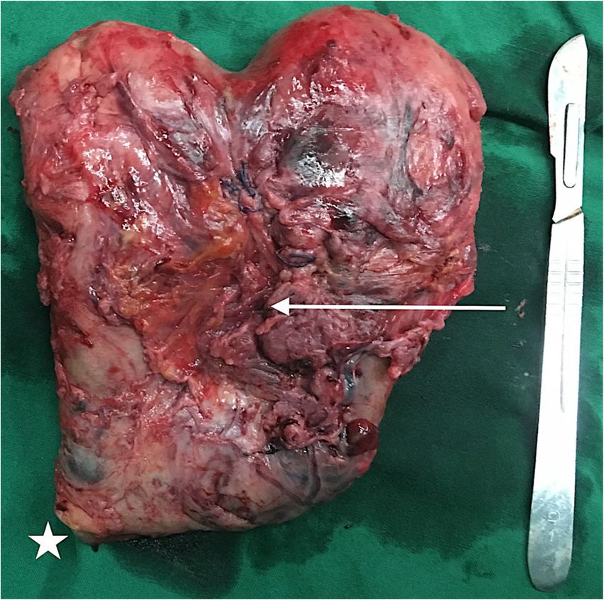
could not be removed. The uterine cavity was filled with Fig. 3 Hysterectomy specimen. The uterus is rigid and non-elastic,
especially in the scar area. The longitudinal scar of the fused uterus
gauze and active bleeding was stopped. In addition to the
is 2 mm thick. The placenta adherent to the anterior wall has
600 ml hemorrhage before the cesarean, the patient lost breached the serosa and adhered to the bladder (white arrow).
another 3,400 ml of blood due to hemorrhage after sur- (White star: cervix)
gery. The patient was transfused with 10 U of red blood
cells, 1,200 ml of plasma, and 1 U of platelets. Subse-
quently, antibiotics were administered to prevent infec- uterus with a single cervix, resulting from incomplete lateral
tion. Over the next 48 hours, the patient had no fever or fusion of the two müllerian ducts. The bicornuate uterus is
bleeding, and her vital signs were stable. defined as partial or complete according to the degree of div-
Massive vaginal bleeding resumed when the gauze was ision in the uterine corpus [7]. Surgical intervention is not
removed under intravenous anesthesia 48 hours after recommended unless the patient has experienced repeatedly
the Caesarean section, with about 2,000 ml of blood loss abnormal pregnancy outcomes (such as abortion, preterm
in only 5 minutes. Meanwhile, the uterus did not re- birth, and infertility) without other causes [8]. The Strassman
spond to massage or oxytocin, hemabate, or carbetocin metroplasty joins the two narrow uterine corpora into one
administration. and tries to reconstruct the normal anatomical structure.
A transabdominal hysterectomy was performed imme- Both abdominal and laparoscopic metroplasty can im-
diately. The uterus adhered closely to the ovary, intes- prove uterine morphology, enlarge cavity volume, reduce
tine, and bladder, and was a rigid saddle shape and intrauterine pressure, and increase blood flow to the
bloated to 16 weeks in pregnancy size with blood clots endometrium and muscle. After surgery, the patient is
inside (Fig. 3). After the hysterectomy, a 2 × 2-cm rup- more likely to achieve conception, and the risk of abor-
ture was found on the bladder, which was repaired and tion and preterm delivery is reduced. The fertility and
required a left ureteral stent. The patient lost 2,000 ml live birth rate associated with metroplasty could reach
and 2,500 ml of blood, respectively, before and during 70–80% [9, 10]. However, there are reports of less satis-
the 5-hour surgery and was transfused with 13 U of factory prognoses for patients who underwent Strassman
RBC, 1,200 ml of plasma, and 1 U of platelets. The post- metroplasty for a complete bicornuate uterus. In a study
operative period was uneventful, including bladder irri- of 11 metroplasty patients, the 4 patients with a partial
gation and the administration of intravenous ceftriaxone bicornuate uterus all had successful pregnancies, while
2 g once daily for 5 days. The patient was discharged none of the 7 patients with a complete bicornuate uterus
after the ureteral stent extraction. Both mother and child achieved conception [11]. In another report of 10 pa-
remained well at follow-up 2 years after delivery. tients with metroplasty for bicornuate uterus, 4 patients
had 5 babies, 3 remained infertile (including 1 case due
to male infertility), and 2 were lost to follow-up. One pa-
Discussion and conclusions tient had placenta previa and lost 2,000 ml of blood dur-
According to the Revised American Fertility Society clas- ing Caesarean section [12]. No cases of uterine rupture
sification, a bicornuate uterus is defined as a double or other intrapartum complications were reported [13].Zhang et al. BMC Pregnancy and Childbirth (2021) 21:95 Page 4 of 5
A bicornuate uterus can cause reduced muscle tissue, poor vessel contraction and does not respond to mas-
abnormal blood flow, and cervical incompetence, all of sage and contraction drugs. Recently, there were two
which may lead to infertility, abortion, preterm delivery, cases reported with successful management of postpar-
IUGR, and malpresentation [14]. A meta-analysis in tum hemorrhage in a bicornuate uterus with balloon
2014 found that a bicornuate uterus was irrelevant to tamponade and no massive bleeding on balloon removal
the fertilization rate and the success rate of assisted re- [18, 19]. Therefore, tamponade of the uterine cavity may
productive technology [15]. be effective in the management of postpartum
In this case, the patient was diagnosed with a complete hemorrhage in the presence of a bicornuate uterus. Due
bicornuate uterus based on findings of laparoscopy com- to the high bleeding risks, uterine artery embolization is
bined with hysteroscopic exploration. The abnormal recommended for these patients after Caesarean section
uterus, endometriosis, and pelvic adhesions contributed and uterine packing, and insertion of an aortic sac and
to infertility and recurrent failed assisted reproductive ureteral stent before gauze removal may also be recom-
technology. Abortion in the first trimester was probably mended. Although we were unable to utilize any of these
caused by inadequate trophoblast blood vessel forma- methods on our patient, in cases such as this one, mea-
tion, which was related to the reduced muscle tissue and sures such as uterine artery embolization, insertion of an
abnormal vasculature in the process of implantation aortic sac before gauze removal, and balloon tamponade,
[16], which might also have led to the lethal placenta might reduce vaginal bleeding and avoid hysterectomy
percreta [17]. Alternatively, the placenta previa and per- after gauze removal.
creta might have been caused by scar tissue, which Regular antenatal care, color flow ultrasound, and MRI
formed during metroplasty and curettage. In Xia’s report may be needed for the evaluation of an adherent pla-
[12], a similar patient underwent laparoscopic Strassman centa in patients with a history of Strassman metro-
metroplasty of a complete bicornuate uterus. Two years plasty. Further research is needed to evaluate the
later, she lost 2,000 ml of blood during surgical delivery optimal methods of treating postpartum hemorrhage
due to placenta previa at 37 weeks of gestation. A mal- with congenital uterine anomalies.
formed uterus is usually combined with limited cavity Women with a bicornuate uterus must be counseled
volume, less elastic muscle, and cervical incompetence, on the severe complications that may occur during preg-
which are relevant to abortion, preterm delivery, and nancy after Strassman metroplasty, which can result in
malpresentation. hysterectomy. Further, a scheduled Caesarean section is
Despite Strassman metroplasty, the uterus, in this case, the recommended mode of delivery due to the risk of
was still morphologically abnormal, and the horns did uterine rupture [20]. Clinicians must monitor patients
not expand during pregnancy. Limited cavity volume is carefully for intrapartum complications including pla-
likely the reason for the massive vaginal bleeding and centa implantation and postpartum hemorrhage.
preterm delivery. Although we did not expect uterine
Abbreviations
rupture and preterm birth in our patient and did not, IVF-ET: In vitro fertilization and embryo transplantation; IUGR: Intrauterine
therefore, implement measures to extend her pregnancy growth restriction; MRI: Magnetic resonance imaging
or prevent uterine rupture, we believe that her prognosis
Acknowledgements
might have been improved by cervical cerclage at 12 to We would like to thank the patient and her family for consenting to publish
14 weeks of gestation. We only scheduled the patient’s this case report.
hospitalization at around 28 weeks of gestation for ad-
Authors’ contributions
ministration of antenatal corticosteroids to enhance fetal CZ, XW, HJ, LH, LZ contributed to conception of the manuscript. LZ
lung maturation and multidisciplinary consultation with contributed to prenatal care of the patient. CZ, XW, HJ, LH, LZ contributed to
the neonatology, anesthesia, and intervention depart- the surgery. CZ, LZ contributed to writing the manuscript. CZ, LH, LZ helped
perform the analysis with constructive discussions. All the authors were in
ments for the surgical planning. However, prior to this charge of the patient and took part in the operation. All authors read and
scheduled appointment, our patient experienced massive approved the final manuscript.
vaginal bleeding, which did not allow us to conduct the
Funding
intended multidisciplinary consultation and led us to
Not applicable.
perform a hysterectomy. The postpartum hemorrhage
after gauze removal probably occurred because the Availability of data and materials
uterus did not contract and respond to oxytocin or pros- All data generated or analyzed during this study are included in this
published article.
taglandin drugs. In the normal uterus, active
hemorrhage seldom occurs after 48-hours of compres- Ethics approval and consent to participate
sion. However, patients with a malformed uterus have a This case report was approved by the Human Ethics Committee of Beijing
Obstetrics & Gynecology Hospital, Capital Medical University (approval
high risk of massive bleeding during gauze removal be- number 2019-KY-014-02). Written informed consent was obtained from the
cause of muscle hypoplasia, which is complicated by patient.Zhang et al. BMC Pregnancy and Childbirth (2021) 21:95 Page 5 of 5
Consent for publication
Written informed consent was obtained from the patient for the publication
of this case report.
Competing interests
The authors declare that they have no competing interests.
Received: 1 November 2020 Accepted: 2 January 2021
References
1. Saravelos SH, Cocksedge KA, Li TC. Prevalence and diagnosis of congenital
uterine anomalies in women with reproductive failure: a critical appraisal.
Hum Reprod Update. 2008;14:415–29.
2. Troiano RN, McCarthy SM. Mullerian duct anomalies: imaging and clinical
issues. Radiology. 2004;233:19–34.
3. Chan YY, Jayaprakasan K, Tan A, Thornton JG, Coomarasamy A, Raine-
Fenning NJ. Reproductive outcomes in women with congenital uterine
anomalies: a systematic review. Ultrasound Obstet Gynecol. 2011;38:371–82.
4. LaHood J, You W. Uterine torsion and subsequent rupture in a gravid
bicornuate uterus associated with an elevated alpha-fetoprotein. BMJ Case
Rep. 2018. doi:https://doi.org/10.1136/bcr-2018-224388.
5. Faria J, Henriques C, do Carmo Silva M, Mira R. Rupture of an unscarred
uterus diagnosed in the puerperium: a rare occurrence. BMJ Case Rep. 2012.
doi:https://doi.org/10.1136/bcr2012006372.
6. Gagnier J, Kienle G, Altman DG, Moher D, Sox H, Riley D, et al. The CARE
guidelines: consensus-based clinical case report guideline development. J
Clin Epidemiol. 2014;67:46–51.
7. American Fertility Society. The American Fertility Society classifications of
adnexal adhesion, distal tubal occlusion, tubal occlusion secondary to tubal
ligation, tubal pregnancies, Mullerian anomalies and intrauterine adhesions.
Fertil Steril. 1988;49:944–55.
8. Lin PC, Bhatnagar KP, Nettleton GS, Nakajima ST. Female genital anomalies
affecting reproduction. Fertil Steril. 2002;78:899–915.
9. Lolis DE, Paschopoulos M, Makrydimas G, Zikopoulos K, Sotiriadis A,
Paraskevaidis E. Reproductive outcome after Strassman metroplasty in
women with a bicornuate uterus. J Reprod Med. 2005;50:297–301.
10. Alborzi S, Asefjah H, Amini M, Vafaei H, Madadi G, Chubak N, et al.
Laparoscopic metroplasty in bicornuate and didelphic uteri: feasibility and
outcome. Arch Gynecol Obstet. 2015;291:1167–71.
11. Nishida M, Otsubo Y, Arai Y, Ichikawa R, Sakanaka M. Difference in
reproductive performance between two subtypes of bicornuate uterus.
Arch Gynecol Obstet. 2016;293:1335–8.
12. Xia EL, Yu D, Huang XW, Ma NX, Yu QJ. Four cases of successful delivery
after hysteroscopy combined with complete bicornuoplasty and literature
review. Zhonghua Fu Chan Ke Za Zhi. 2015;50:777–9.
13. Rechberger T, Monist M, Bartuzi A. Clinical effectiveness of Strassman
operation in the treatment of bicornuate uterus. Ginekol Pol. 2009;80:88–92.
14. Reichman DE, Laufer MR. Congenital uterine anomalies affecting
reproduction. Best Pract Res Clin Obstet Gynaecol. 2010;24:193–208.
15. Venetis CA, Papadopoulos SP, Campo R, Gordts S, Tarlatzis BC, Grimbizis GF.
Clinical implications of congenital uterine anomalies: a meta-analysis of
comparative studies. Reprod Biomed Online. 2014;29:665–83.
16. Crocker IP, Wareing M, Ferris GR, Jones CJ, Cartwright JE, Baker PN, et al. The
effect of vascular origin, oxygen, and tumour necrosis factor alpha on
trophoblast invasion of maternal arteries in vitro. J Pathol. 2005;206:476–85.
17. Ngichabe S, Sura M. Placenta percreta in a gravid bicornuate unicollis uterus.
Case Rep Obstet Gynecol. 2017. doi:https://doi.org/10.1155/2017/4082182.
18. Abraham C. Bakri balloon placement in the successful management of
postpartum hemorrhage in a bicornuate uterus: A case report. Int J Surg
Case Rep. 2017;31:218–20.
19. Khan ES, Basharat A. Successful use of balloon tamponade in the management
of postpartum hemorrhage in a case of bicornuate uterus. SAGE Open Med
Case Rep. 2018. doi:https://doi.org/10.1177/2050313X18776174.
20. Letterie GS. Management of congenital uterine abnormalities. Reprod
Biomed Online. 2011;23:40–52.
Publisher’s Note
Springer Nature remains neutral with regard to jurisdictional claims in
published maps and institutional affiliations.You can also read