Slide-free MUSE Microscopy to H&E Histology Modality Conversion via Unpaired Image-to-Image Translation GAN Models - arXiv
←
→
Page content transcription
If your browser does not render page correctly, please read the page content below
Slide-free MUSE Microscopy to H&E Histology Modality Conversion
                                                           via Unpaired Image-to-Image Translation GAN Models

                                                        Tanishq Abraham 1 Andrew Shaw 2 3 Daniel O’Connor 2 Austin Todd 4 Richard Levenson 5

                                                                     Abstract                                  tissue slides; these dyes color cell nuclei blue and cytoplasm
arXiv:2008.08579v1 [eess.IV] 19 Aug 2020

                                                                                                               pink, respectively. MUSE dyes, on the other hand, typically
                                                  MUSE is a novel slide-free imaging technique                 involve DAPI or Hoechst dyes for nuclei, and rhodamine
                                                  for histological examination of tissues that can             for the other tissue components (Fereidouni et al., 2017).
                                                  serve as an alternative to traditional histology.            The resulting images thematically resemble H&E, but the
                                                  In order to bridge the gap between MUSE and                  colors generated by the UV excitation light impinging on
                                                  traditional histology, we aim to convert MUSE                these dyes are dramatically different from the traditional
                                                  images to resemble authentic hematoxylin- and                brightfield hues.
                                                  eosin-stained (H&E) images. We evaluated four
                                                  models: a non-machine-learning-based color-                  In order to bridge the gap between MUSE imaging and tra-
                                                  mapping unmixing-based tool, CycleGAN, Dual-                 ditional histological examination, it is possible to digitally
                                                  GAN, and GANILLA. CycleGAN and GANILLA                       modify the MUSE images to match H&E images. In (Fer-
                                                  provided visually compelling results that appropri-          eidouni et al., 2017), a spectral unmixing color mapping
                                                  ately transferred H&E style and preserved MUSE               model was used, but it required user input of expected colors
                                                  content. Based on training an automated critic               and is limited to conversion of nuclear and cytoplasm colors,
                                                  on real and generated H&E images, we deter-                  failing to handle cases in which a larger gamut of colors
                                                  mined that CycleGAN demonstrated the best per-               are generated. Therefore, we aim to utilize deep learning
                                                  formance. We have also found that MUSE color                 methodologies in order to learn the appropriate transforma-
                                                  inversion may be a necessary step for accurate               tion for generating visually convincing virtual H&E images
                                                  modality conversion to H&E. We believe that our              that works well on a variety of tissue and cell types.
                                                  MUSE-to-H&E model can help improve adoption                  Deep learning has been used successfully in microscopy
                                                  of novel slide-free methods by bridging a percep-            modality conversion tasks like the one we present here,
                                                  tual gap between MUSE imaging and traditional                (Rivenson et al., 2019a; Borhani et al., 2019; Rivenson et al.,
                                                  histology.                                                   2019b). These modality conversion algorithms often use a
                                                                                                               generative adversarial network (GAN) framework (Good-
                                                                                                               fellow et al., 2014). In this case, the generator needs to be
                                           1. Introduction                                                     trained with the input modality image and a corresponding
                                                                                                               output modality image. Therefore, paired image datasets
                                           Microscopy with ultraviolet surface excitation (MUSE) is
                                                                                                               are required for modality-converting GANs. Unfortunately,
                                           a novel non-destructive, slide-free tissue imaging modality
                                                                                                               it is not possible to obtain exact pixel-aligned H&E and
                                           for histology (Fereidouni et al., 2017). Using MUSE instead
                                                                                                               MUSE images. Therefore, we investigated unpaired image-
                                           of conventional histology processing eliminates the need for
                                                                                                               to-image translation methods that eliminate the need for
                                           fixing and thin-sectioning the tissue. While MUSE has been
                                                                                                               precisely paired datasets.
                                           evaluated for many purposes, the current gold standard in
                                           medicine and biological research for tissue sample analysis         We propose a framework for training and applying an image-
                                           is still based mainly on brightfield imaging of H&E-stained         to-image translation GAN-based algorithm for successful
                                              1
                                                                                                               conversion of MUSE images to virtual H&E images. We
                                                Department of Biomedical Engineering, University of Califor-   evaluated CycleGAN (Zhu et al., 2017), DualGAN (Yi et al.,
                                           nia, Davis 2 University of San Francisco Data Institute 3 Wicklow
                                           AI in Medicine Research Initiative 4 University of Texas, San An-   2017), and GANILLA (Hicsonmez et al., 2020) for MUSE-
                                           tonio Health 5 Department of Pathology, University of Califor-      to-H&E conversion. We hope that our framework will help
                                           nia, Davis. Correspondence to: Richard Levenson .                                                   of the pathologists workflow.
                                           Presented at the ICML 2020 Workshop on Computational Biology
                                           (WCB). Copyright 2020 by the author(s).
MUSE Microscopy to H&E Histology Modality Conversion

2. Methodology                                                  2.3. GANILLA
We define two image domains, one for MUSE images (X),           GANILLA (Hicsonmez et al., 2020) is a variant architecture
and one for H&E images (Y ). We attempt to determine the        for the CycleGAN model designed to appropriately transfer
transformation G : X → Y . In our framework, we have two        the content to the stylized image. See (Hicsonmez et al.,
tasks. One task is to learn a generator GX : X → Y that         2020) for generator architecture details. The discriminator is
maps x ∈ X to y ∈ Y . The auxiliary task is to learn a gen-     a 70x70 PatchGAN. The model was trained for 200 epochs
erator GY : Y → X. Additionally, we have the adversarial        in an identical manner as the CycleGAN (Section 2.1).
discriminators DX and DY . DX discriminates between
the fake outputs of GX and real images from domain Y .          2.4. Color mapping tool
Conversely, DY discriminates between the fake outputs of
GY and real images from domain X. These two GANs                As a baseline, we used a color mapping tool using spectral
form the training framework for MUSE-to-H&E conver-             unmixing algorithms, as previously described in (Fereidouni
sion. CycleGAN, DualGAN, and GANILLA all follow this            et al., 2017).
framework and only differ slightly in model architectures,
loss functions, and training procedures.                        2.5. Tiled inference
                                                                We performed GAN model inference on overlapping tiles
2.1. CycleGAN                                                   with stride 256. The generator was applied to each patch,
CycleGAN exploits the cycle-consistency property that           yielding a 19x19 array of overlapping predicted H&E
GY (GX (x)) ≈ x and GX (GY (y)) ≈ y. This constraint            patches. A 5120x5120 generated H&E montage was then
can be expressed as the following loss:                         constructed, with each pixel intensity value in the montage
                                                                being a weighted average of intensity values from the H&E
                                                                patches which overlapped at the given pixel location. The
                                                                pixel intensity values from each contributing patch were
 Lcycle (GX , GY ) =Ex∼pdata (x) [kGY (GX (x)) − xk1 ]
                                                                weighted proportionally to exp(−d2 /2σ 2 ) where d is the
                      + Ey∼pdata (y) [kGX (GY (y)) − yk1 ]      distance from the given pixel location to the center of the
                                                                contributing patch. The weights in the weighted average
where k · k1 is the L1 norm. Additionally, the GANs are         were normalized to sum to 1. The parameter σ was set to
trained with the traditional adversarial losses (Zhu et al.,    128 pixels.
2017). Finally, for regularization, we impose an identity
constraint:                                                     2.6. Quantitative evaluation of the models
                                                                An external critic model (70x70 PatchGAN with a fully
                                                                connected layer) was trained to quantitatively evaluate how
     Lidentity (GX , GY ) = Ey∼pdata (y) [kGX (y) − yk1 ]       real” the outputs of the various models look. We used ac-
                         + Ex∼pdata (x) [kGY (x) − xk1 ]        curacy and a binary cross-entropy loss from the critic as
                                                                quantitative measures to compare the quality of the genera-
The generator architecture is a ResNet-based fully convo-       tive models. We trained a separate critic on the predictions
lutional network described in (Zhu et al., 2017). A 70x70       for each model to keep results independent. Each critic
PatchGAN (Isola et al., 2017) is used for the discriminator.    were trained for 20 epochs with a 0.001 learning rate (one-
The same loss function and optimizer as described in the        cycle learning rate schedule). Each dataset consisted of
original paper (Zhu et al., 2017) was used. The learning rate   fake” H&E images generated from the test set and real H&E
was fixed at 2e-4 the first 100 epochs and linearly decayed     images from the train set. It was a balanced dataset with an
to zero in the next 100 epochs, like (Zhu et al., 2017).        80/20 dataset split.

2.2. DualGAN                                                    2.7. Datasets and implementation

DualGAN (Yi et al., 2017) also solves the same task as          The H&E data came from a region in a single whole-slide
CycleGAN, while using Wasserstein GANs (Arjovsky et al.,        image of human kidney with urothelial cell carcinoma. The
2017). DualGAN also uses a reconstruction loss, which is        MUSE data came from a single surface image of similar
similar to CycleGANs cycle-consistency loss. The generator      tissue. We obtained 512x512 tiles from the images, resulting
architecture is a U-net (Ronneberger et al., 2015), and the     in 344 H&E tiles and 136 MUSE tiles. The tiles were
discriminator is a 70x70 PatchGAN (Isola et al., 2017). The     randomly cropped into 256x256 images when loaded into
model was trained with the Adam optimizer for 200 epochs        the model. Code was implemented with PyTorch 1.4.0
similar to the CycleGAN (Section 2.1).                          (Paszke et al., 2019), and fastai (Howard & Gugger, 2020).
MUSE Microscopy to H&E Histology Modality Conversion
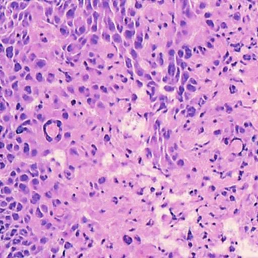
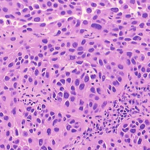
3. Results Example H&E Inverted MUSE Color-mapped
3.1. Training on unprocessed MUSE images
In Figure 1, results on the test dataset after training a Cycle-
GAN on MUSE and H&E images are shown. With close
inspection, it is evident that the generated H&E images do
not appropriately transfer the content of the original MUSE
CycleGAN DualGAN GANILLA
image. Bright in-focus nuclei are converted to white spaces
in the virtual H&E image (boxed in red). On the other hand,
the darker regions are converted to nuclei in the H&E image
(boxed in yellow). The overall trend that the CycleGAN fol-
lowed was converting brighter regions to background white
spaces, and darker regions to nuclei. We have observed that
using color- and intensity-inverted MUSE images greatly
improves training and subsequent models were trained on Figure 2. Comparison of MUSE-to-H&E translation models
inverted MUSE images.
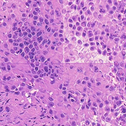
3.2. MUSE-to-H&E translation into the model due to memory constraints. While the models
performed well on individual 512x512 patches, we observed
We trained a CycleGAN, DualGAN, and GANILLA model (Figure 3) that the montage had artifacts near the edges of
on the MUSE and H&E image dataset (Section 2.7), and per- the individual patches (tiling artifacts), and the predictions
formed inference on the test dataset, which is a 5120x5120 are inconsistent in color and style between tiles.
image. Individual 512x512 tiles were inputted into the
In order to resolve these problems, we performed model
model.
inference on overlapping tiles with stride 256 as explained
In Figure 2, we can see that the CycleGAN and GANILLA in Section 2.5. Figure 3 demonstrates how this blending ap-
models provided visually compelling results that appropri- proach suppressed the emergence of tiling artifacts. Figure
ately transfer style and content. The model successfully 4 shows that the final generated montages were much more
converted MUSE representations of cancer cells, inflamma- consistent in style and color throughout the montage.
tory cells, and connective tissue to the corresponding H&E
representations. However, DualGANs performed poorly, 3.4. Critic training
with weak transfer of style, and many artifacts. Finally,
CycleGAN and GANILLA performed better than the tradi- Using the H&E generated results of the CycleGAN,
tional color-mapping baseline. GANILLA and DualGAN models, we trained three sep-
arate external critics to objectively measure the quality of
3.3. Inference the generated images. A fourth critic was trained on images
from the color mapping tool as a baseline comparison. In
We have tested inference with a single 5120x5120 image. this experiment, we would expect the critic model to fail
As the generators are fully convolutional networks, variable more often if the model outputs are higher quality, that is,
sizes are allowed for these models (though the scale must resemble H&E images more closely.
remain same). However, the full region cannot be inputted
Figure 5 shows the graphs of the validation loss and accu-
Inference on tile without overlap Inference on tile with overlap (stride=256)
Figure 1. Training CycleGAN on unprocessed MUSE images Figure 3. Demonstration of tiling artifacts
MUSE Microscopy to H&E Histology Modality Conversion
Inverted MUSE CycleGAN DualGAN GANILLA
Table 1. Negative Critic Accuracy (%)
Epoch #
GAN Loss 1 5 10 20
CycleGAN 56.3 54.1 67.7 71.9
GANILLA 56.3 63.5 81.2 84.4
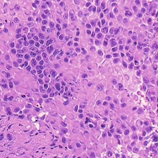
Figure 4. Montages generated from predictions on overlapping
512x512 tiles with stride 256. DualGAN 58.1 94.2 100 100
Color Mapper 56.3 100 100 100
racy while Table 1 presents the accuracy from critic training.
translation models is the lack of quantitative metrics. Most
They show that the critic performed more poorly on Cy-
approaches for quantifying model performance relied on
cleGAN and GANILLA images. The DualGAN was not
crowdsourcing approaches (e.g. Amazon Mechanical Turk)
able to fool the critic because of its poor performance in
to rate quality of the produced images. However, with
producing a convincing color conversion. Interestingly, Du-
difficult-to-interpret histological images, this is not an op-
alGAN performed worse than the color mapper baseline.
tion. Most microscopy modality conversion studies (Riven-
Overall, the critic had the hardest time identifying the Cy-
son et al., 2019a; Borhani et al., 2019; Rivenson et al.,
cleGAN model as fake, which seems to suggest this model
2019b) had paired data and therefore quantitatively eval-
produced the most realistic images. These results supports
uated via structural and perceptual similarity metrics like
the conclusions from qualitative analysis in Section 3.2.
SSIM and PSNR (Wang et al., 2004). However, there are
some key structural differences between MUSE and H&E
4. Discussion images. This would mean that visually compelling virtual
H&E images that also preserve structural content may not
In this study, MUSE modality conversion using unpaired
have high perceptual similarity scores. Instead, we relied on
image-to-image translation techniques was performed in
an independently trained critic model to estimate image qual-
order to generate virtual H&E images. We qualitatively
ity and perceptual similarity. While we found the results to
observed that the GAN-based models studied here produce
be very consistent with our visual inspection, it is important
visually compelling results, with CycleGAN providing the
to note that it is not a perfect metric and does not account for
best results.
GAN hallucinations” or preservation of content. The best
For proper training and inference of the models tested here, metric is still visual inspection by human beings. Future
inverting the MUSE images was required. This is likely be- work will quantitatively evaluate image-to-image translation
cause the CycleGAN cannot learn the association between models with pathologist ratings and interpretation.
brighter nuclei in MUSE to darker nuclei in H&E. It as-
Another key consideration during the development of these
sumed all bright objects in MUSE must be background in
models is model inference. We expect users to be able to
H&E, while dark background objects in MUSE must be
select regions of interest from a whole image to convert to
tissue in H&E. We found this content preservation problem
virtual H&E almost instantly. Currently, this is still a chal-
especially prevalent in DualGANs. In future work, addi-
lenge that needs to be addressed (CycleGAN on 5120x5120
tional constraints, such as the saliency constraint introduced
with stride 512 took 12.7 s on NVIDIA TITAN RTX). Fu-
in (Li et al., 2019), may be tested in order to directly convert
ture work will analyze how model inference can be sped up
unprocessed MUSE images to virtual H&E images.
while minimizing the trade-off regarding montage consis-
A major challenge with training unpaired image-to-image tency.
5. Conclusion
External Critic Loss External Critic Accuracy (%)
0.8
100%
0.7
In conclusion, we have tested three unpaired image-to-
0.6 80%
0.5
image translation models for MUSE-to-H&E conversion.
60%
0.4
CycleGAN CycleGAN
We observed that color- and intensity-inversion is an essen-
0.3 40%
0.2
GANILLA
DualGAN
GANILLA tial preprocessing step when training with MUSE images.
DualGAN
ColorMapper
0.1
20%
ColorMapper Additionally, we used a semi-quantitative method of eval-
0
0 2 4 6 8 10 12
Epoch number
14 16 18
0%
0 2 4 6 8 10 12
Epoch number
14 16 18 uating the models and determined CycleGANs obtain the
best results. We hope our framework can help improve the
Figure 5. Quantitative evaluation of the models via real/fake H&E
efficiency of the pathologist workflow by bridging the gap
critic training between MUSE and traditional histology.
MUSE Microscopy to H&E Histology Modality Conversion

References                                                         L., Bai, J., and Chintala, S. PyTorch: An Imperative
                                                                   Style, High-Performance Deep Learning Library. In Wal-
Arjovsky, M., Chintala, S., and Bottou, L. Wasserstein GAN.
                                                                   lach, H., Larochelle, H., Beygelzimer, A., Alch-Buc, F. d.,
  arXiv:1701.07875 [cs, stat], December 2017. arXiv:
                                                                   Fox, E., and Garnett, R. (eds.), Advances in Neural Infor-
  1701.07875.
                                                                   mation Processing Systems 32, pp. 8026–8037. Curran
Borhani, N., Bower, A. J., Boppart, S. A., and Psaltis, D.         Associates, Inc., 2019.
  Digital staining through the application of deep neural net-
                                                                 Rivenson, Y., Liu, T., Wei, Z., Zhang, Y., de Haan, K.,
  works to multi-modal multi-photon microscopy. Biomedi-
                                                                   and Ozcan, A. PhaseStain: the digital staining of
  cal Optics Express, 10(3):1339–1350, March 2019. ISSN
                                                                   label-free quantitative phase microscopy images using
  2156-7085. doi: 10.1364/BOE.10.001339. Publisher:
                                                                   deep learning. Light: Science & Applications, 8(1):1–
  Optical Society of America.
                                                                   11, February 2019a. ISSN 2047-7538. doi: 10.1038/
Fereidouni, F., Harmany, Z. T., Tian, M., Todd, A., Kint-          s41377-019-0129-y. Number: 1 Publisher: Nature Pub-
  ner, J. A., McPherson, J. D., Borowsky, A. D., Bishop,           lishing Group.
  J., Lechpammer, M., Demos, S. G., and Levenson, R.             Rivenson, Y., Wang, H., Wei, Z., de Haan, K., Zhang,
  Microscopy with ultraviolet surface excitation for rapid         Y., Wu, Y., Gnaydn, H., Zuckerman, J. E., Chong, T.,
  slide-free histology. Nature Biomedical Engineering, 1           Sisk, A. E., Westbrook, L. M., Wallace, W. D., and
  (12):957–966, December 2017. ISSN 2157-846X. doi:                Ozcan, A. Virtual histological staining of unlabelled
  10.1038/s41551-017-0165-y. Number: 12 Publisher: Na-             tissue-autofluorescence images via deep learning. Na-
  ture Publishing Group.                                           ture Biomedical Engineering, 3(6):466–477, June 2019b.
Goodfellow, I., Pouget-Abadie, J., Mirza, M., Xu, B.,              ISSN 2157-846X. doi: 10.1038/s41551-019-0362-y.
 Warde-Farley, D., Ozair, S., Courville, A., and Bengio, Y.        Number: 6 Publisher: Nature Publishing Group.
  Generative Adversarial Nets. In Ghahramani, Z., Welling,       Ronneberger, O., Fischer, P., and Brox, T. U-Net: Convo-
  M., Cortes, C., Lawrence, N. D., and Weinberger, K. Q.           lutional Networks for Biomedical Image Segmentation.
 (eds.), Advances in Neural Information Processing Sys-            In Navab, N., Hornegger, J., Wells, W. M., and Frangi,
  tems 27, pp. 2672–2680. Curran Associates, Inc., 2014.           A. F. (eds.), Medical Image Computing and Computer-
                                                                   Assisted Intervention MICCAI 2015, Lecture Notes in
Hicsonmez, S., Samet, N., Akbas, E., and Duygulu, P.
                                                                   Computer Science, pp. 234–241, Cham, 2015. Springer
  GANILLA: Generative adversarial networks for image
                                                                   International Publishing. ISBN 978-3-319-24574-4. doi:
  to illustration translation. Image and Vision Comput-
                                                                  10.1007/978-3-319-24574-4 28.
  ing, 95:103886, March 2020. ISSN 0262-8856. doi:
  10.1016/j.imavis.2020.103886.                                  Wang, Z., Bovik, A., Sheikh, H., and Simoncelli, E. Image
                                                                  quality assessment: from error visibility to structural
Howard, J. and Gugger, S. Fastai: A Layered API for Deep
                                                                  similarity. IEEE Transactions on Image Processing, 13
  Learning. Information, 11(2):108, February 2020. doi:
                                                                  (4):600–612, April 2004. ISSN 1941-0042. doi: 10.1109/
 10.3390/info11020108. Number: 2 Publisher: Multidisci-
                                                                  TIP.2003.819861. Conference Name: IEEE Transactions
  plinary Digital Publishing Institute.
                                                                  on Image Processing.
Isola, P., Zhu, J.-Y., Zhou, T., and Efros, A. A. Image-
                                                                 Yi, Z., Zhang, H., Tan, P., and Gong, M. DualGAN: Un-
   to-Image Translation with Conditional Adversarial Net-
                                                                   supervised Dual Learning for Image-to-Image Transla-
  works. In 2017 IEEE Conference on Computer Vision
                                                                   tion. In 2017 IEEE International Conference on Com-
   and Pattern Recognition (CVPR), pp. 5967–5976, July
                                                                   puter Vision (ICCV), pp. 2868–2876, October 2017. doi:
   2017. doi: 10.1109/CVPR.2017.632. ISSN: 1063-6919.
                                                                   10.1109/ICCV.2017.310. ISSN: 2380-7504.
Li, X., Zhang, G., Wu, J., Xie, H., Lin, X., Qiao, H., Wang,     Zhu, J.-Y., Park, T., Isola, P., and Efros, A. A. Unpaired
  H., and Dai, Q. Unsupervised content-preserving im-              Image-to-Image Translation Using Cycle-Consistent Ad-
  age transformation for optical microscopy. bioRxiv, pp.          versarial Networks. In 2017 IEEE International Confer-
  848077, November 2019. doi: 10.1101/848077. Pub-                 ence on Computer Vision (ICCV), pp. 2242–2251, Oc-
  lisher: Cold Spring Harbor Laboratory Section: New               tober 2017. doi: 10.1109/ICCV.2017.244. ISSN: 2380-
  Results.                                                         7504.
Paszke, A., Gross, S., Massa, F., Lerer, A., Bradbury, J.,
  Chanan, G., Killeen, T., Lin, Z., Gimelshein, N., Antiga,
  L., Desmaison, A., Kopf, A., Yang, E., DeVito, Z., Rai-
  son, M., Tejani, A., Chilamkurthy, S., Steiner, B., Fang,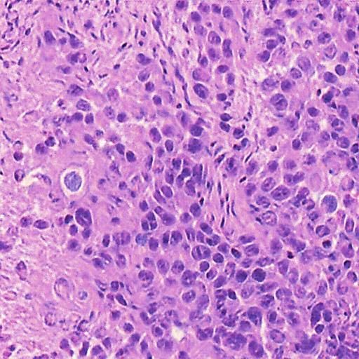
You can also read