Journal of Clinical Images and Medical Case Reports
←
→
Page content transcription
If your browser does not render page correctly, please read the page content below
www.jcimcr.org

                                  Journal of
                                  Clinical Images and Medical Case Reports
                                                                                                                   ISSN 2766-7820

Case Report
Open Access, Volume 2

Rapidly progressive labyrinthitis ossificans in an
immunocompromised pediatric patient
Sean Holmes1*; Katherine Babin2; Avery Bryan2; Gauri Mankekar1
1
 LSU Shreveport Dept of Otolaryngology/Head and Neck Surgery, LSU Health Shreveport, 1501 Kings Highway, Shreveport, LA,
71103, USA.
2
 School of Medicine, LSU Health Shreveport, 1501 Kings Highway, Shreveport, LA, 71103, USA.

                                                               Abstract
   *Corresponding Authors: Sean Holmes
                                                                   In this report, we present a case of rapid otic capsule oblitera-
 Otolaryngology Resident, Department of Otolaryngol-
                                                               tion within an exceedingly short timeframe in the setting of Chronic
 ogy/HNS, LSU Health Shreveport, 1501 Kings Highway,           Suppurative Otitis Media (CSOM) in an immunocompromised pedi-
 Shreveport, LA 71103.                                         atric patient with Down Syndrome. Following maximal therapy for
 Email: sholm6@lsuhsc.edu                                      a right sided cholesteatoma, the patient developed a multi-drug re-
                                                               sistant infection that cause CSOM, which within 6 weeks progressed
                                                               to complete obliteration of the right cochlea and otic capsule. The
                                                               possibility of congenital temporal bone microscopic dehiscence al-
Received: Apr 22, 2021                                         lowing infection propagation cannot be excluded. Nonetheless, this
                                                               case highlights the importance of appreciating how quickly chronic
Accepted: May 12, 2021
                                                               middle ear disease can progress to involve the labyrinth and cause
Published: May 17, 2021                                        intracranial complications, even with adequate concurrent medical
Archived: www.jcimcr.org                                       therapy in the form of antibiotics and surgical therapy. A greater
Copyright: © Holmes S (2021).                                  awareness as physicians should be made on management of refrac-
                                                               tory chronic middle ear disease to better treat their potential com-
                                                               plications, which is made apparent in this case report.

                                                               Keywords: Otic capsule; Chronic suppurative otitis media; Otic cap-
                                                               sule; Tympanomastoidectomy; Multi-drug resistance.
                                                               Abbreviations: CSOM: Chronic Suppurative Otitis Media; MDR:
                                                               Multi-Drug Resistant; CT: Computed Tomography; MRI: Magnetic
                                                               Resonance Imaging; ID: Infectious Disease.

   Introduction/background                                              Chronic Suppurative Otitis Media (CSOM), once common,
                                                                    has become a rarity in the medical world today. The develop-
    The otic capsule, described as the bony labyrinth that sur-     ment of antibiotics along with culture driven therapy and sur-
rounds the membranous labyrinth of the inner ear, is composed       gical intervention has drastically reduced the development of
of the cochlea, vestibule, and semi-circular canals. Partial cap-   complications of CSOM, dropping the intracranial complication
sular erosion can occur secondary to a variety of causes, includ-   rate from 2.3-4% to 0.15-0.04%. These complications include
ing Chronic Suppurative Otitis Media (CSOM), meningitis, aber-      both intracranial and extracranial pathology and can include the
rant arterial supply, schwannomas, cochlear device failures, and    following conditions: mastoiditis, facial nerve palsy, extratem-
Langerhans cell histiocytosis to name a few [1-6]. This rare case   poral abscesses, lateral sinus thrombosis, brain abscesses, cere-
presentation of rapidly progressive capsular obliteration high-     bellar abscesses, labyrinthitis, labyrinthine fistulase, meningitis,
lights the importance of aggressive treatment of chronic middle     extradural abscesses, cochlear erosion, subdural empyemas,
ear disease in children.                                            petrositis, and ossicular erosion [7]. Although medical and sur-
Citation: Holmes S, Babin K, Bryan A, Mankekar G. Rapidly progressive labyrinthitis ossificans in an immunocompromised
pediatric patient. J Clin Images Med Case Rep. 2021; 2(3): 1149.
gical therapies can mitigate these now rare complications, we
must remain aware of the potential for these to arise, and be
up to date on the knowledge of how to treat these conditions.
Suppurative labyrinthitis, a bacterial infection of the inner
ear is relatively uncommon today. There is a radiological classifi-
cation illustrating the four stages of suppurative labyrinthitis: 1)
serous, 2) purulent, 3) fibrous and 4) osseous. The serous stage
involves production of Ig rich exudate in the perilymph, which
then progresses to the purulent stage of bacterial and leuko-
cyte invasion of the perilymphatic scala-end organ necrosis.
The serous and purulent stages together are considered acute
labyrinthitis. The fibrous and osseus stages are together known
as chronic labyrinthitis. Clinical features of acute suppurative
labyrinthitis include severe vertigo with nausea, vomiting, and
hearing loss. The fibrous stage is characterized by fibroblast
Figure 1: Initial Imaging Studies; (IA) Coronal CT: Soft tissue den-
proliferation with granulation tissue in the perilymph. This leads
sity occupying the antrum mastoideum, middle ear, and external
to the osseous stage, which describes new bone deposition in auditory canal with interval severe destruction of the facial canal.
the involved labyrinth [8]. Bony external auditory canal shows evidence consistent with a se-
vere inflammatory process with possible petrositis, osteomyelitis,
In the case of our patient presented below, he originally
labyrinthitis, and otosclerosis. The scutum and ossicular chain are
presented with a right sided cholesteatoma with initiation of
eroded; (IB) Axial CT: Right cochlea and intact, bony labyrinth with
maximal therapy. Despite medical and surgical intervention, the inflammatory changes. Further findings noted above; (IC) Axial T2
patient developed CSOM with a multi-drug resistant bacteria MRI w/ contrast: enhancement of the right petrous bone, involve-
that eventually obliterated the right cochlea and otic capsule in ment of internal auditory canal and cochlea. Severe inflammatory
a matter of 6 weeks. This case adequately demonstrates the im- process involving the right middle ear, internal auditory canal.
portance of awareness of refractory chronic middle ear disease
and its complications as physicians. Following imaging, the patient underwent right canal wall-
down tympanomastoidectomy with facial nerve decompres-
Case presentation sion. Intraoperatively, the cholesteatoma matrix was noted to
A 14-year-old male with history of Down’s Syndrome, Acute be within the epitympanum, extending into the antrum, cover-
Myeloid Leukemia in remission, several previous ear tube place- ing the facial nerve, and without identification of the ossicles.
ments, and bilateral mixed hearing loss presented to our facility The cholesteatoma matrix was resected, and the facial nerve
for evaluation of chronic otitis media referred by a local oto- was stimulated at end of case with adequate response. The
laryngologist. Upon presentation, his parents noted bilateral patient initially did well post-operatively; however, weeks after
otorrhea worse on the right as compared to the left, right-sid- surgery, he developed recurrent right-sided otorrhea for which
ed otalgia, and right-sided facial weakness. Physical examina- further interventions were required.
tion revealed purulent otorrhea bilaterally from patent and The patient returned to clinic for subsequent follow up visits in
functional ear tubes, along with a right sided grade IV House- 2-3 week intervals. The patient’s mom reported persistent right-
Brackmann facial weakness. His initially presenting left sided sided thick otorrhea, although patient continued to be afebrile.
otorrhea resolved after initial topical therapy and no further in- On physical examination, mucoid debris was noted in the
tervention was required for the left ear at the time. The left ear mastoid cavity with no visualization of the tympanic membrane
tube remains in place and functional at this time. at all three visits. At this time, an exam under anesthesia was
Empiric broad-spectrum IV antibiotic therapy was initiated scheduled for further evaluation. This subsequently revealed
for treatment of his right ear symptoms and physical examina- right mastoid cavity with thickened yellow secretions and
tion, and an initial diagnostic work-up with CT and MRI was ob- extensive granulation tissue to anterior and posterior aspects
tained which are described below. of medial mastoid cavity which was removed up to the medial
aspect of the mastoid cavity as to avoid any inadvertent injuries
Computer Tomography (CT) showed a right-sided soft tissue to inner ear structures. Intra-operative cultures were obtained
density occupying the mastoid antrum, middle ear, and external which revealed subsequent growth of Multi-Drug Resistant
auditory canal with severe destruction of facial canal and otic (MDR) Escherichia Coli (E. Coli). He completed 21 days of
capsule with erosion of the cochlea and vestibule and sclerosis culture sensitivity directed IV antibiotics with cefuroxime, as per
of the semicircular canals along with evidence of petrositis and recommendations from the Infectious Disease (ID) department.
erosion of the ossicular chain with the tegmen tympani intact
(Figure 1A,B). Magnetic Resonance Imaging (MRI) revealed re- Auditory Brainstem Response (ABR) was performed after
markable enhancement of the right petrous bone with involve- right-sided canal wall down tympanomastoidectomy, during
ment of the internal auditory canal including the right cochlea, which the patient was roughly half-way through completion
as well as extension to the middle ear and external auditory of his course of IV antibiotics. Results revealed no repeatable
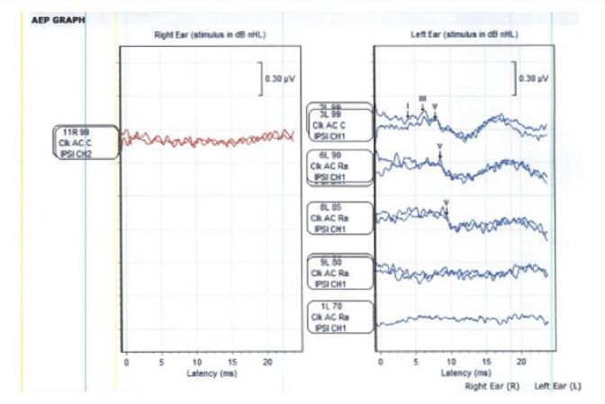
canal. Erosion of the ossicular chain was observed (Figure 1C). waveforms at 99dB nHL for right ear, and moderately severe
mixed hearing loss for left ear (Figure 2).
www.jcimcr.org Page 2Figure 3: ABR tracings- Right ear with no repeatable waveforms at
99 dB nHL, and left ear with moderately severe mixed hearing loss.
Figure 2: Follow-up Imaging Studies; (2A) Coronal CT: Interval ca-
nal wall down mastoidectomy with the mastoid bowl occupied by
The patient clinically improved initially after culture-directed
fluid, including the middle ear and external auditory canal. Pro-
antibiotic therapy following surgical intervention. However, gression of the erosion of the otic capsule with communication of
when he returned to clinic for a second follow-up visit despite the vestibular cochlear system with the internal auditory canal.
51 days of IV Vancomycin and Ceftriaxone, he had right sided The ossicles are now absent. Facial canal intact. The tegmen tym-
persistent otorrhea. Cultures were obtained at this time and pani, regular foramina mastoideum, and carotid canal are intact;
revealed MDR Corynebacterium sensitive to meropenem. The however, the bone surrounding the jugular foramina and appears
patient received 31 days of PO meropenem. At his third follow eroded. The geniculate are ganglion and cochlear segment of the
up visit, the patient had complete resolution of otorrhea and no facial canal appear widened and is suspicious for intracranial ex-
clinical signs of otitis media. tension of the inflammatory process. Bone surrounding the jugu-
lar foramina and appears eroded. Otic capsule shows decreased
Follow up imaging demonstrated complete obliteration of density throughout. Consistent with R temporal bone osteomy-
the right otic capsule. CT imaging of the right temporal bone elitis and high suspicion of intracranial extension of inflammatory
revealed erosion of bone around jugular foramina and widen- process. Additional sclerosis of semicircular canals. (2B) Axial CT:
ing of the geniculate ganglion along with the cochlear segment Complete otic capsule obliteration present, further findings noted
of the facial canal suggesting intracranial extension consistent above in 2A. (2C) Axial T2 MRI w/ contrast: Status-post right sided
mastoidectoimy with persistent inflammatory changes of the right
with complete otic capsular obliteration (Figure 3A,B). MRI re-
temporal bone. Destruction of the otic capsule including osteomy-
vealed persistent inflammatory changes of the right temporal
elitis and labyrinthitis with intracranial extension through the inte-
bone with destruction of the otic capsule including osteomy- mal auditory canal with meningitis also noted with involvement of
elitis and labyrinthitis with intracranial extension through the the 7th and 8th cranial nerves.
internal auditory canal with meningitis. It additionally shows
involvement of the 7th and 8th cranial nerves (Figure 3C).
complete, rapid otic capsular obliteration secondary to CSOM.
We continued to follow our patient monthly to monitor for
Madana et al. have defined CSOM as an insidious and chronic
recurrence of CSOM. Our patient showed no clinical symp-
intractable inflammation of mucosa, submucosa with destruc-
toms of recurrence after completion of the final course of IV
tion of bone of the middle ear cleft characterized by persistent
antibiotic therapy with Meropenem. At the third symptom free
perforation of the tympanic membrane and recurrent otorrhea
monthly follow up visit, we scheduled routine visits every three
[9]. Chronic suppurative otitis media, while common in develop-
months. Our patient is one year removed from the infection and
ing countries, has become a rarity in developed countries, with
has not yet had a recurrence. He now has full resolution of fa-
mortality rates falling from 35% to 5% with the introduction of
cial function with right-sided House-Brackman grade I examina-
antibiotics and advances in health care [9]. Complications of
tion. Due to his history of Down’s Syndrome and intermittent
Chronic Suppurative Otitis Media (CSOM) include mastoiditis,
otorrhea due to ear tubes, methods of amplification have been
facial palsy, extratemporal abscesses, lateral sinus thrombosis,
difficult thus far. In the future we may consider ossicular chain
brain abscess, cerebellar abscess, labyrinthitis, labyrinthine fis-
reconstruction if no disease recurrence becomes evident versus
tula, meningitis, extradural abscess, cochlear erosion, subdural
bone conduction device, as this patient is a poor cochlear im-
empyema, petrositis, ossicular erosion [5,6,10-13]. These com-
plant candidate due to otic capsule obliteration.
plications are typically insidious in nature [5], and are primar-
Discussion ily documented in developing countries [6,14-17]. There have
been few reports of complications occurring in developed coun-
The bony labyrinth that surrounds the membranous labyrinth tries in the last several decades including a case of petrositis
of the inner ear is otherwise known as the otic capsule. The otic and cerebellar abscess complicating chronic otitis media[18].
capsule comprises of 3 parts: The vestibule, semicircular canals, This resolved with a combination of oral and IV antibiotics with
and cochlea. Partial otic capsular invasion has been reported in no hearing deficit after resolution of infection. Our patient has
literature due to aberrant internal carotid artery [1], Langerhans Trisomy 21, and is in remission from AML which further predis-
histiocytosis [2], facial nerve schwannomas [3], cochlear im- poses him to otitis media due to compromised immunity, mid-
plant device failures [3,4], and chronic suppurative otitis media face hypoplasia with malformation of the eustachian tube, a
[5,6]. Although partial otic capsule erosion has been reported, shortened palate, macroglossia, and narrowing of the orophar-
there are no reports of complete cochlear, vestibular, and semi- ynx and nasopharynx [19].
circular canal obliteration. We are reporting the first case of
www.jcimcr.org Page 3Two types of COSM have been described, non-cholestea- 7. Sharma N, Jaiswal AA, Banerjee PK, Garg AK. Complications of
tomatous/tubotympanic and cholestatomatous or atticoantral Chronic Suppurative Otitis Media and Their Management: A
[9,14]. CSOM complications such as intracranial abscess, facial Single Institution 12 Years Experience. Indian J Otolaryngol Head
nerve palsy, meningitis, petrositis, and mastoiditis, and lateral Neck Surg. 2015; 67: 353-360.
sinus thrombophlebitis are more commonly found in the cho- 8. Paparella MM, Shumrick DA, Gluckman JL, Meyerhoff WL. (Eds.).
lesteotomatous type of COSM [9]. Mostafa et al describe 422 Otolaryngology: Volume II Otology and Neurootology. (3rd ed.).
patients with CSOM complications [5]. Of these 422 patients, Philadelphia: W.B. Saunders Company. 1991.
8% had cochlear erosion. Haider et al found that 66 of 279
9. Madana J, Yolmo D, Kalaiarasi R, et al. Microbiological profile
(23.66%) of patients who underwent surgery for CSOM exhib-
with antibiotic sensitivity pattern of cholesteatomatous chronic
ited ossicular chain erosion [9]. Partial ossicular chain interrup- suppurative otitis media among children. Int J Pediatr Otorhino-
tion was found in 69.3% of patients with cholesteatomas CSOM laryngol. 2011; 75: 1104–1108.
vs 13.9% of patients with non-cholesteatomas CSOM [5].
10. Haidar H, Sheikh R, Larem A, et al. Hiader.pdf. Otolaryngol Open
Our patient initially had cholesteatoma for which he under- Access. 2015; 5: 2.
went had right canal wall down tympanomastoidectomy with
facial nerve decompression, and then subsequently has resur- 11. Varshney S, Nangia A, Bist SS, et al. Ossicular Chain Status in
Chronic Suppurative Otitis Media in Adults. Indian J Otolaryngol
gence of MDR bacterial infection consistent with CSOM which
Head Neck Surg. 2010.
progressed to full blown osteomyelitis of the right temporal
bone and obliteration of the vestibule, cochlea, and semicircu- 12. Smith JA, Danner CJ. Complications of Chronic Otitis Media and
lar canals. Congenital temporal bone dehiscence, although rare, Cholesteatoma. Otolaryngol. Clin. North Am. 2006.
could explain the rapid invasion of infection into the inner ear
13. Lin YS, Lin LC, Lee FP, et al. The prevalence of chronic otitis media
leading to purulent labyrinthitis. This congenital pathway for and its complication rates in teenagers and adult patients. Oto-
spread of infection has been reported as a common cause of laryngol - Head Neck Surg Published Online First: 2009.
pediatric meningitis [20,21].
14. Wakode PT, Joshi S V., Gawarle SH. Chronic suppurative otitis
The difficulty in treatment for this case presentation was media in school going children. Indian J Otolaryngol Head Neck
highlighted by the results of bacterial culture including MDR Surg. 2006; 58: 152–155.
E. Coli sensitive to amikacin and ceftriaxone and MDR Coryne-
15. Migirov L, Bendet E, Kronenberg J. Cholesteatoma invasion into
bacterium sensitive to meropenem. This exemplifies the impor-
the internal auditory canal. Eur Arch Oto-Rhino-Laryngology.
tance of culture of ear drainage for culture directed IV antibiotic 2009; 266: 657–662.
therapy which is what ultimately led to resolution for this pa-
tient. Although treated with appropriate culture directed anti- 16. Kangsanarak J, Fooanant S, Ruckphaopunt K, et al. Extracranial
biotic therapy, our patient had refractory infection. With the an- and intracranial complications of suppurative otitis media. Re-
tibiotic resistance crisis, developed countries could potentially port of 102 cases. J Laryngol Otol. 1993.
see a steady increase of CSOM complications that were once 17. Oberdorfer P, Kongthavonsakul K, Intachumpoo J, et al. A
nearly ameliorated with antibiotics. 14-year-old girl with tuberculous otitis media and brain abscess.
BMJ Case Rep. 2012; 2–4.
References
18. Trimis G, Mostrou G, Lourida A, et al. Petrositis and cerebel-
1. Yao W, Benjamin LC, Korzec K. Aberrant Internal Carotid Artery
lar abscess complicating chronic otitis media. J Paediatr Child
Causing Erosion of the Otic Capsule: An Unusual Cause of Pulsa-
Health. 2003.
tile Tinnitus. Otolaryngol Neck Surg 1998; 118: 678–679.
19. Mitchell R, Call E, Kelly J. Ear, nose and throat problems in chil-
2. Blumberg JM, Malhotra A, Wu X, et al. Langerhans Cell Histiocy-
dren with down syndrome. Br J Hosp Med. 2005; 66: 504–506.
tosis of the Temporal Bone with Otic Capsule Involvement. Clin
Neuroradiol 2017; 27: 163–168. 20. Harrington JW, Birck HG. Recurrent Meningitis Due to Congeni-
tal Petrous Fistula: A Case Report. Arch Otolaryngol 1967; 85:
3. Loos E, Wuyts L, Puls T, et al. Cochlear erosion due to a facial
572–575.
nerve schwannoma. J Int Adv Otol. 2019; 15: 330–332.
21. Kimitsuki T, Inamitsu M, Komune S, et al. Congenital malforma-
4. Doherty JK, Linthicum FH. Cochlear endosteal erosion with focal
tion of the inner ear associated with recurrent meningitis. Eur
osteomyelitis induced by cochlear implantation. Otol Neurotol.
Arch Oto-Rhino-Laryngology. 1999.
2004; 25: 1029–1030.
5. Mostafa BE, El Fiky LM, El Sharnouby MM. Complications of sup-
purative otitis media: Still a problem in the 21st century. ORL
Published Online First. 2009.
6. Osma U, Cureoglu S, Hosoglu S. The complications of chronic oti-
tis media: Report of 93 cases. J Laryngol Otol Published Online
First. 2000.
www.jcimcr.org Page 4You can also read