The role of preoperative 3D-ultrasound in intraoperative judgement for hysteroscopic adhesiolysis
←
→
Page content transcription
If your browser does not render page correctly, please read the page content below
Original Article on Intrauterine Adhesion
                                                                                                                                     Page 1 of 11

The role of preoperative 3D-ultrasound in intraoperative
judgement for hysteroscopic adhesiolysis
Arvind Burjoo1#, Xingping Zhao1#, Lingxiao Zou1, Xinyi Liu1, Lei Lei1, Baiyun Zhang2, Dabao Xu1
1
    Department of Obstetrics and Gynecology, Third Xiangya Hospital of Central South University, Changsha 410013, China; 2Department of
Ultrasound, Hunan Guangxiu Hospital, Changsha 410013, China
Contributions: (I) Conception and design: D Xu, A Burjoo; (II) Administrative support: D Xu; (III) Provision of study materials or patients: D Xu,
B Zhang; (IV) Collection and assembly of data: X Zhao, X Liu; (V) Data analysis and interpretation: X Zhao, L Zou; (VI) Manuscript writing: All
authors; (VII) Final approval of manuscript: All authors.
#
These authors contributed equally to this work as co-first authors.
Correspondence to: Dabao Xu, MD. Department of Obstetrics and Gynecology, Third Xiangya Hospital of Central South University, 138 Tongzipo
Rd., Changsha 410013, China. Email: dabaoxu@yahoo.com; Baiyun Zhang, MD. Department of Ultrasound, Hunan Guangxiu Hospital, No. 8,
Luyun Road, Changsha 410013, China. Email: 158478968@qq.com.

                  Background: Hysteroscopic adhesiolysis (HA) remains the mainstay of treatment for intrauterine
                  adhesions (IUA). In cases of moderate or severe IUA, the assistance of various adjunctive aids are usually
                  sought to improve HA’s success rate. Among these, intraoperative transabdominal ultrasound (TAS) is the
                  most common; however, it has certain limitations. Preoperative three-dimensional transvaginal ultrasound
                  (3D-TVUS) has been accepted as a non-invasive way to provide accurate information about the uterine
                  cavity. This prospective, non-randomized controlled study will assess the effects of pre-operative 3D-TVUS
                  prior to HA in improving the surgeon’s intraoperative judgement.
                  Methods: A total of 362 patients, who met the inclusion criteria, aged between 18 and 45 years and
                  diagnosed with moderate or severe IUA underwent HA at our hospital from March 2018 to December
                  2018. Participants were divided into 2 groups; the study group; n=182 performed 3D-TVUS evaluation
                  prior to HA, and the control group; n=180 underwent HA without preoperative 3D-TVUS evaluation. The
                  following basic information were collected prospectively for both groups: age, parity, history of abortion,
                  degree of IUA, surgical complications and number of hysteroscopic interventions. The data obtained
                  from 3D-TVUS in the study group was carefully studied at the preoperative stage by the operator and was
                  integrated into intraoperative findings, further assisting with intraoperative decisions. The guiding value of
                  preoperative 3D-TVUS for HA was evaluated by comparing and analyzing the postoperative exposure rate
                  of clearly visible tubal ostia between the groups.
                  Results: Based on the basic information (P>0.05) collected preoperatively, there were no statistically
                  significant differences between the groups. Postoperatively, the study group had a better surgical success rate
                  with a more significant AFS score reduction (4.71±2.05; P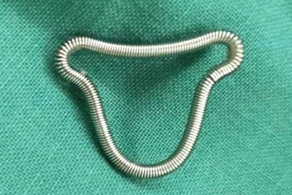
Page 2 of 11                                       Burjoo et al. The role of preoperative 3D-ultrasound in intraoperative judgement for HA

                Submitted Nov 29, 2019. Accepted for publication Dec 30, 2019.
                doi: 10.21037/atm.2020.01.06
                View this article at: http://dx.doi.org/10.21037/atm.2020.01.06

Introduction                                                               outcome. Filmy adhesions (especially central cavity lesions)
                                                                           can be bluntly lysed with cavity distension and by the tip
Intrauterine adhesions (IUA) refer to partial or
                                                                           of the hysteroscope or blunt dissecting forceps. In cases of
complete adhesions which occur between the uterine
                                                                           severe adhesions where various segments are inaccessible
walls and may result in several clinical manifestations
                                                                           or at least one of the ostia cannot be seen or retrieved, or
such as hypomenorrhea, amenorrhea, dysmenorrhea,
                                                                           in cases of cervical stenosis, however, HA proves to be very
low abdominal pain, sub-fertility/infertility, recurrent
                                                                           challenging, and can result in difficulties with intraoperative
abortions, premature delivery, and abnormal placental
                                                                           judgement and surgical complications. When the severity of
implantation. IUA can be either primary, after pregnancy-
                                                                           the adhesion hinders the procedure, the assistance of TAS
related curettage or hysteroscopic surgery, or secondary,
                                                                           is usually sought. In cases of severe IUA, intraoperative
re-occurring after adhesiolysis has been performed (1,2).
                                                                           ultrasound monitoring is very important for HA; however,
Dilation and curettage (D & C) after miscarriage accounts
                                                                           the timing for ultrasound is not ideal in the proliferative
for 93% of IUA (3). IUA are significant but usually (partly)               phase, as the endometrium is very thin during this particular
correctable cause of infertility. When they are treated,                   period. Furthermore, the imaging quality of portable TAS is
fertility outcomes can be improved and symptoms can                        usually not very appreciable and is unable to provide precise
be relieved or resolved. Accurate identification of the                    information intraoperatively, unlike preoperative three-
extent and character of adhesions and reliable diagnostic                  dimensional transvaginal ultrasound (3D-TVUS), which
tools for assessment of the uterine cavity is a necessary                  allows the operator to place a high-frequency endocavitary
first step in improving adhesiolysis success rates. Several                ultrasound transducer in close proximity to target pelvic
diagnostic modalities have been proposed for the diagnosis                 organs, thus improving image resolution. With the advent
of IUA: hysterosalpingography (HSG), saline infusion/                      of high-resolution vaginal probes, 3D-TVUS has recently
contrast sonohysterography (SHG), 3-D ultrasonography,                     been adopted in the gynecological sciences. By enabling
diagnostic hysteroscopy and magnetic resonance imaging                     multiplanar displays, which simultaneously visualize
(MRI). Diagnostic hysteroscopy is considered the gold                      the three orthogonal scan planes, 3D-TVUS boasts the
standard among these studies as it more accurately confirms                additional advantage of being able to obtain anatomical
the presence, extent, and morphological characteristics                    views which are often unattainable by TAS or 2D-TVUS
of adhesions, as well as the quality of the endometrium,                   and also due to the fact that the coronal plane is easily
and helps in classification and concurrent treatment                       accessible. The coronal views show the relationship between
of IUA (4). However, there are disadvantages with                          the endometrium and the myometrium at the uterine
diagnostic hysteroscopy, the most important of which is                    fundus, delineate the entire cervical canal and visualize
its inability to access and assess the intrauterine cavity                 the cornual angles. Intraoperative 2D-TVUS/3D-TVUS,
in cases of severe cervical stenosis and severe IUA. This                  however, would lengthen the duration of HA, potentially
is a particular problem with lower segment obliteration,                   resulting in fluid overload. Instead, preoperative 3D-TVUS
when the hysteroscope is unable to reach the cavity                        during the mid-menstrual phase would be more informative
beyond the point of obliteration. Moreover, it is associated               and accurate as it can be performed for a longer period with
with complications such as cervical laceration, uterine                    better image quality. Ultrasonographic data can be rapidly
perforation, bleeding, reactions to the distention media,                  acquired preoperatively and stored for retrospective analysis
and anesthesia. Surgery is considered as the main therapy                  with no loss of information. One can also “scroll” in real-
for IUA, with no role for medical management. Lysis of                     time through the acquired volume that can be rotated and
IUA under direct hysteroscopic visualization is considered                 magnified. 3D-TVUS can give the surgeon preoperative
as the treatment of choice for IUA (4). HA aims to restore                 detailed information about the cavity including which
a normal uterine cavity, prevent recurrence of adhesions,                  segments are obliterated, the extent of obliteration and
normalize menstrual flow and improve reproductive                          the functional state of the endometrium. 3D-TVUS is

© Annals of Translational Medicine. All rights reserved.                Ann Transl Med 2020;8(4):55 | http://dx.doi.org/10.21037/atm.2020.01.06
Annals of Translational Medicine, Vol 8, No 4 February 2020                                                                    Page 3 of 11

considered as an optimal diagnostic test as it is non-invasive,         (IV) Surgical intolerance or inability to follow the doctor’s
safe, painless, widely available, inexpensive and applicable                 advice for review or follow-up.
to all women regardless of their pretest probability of                 (V) Congenital malformation of uterus.
having a particular condition of interest. Most importantly,
3D-TVUS can explore the areas where a hysteroscope may
                                                                        3D-TVUS examination
have difficulties reaching, avoiding possible errors by the
surgeon which could lead to failure to retrieve the fallopian           GE VOLUSON E8 ultrasound instrument (GE Healthcare
tube ostia, create a false passage or even cause uterine                GmbH & Co OG, Tiefenbach, Styria, Austria) with the
perforation. To the best of our knowledge, there has been               two-dimensional volume probe in the cavity and the real-
no published literature focusing on the use of preoperative             time three-dimensional volume probe were used for the
3D-TVUS in intraoperative judgement during HA in IUA                    preoperative three-dimensional ultrasound examination.
patients. In this study, we used the data obtained from                 Patients from the study group underwent preoperative
3D-TVUS to make proper surgical planning, informed the                  transvaginal 3D-TVUS during the secretory phase of
patients about the current condition of their endometrial               the menstrual cycle, using 7.5 MHz IC5-9D vaginal
cavity and the potential course of their treatment, and                 probe. During the examination, the patients emptied
capitalized on the information retrieved for intraoperative             their bladder and were placed in the lithotomy position.
judgement while also minimizing risks and complications                 Routine two-dimensional ultrasonographic examination
during HA.                                                              was performed first. During 2D-ultrasound, the integrity of
                                                                        the endometrial layer was assessed to look for disruptions
                                                                        of the endometrial–myometrial junction. Adhesions on
Methods                                                                 ultrasound are seen as bands of myometrial tissue traversing
Patients                                                                the endometrial cavity and adjoining the opposing uterine
                                                                        walls. After rotating the real-time three-dimensional
This study was designed as a prospective non-randomized                 volume probe, the panoramic technology was used to obtain
controlled study. Patients were collected from March, 2018              the overall image information and select the target area.
to December, 2018. After strict screening, 362 patients                 During the 3D-ultrasound examination, IUA are seen with
with IUA who met the inclusion criteria were enrolled.                  the characteristic appearance of hyperechoic areas within
Of these, 182 patients received transvaginal 3D-TVUS                    the endometrium. Finally, the information was stored on a
examination during the secretory phase of their menstrual               removable hard disk for further evaluation and calculation.
cycle before their operation (study group), and 180 patients
did not receive transvaginal 3D-TVUS examination before
their operation (control group). All 3D-TVUS images were                Surgical procedure
reconfirmed by the same senior and experienced doctor.                  Hysteroscopic adhesiolysis (HA) was performed within
                                                                        3–7 days following menstruation, with the patient
Inclusion criteria                                                      placed in the lithotomy position and given intravenous
(I) Aged 18–45 years, fertility seeking patients or patients            anaesthesia. Patients fasted for 6–8 hours before surgery.
      with menstrual outflow obstruction.                               A sterile saline solution was used to distend the uterus.
(II) Diagnosed as IUA by diagnostic hysteroscopy with                   Distension pressure was 110–120 mmHg with a flow
      AFS score ranging from 5 to 12.                                   rate of 300–350 mL/min. The operation was monitored
(III) At least one unilateral fallopian tube ostium is not visible,     by transabdominal ultrasound (TAS). After routine
      confirmed by preoperative diagnostic hysteroscopy.                disinfection and draping, hysteroscopy was carried out
                                                                        using an operative hysteroscope with an outer sheath
Exclusion criteria                                                      diameter of 5.4 mm and a 5-Fr working channel (KARL
(I) Cervical or endometrial lesions.                                    STORZ SE & Co. KG, Tuttlingen, Baden-Württemberg,
(II) Serious heart, liver or renal insufficiency.                       Germany). The hysteroscope was introduced into the
(III) Patients with serious nervous system diseases, who                cervical canal through the cervix with the aim of reaching
      are unable to take care of themselves in daily life or            the intrauterine cavity. The adhesions located in the
      unable for undergo relevant treatment.                            central part of the uterine cavity were usually dissected

© Annals of Translational Medicine. All rights reserved.              Ann Transl Med 2020;8(4):55 | http://dx.doi.org/10.21037/atm.2020.01.06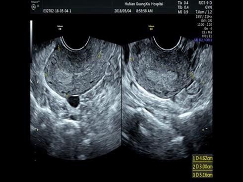
Page 4 of 11 Burjoo et al. The role of preoperative 3D-ultrasound in intraoperative judgement for HA
technique” (6) until the entire uterine cavity had been
opened successfully with clearly visible bilateral fallopian
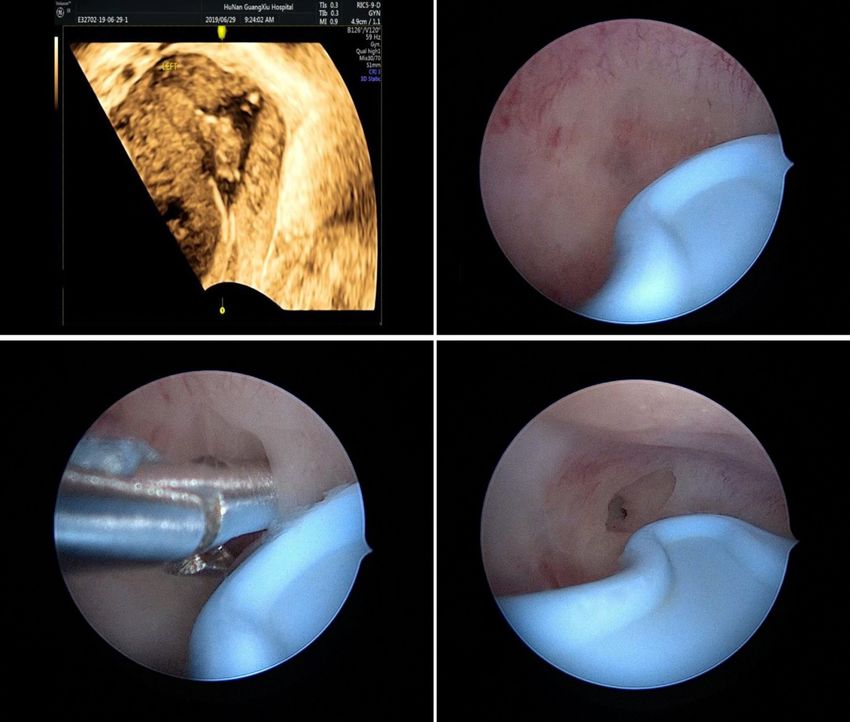
tube ostia. A uterine-shaped stainless-steel intrauterine
device (IUD) (Figure 1) was then inserted into the uterine
cavity, with its position checked via hysteroscopy to ensure
that the size of the IUD matched the uterine cavity size
and that the IUD was correctly positioned (7). A double
channel, 12-Fr Foley catheter balloon, with the top
catheter portion beyond the balloon removed (Figure 2),
Figure 1 A uterine-shaped stainless-steel IUD. IUD, intrauterine
was inserted into the uterine cavity and distended using
device.
2.5 mL of sterile saline with the balloon in the center
of the uterine-shaped IUD. Three mL hyaluronic acid
gel was then injected into the uterine cavity through the
catheter. Postoperatively, the intrauterine balloon was kept
in place for 7–14 days for patients with severe IUA and for
2–3 days for patients with mild and moderate IUA. For
patients with only lower segment IUA, no IUD was used,
and instead, a distended Foley catheter balloon was left in
situ for up to 3 weeks.
Postoperative follow up hysteroscopy recommendation:
For patients with an AFS score ≥8, a two-time follow-
up strategy was implemented (the first follow-up is done
one month after initial surgery, and the second one, 3
months after the first follow up hysteroscopy). For those
with an AFS score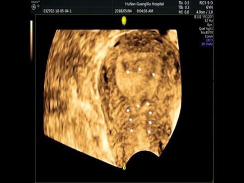
Annals of Translational Medicine, Vol 8, No 4 February 2020                                                                       Page 5 of 11

 Table 1 Analysis of variables between groups with unilaterally            the study group and the control group, respectively
 invisible tubal ostium                                                    (P=0.2674). Preoperative mean AFS scores were 9.12±1.59
 Variables                    Control group Study group P value            and 8.58±1.68 for the study group and the control group,
 Age                                                           0.2674      respectively (P=0.0969). There were no statistically
   Mean ± SD                   32.47±4.71      31.60±4.86                  significant differences between the groups in terms
                                                                           of pregnancy history, menstruation and uterine cavity
   Median [min, max]           32.0 [23, 43]   31.0 [22, 46]
                                                                           operations preoperatively (P>0.05), but for the study group,
 Gravidity                                                     0.7429
                                                                           the cornual adhesions were more serious than those of the
   Mean ± SD                    3.38±1.75       3.32±1.76                  control group (P
Page 6 of 11 Burjoo et al. The role of preoperative 3D-ultrasound in intraoperative judgement for HA
Table 2 Multivariate logistic regression analysis of variables affecting retrieval rate of unilaterally invisible fallopian tube ostium
Effect Degree of freedom Estimate Standard error Wald Chi-square 95% CI P value
Type 3 effect analysis
Menstruation 2 1.2850 0.5260
Preoperative AFS score 1 3.6986 0.0545
Group 1 6.2979 0.0121
Maximum Likelihood estimate
Menstruation
Hypomenorrhea 1 0.9046 0.8339 1.1768 0.2780
Amenorrhea 1 1.4542 1.3867 1.0997 0.2943
Preoperative AFS score 1 −0.3528 0.1835 3.6986 0.0545
Group: study group 1 1.1849 0.4722 6.2979 0.0121
Odds ratio estimate
Menstruation
Hypomenorrhea vs. normal menses 2.471 0.482–12.666
Amenorrhea vs. normal menses 4.281 0.283–64.845
Preoperative AFS score 0.703 0.490–1.007
Group: study group vs. control group 3.271 1.296–8.251
and preoperative cornual adhesions were the main clearly visible fallopian tube ostia: (details in Table 5). Rank
factors that affect postoperative exposure rate of clearly variance analysis showed that preoperative transvaginal 3-D
visible tubal ostia (group: PAnnals of Translational Medicine, Vol 8, No 4 February 2020 Page 7 of 11
Table 3 Analysis of variables between groups with bilaterally balloon-aided cervical dilation has also been proposed for
invisible tubal ostia treatment of IUA. But both of the aforesaid techniques are
Variables Control group Study group P value useful only for treatment of mild adhesions and have low
Age 0.8783 efficiency in treatment of moderate to severe adhesions.
Mean ± SD 32.38±4.83 32.27±5.02 For moderate to severe IUA, the myometrial scoring
Median [min, max] 32.5 [23, 43] 33.0 [20, 42] method was proposed, in which a series of 6–8 incisions are
Gravidity 0.6702 made on the myometrium, from the fundus to the isthmus
Mean ± SD 3.40±1.84 3.46±2.34
to a depth of 4 mm using a Collins knife electrode (11).
However, as electrosurgical energy poses a risk of
Median [min, max] 3.0 [1, 10] 3.0 [0, 12]
endometrial destruction and there is a tendency for IUA
Parity 0.4534
to reoccur, its use is not recommended for fertility-seeking
Mean ± SD 0.63±0.69 0.72±0.79
patients (6). Roy et al., in 2010, reported on 89 infertile
Median [min, max] 1.0 [0, 4] 1.0 [0, 5]
patients with IUA who underwent HA with concomitant
Abortion 0.5179 laparoscopy (12). They stated that concurrent laparoscopy
Mean ± SD 2.57±1.72 2.53±2.04 is helpful for confirming tubal patency and ruling out
Median [min, max] 2.0 [0, 9] 2.0 [0, 11] other pelvic pathologies to elucidate the boundaries of
Menstruation 0.5164 adhesiolysis by observing the transillumination. However,
Normal menses 0 (0.00%) 4 (3.74%) they encountered 2 cases of uterine perforation during the
Hypomenorrhea 79 (74.53%) 77 (71.96%) procedure. Thomson et al., in 2007 reported on 30 patients
Amenorrhea 27 (25.47%) 26 (24.30%) with IUA who underwent HA under fluoroscopic guidance,
Total 106 (100.00%) 107 (100.00%) which allows the surgeon to view islands of endometrium
Preoperative cornual adhesions 0.0196
behind scar tissue in an obliterated uterine cavity. The use
of a Tuohy needle is used in parallel to hysteroscopy (13,14)
No adhesion 25 (23.58%) 10 (9.35%)
through which a radiopaque dye is injected into an area
Unilateral adhesions 6 (5.66%) 7 (6.54%)
of dense scar at the point where the cavity is obliterated.
Bilateral adhesions 75 (70.75%) 90 (84.11%)
Any pockets of endometrium beyond the adhesive area can
Total 106 (100.00%) 107 (100.00%)
then be identified using fluoroscopy and this area can be
Preoperative AFS score 0.8962 opened up by sharp dissection under hysteroscopic view (13).
Mean ± SD 10.02±1.59 10.07±1.43 However, fluoroscopy can result in relatively high doses of
Median [min, max] 10.0 [5, 12] 10.0 [6, 12] radiation, especially for complex interventional procedures
Causes of IUA 0.8150 which require its administration for a longer period of time.
Dilatation and curettage 90 (84.91%) 88 (82.24%) Another limitation of this technique is that it involves the
Other uterine cavity 16 (15.09%) 19 (17.76%) use of an image intensifier and requires the presence of a
operation radiographer throughout the HA procedure. Studies have
Total 106 (100.00%) 107 (100.00%) shown that HA under laparoscopic or fluoroscopic guidance
IUA, intrauterine adhesion. is not necessary to reduce the risk of uterine perforation (15).
Rather, the use of TAS during HA is a preferred and
current option as it is non-invasive. TAS has been described
attempt of entry might lead to failure to retrieve the cavity as a technique to guide hysteroscopic division of IUA
and ostia, creation of false passages, uterine perforation and (16,17) and the availability and familiarity of sonography to
entail several other risks. gynecologists makes this option easy to implement. In our
Several techniques have been proposed to lyse adhesions. study, concomitant TAS was performed on every patient
Coccia et al. in 2001 reported that IUA patients can be intraoperatively to guide the extent of adhesiolysis.
treated by Pressure Lavage under Ultrasound Guidance However, TAS has its limitations; the 2D imaging
(PLUG) method using the distention medium used quality of portable TAS is not very accurate or informative
for hysteroscopy for adhesiolysis (9). The Seldinger during the proliferative phase of the menstrual cycle;
technique (10) under ultrasound (US) guidance followed by during which the endometrium is very thin. Moreover,
© Annals of Translational Medicine. All rights reserved. Ann Transl Med 2020;8(4):55 | http://dx.doi.org/10.21037/atm.2020.01.06Page 8 of 11 Burjoo et al. The role of preoperative 3D-ultrasound in intraoperative judgement for HA Table 4 Multivariate logistic regression analysis of variables affecting retrieval rate of the bilaterally invisible fallopian tube ostia Effect Degree of freedom Estimate Standard error Wald Chi-square 95% CI P value Type 3 effect analysis Group 1 57.7457
Annals of Translational Medicine, Vol 8, No 4 February 2020                                                                       Page 9 of 11

 Table 6 Rank variance analysis of AFS score reduction (part I)            uterine perforation was reported in as many as 5% of cases
 Variables                     Control group Study group       P value     (17-19) when TAS was used as the sole adjunctive aid
 Preoperative AFS score                                        0.0969
                                                                           during HA. This led us to adopt a different non-invasive
                                                                           approach; through preoperative 3D-TVUS as it provided
   Mean ± SD                    8.58±1.68      9.12±1.59
                                                                           detailed information about the location, extent and degree
   Median [min, max]            8.0 [5, 12]    10.0 [6, 12]                of adhesions within the uterine cavity. Depending on the
 Postoperative AFS
Page 10 of 11 Burjoo et al. The role of preoperative 3D-ultrasound in intraoperative judgement for HA
A B
Figure 4 3D-TVUS guides and informs the operator about the condition of the endometrium and the uterine cavity beyond the point of
obliteration. (A) 2D-TVUS showing a normal sized uterus with normal contour, a smooth surface and uneven endometrial thickness; (B)
3D-TVUS showing that the lower part of the uterine cavity is closed, the middle section is narrowed, and the endometrium is continuously
interrupted. However, both cornual angles are clear. 3D-TVUS, three-dimensional transvaginal ultrasound.
reveal the left cornual cavity (Figure 3A,B,C,D). Apart would recommend for all patients to undergo routine
from the size and contour of the uterus, 3D-TVUS is 3D-TVUS examinations prior to HA and a diagnostic
indicative of the distance between the uterine isthmus and hysteroscopy would less likely be needed.
fundus, the intercornual distance, the presence or absence
of healthy endometrium in different segments of the
Conclusions
uterine cavity. Hysteroscopists are often blinded by severe
lower segment adhesions/obliteration and not only face Preoperative 3D-TVUS has proved to have a very
difficulties in retrieving tubal ostia or reaching the uterine important role in intraoperative judgement and also in
cavity but are also unaware what to expect in the middle improving HA success rates.
and upper segments. 3D-TVUS guides and informs the
operator about the condition of the endometrium and the
Acknowledgments
uterine cavity beyond the point of obliteration and can
also indicate the distance between the point of obliteration Funding: Natural Science Foundation of China (Grant No.
and ostium (Figure 4A,B). 3D-TVUS can analyze and 81671492), the Hunan Science and Technology Department
explore areas where the hysteroscope cannot reach. It can (Grant No. 2018SK2102).
not only demonstrate the angular cavity clearly but can
also inform the hysteroscopist about its condition. From
Footnote
time to time, in cases of lower segment uterine cavity
obliteration or severe IUA, a false passage may be created. Conflicts of Interest: The authors have no conflicts of interest
When the latter is diagnosed preoperatively with the aid to declare.
of 3D-TVUS, the hysteroscopist knows exactly where to
expect the false passage and necessary precautions can be Ethical Statement: The authors are accountable for all
taken intraoperatively so as to recognize it and prevent aspects of the work in ensuring that questions related
perforation. to the accuracy or integrity of any part of the work are
In our study, the study group consisted of patients appropriately investigated and resolved. The study was
with higher mean AFS scores than those in the control approved by the Institutional Review Board (IRB) of Third
group, yet we noted that, postoperatively, their mean Xiangya Hospital and Xiangya Hospital, Central South
AFS score and mean AFS score reduction were better; University (No. 2019-S455).
4.97±2.53 and 4.71±2.05, respectively, with P valueAnnals of Translational Medicine, Vol 8, No 4 February 2020 Page 11 of 11
2001;7:567-76. Suppl (Stockholm) 2008;434:47-52.
2. Hellebrekers BW, Trimbos-Kemper TC, Trimbos 11. Protopapas A, Shushan A, Magos A. Myometrial Scoring:
JB, et al. Use of fibrinolytic agents in the prevention A new technique for the management of severe Asherman’s
of postoperative adhesion formation. Fertil Steril syndrome. Fertil Steril 1998;69:860-4.
2000;74:203-12. 12. Roy KK. Reproductive outcome following
3. Yang JH, Chen MJ, Chen CD, et al. Optimal waiting hysteroscopic adhesiolysis in patients with infertility
period for subsequent fertility treatment after various due to Asherman’s syndrome. Arch Gynecol Obstet
hysteroscopic surgeries. Fertil Steril 2013;99:2092-6.e3. 2010;281:355-61.
4. Magos A. Hysteroscopic treatment of Asherman’s 13. Broome JD, Vancaillie TG. Fluoroscopically guided
syndrome. Reprod BioMed Online 2002;4:46-51.
hysteroscopic division of adhesions in severe Asherman
5. Huang H, Cheng C, Johnson G, et al. Hysteroscopic
syndrome. Obstet Gynecol 1999;93:1041-3.
Intrauterine Adhesiolysis Using a Blunt Spreading
14. Duffy S, Reid P, Sharp F. In-vivo studies of uterine
Dissection Technique With Double-action
electrosurgery. Br J Obstet Gynaecol 1992;99:579-82.
Forceps. Journal of Minimally Invasive Gynecology
15. Fedele L, Bianchi S, Frontino G. Septums and synechiae:
2018;25:583-4.
approaches to surgical correction. Clin Obstet Gynecol
6. Simsir C, Var T, Kalem MN, et al. Hysteroscopic
2006;49:767-88.
treatment of Asherman's Syndrome. Cumhuriyet Medical
16. Yu D, Wong YM, Cheong Y, et al. Asherman syndrome:
Journal 2019;41:443-9.
one century later. Fertil Steril 2008;89:759-79.
7. Salma U, Xue M, Sayed AS, et al. Efficacy of Intrauterine
Device in the Treatment of Intrauterine Adhesions. 17. Protopapas A, Shushan A, Magos A. Myometrial scoring:
Biomed Res Int 2014;2014:589296. a new technique for the management of severe Asherman’s
8. Berman JM. Intrauterine adhesions. Semin Reprod Med syndrome. Fertil Steril 1998;69:860-4.
2008;26:349-55. 18. Zikopoulos KA. Live delivery rates in subfertile women
9. Coccia ME, Becattini C, Bracco GL, et al. Pressure Lavage with Asherman’s syndrome after hysteroscopic adhesiolysis
under ultrasound guidance: A new approach for outpatient using the resectoscope or the Versapoint system. Reprod
treatment of intrauterine. Fertil Steril 2001;75:601-6. Biomed Online 2004;8:720-5.
10. Seldinger SI. Catheter replacement of the needle in 19. McComb PF, Wagner BL. Simplified therapy for
percutaneous arteriography: a new technique. Acta Radiol Asherman’s syndrome. Fertil Steril 1997;68:1047-50.
Cite this article as: Burjoo A, Zhao X, Zou L, Liu X, Lei L,
Zhang B, Xu D. The role of preoperative 3D-ultrasound in
intraoperative judgement for hysteroscopic adhesiolysis. Ann
Transl Med 2020;8(4):55. doi: 10.21037/atm.2020.01.06
© Annals of Translational Medicine. All rights reserved. Ann Transl Med 2020;8(4):55 | http://dx.doi.org/10.21037/atm.2020.01.06You can also read