Global sagittal alignment after surgery of right thoracic idiopathic scoliosis in adolescents and adults with and without thoracic hypokyphosis
←
→
Page content transcription
If your browser does not render page correctly, please read the page content below
www.nature.com/scientificreports

                OPEN             Global sagittal alignment
                                 after surgery of right
                                 thoracic idiopathic scoliosis
                                 in adolescents and adults
                                 with and without thoracic
                                 hypokyphosis
                                 Kwong Hang Yeung1, Gene Chi Wai Man2, Wafa Skalli3, Zongshan Hu2, Vivian Wing Yin Hung2,
                                 Alec Lik Hang Hung2, Tsz Ping Lam2, Bobby Kin Wah Ng2, Jack Chun Yiu Cheng2,
                                 Claudio Vergari3* & Winnie Chiu Wing Chu1

                                 This study aimed to characterize global sagittal alignment in adolescent idiopathic scoliosis (AIS)
                                 with normal kyphosis (NTK, kyphosis > 10°) and with thoracic hypokyphosis (THK, kyphosis < 10°),
                                 before and after posterior spinal fusion, and compare them with asymptomatic controls. 27 AIS
                                 girls and young adults with right thoracic curves were included (seventeen with age ≤ 18 years, then
                                 age > 21). Biplanar radiographies were acquired at baseline, immediate post-operatively, 1-year
                                 and 2-year follow-up, and 3D reconstruction of the spine and pelvis was performed. NTK and THK
                                 showed different global sagittal alignment, as well as differences compared to controls. AIS with THK
                                 at baseline had higher SVA/SFD (2.0 ± 2.9 vs − 0.4 ± 1.9; P < 0.05) and OD-HA (0.2 ± 1.4° vs − 1.3 ± 1.6°;
                                 P < 0.05) than controls, indicating that THK had compensated balance with unusual forward leaning
                                 posture. Immediately post-operation, SVA/SFD remained high (1.3 ± 3.0) while OD-HA reversed
                                 (− 1.2 ± 1.7°), indicating that THK patients had found partially compensated balance. After 2-yeas,
                                 both SVA/SFD (− 1.3 ± 2.1) and OD-HA (− 1.4 ± 0.9°) were normalized. The changes in global sagittal
                                 alignment and mechanism of balance are different in AIS with or without THK. As the head plays a
                                 critical role on balance during immediate and delayed post-operation, OD-HA can be complementary
                                 parameter for assessing global balance during post-operative follow-up of AIS patients with THK.

                                 Abbreviations
                                 AIS	Adolescent idiopathic scoliosis
                                 CL	Cervical lordosis
                                 LL	Lumbar lordosis
                                 NTK	Normal thoracic kyphosis
                                 OD-HA	Odontoid-hip axis angle
                                 PI	Pelvic incidence
                                 PT	Pelvic tilt
                                 SFD	Sacro-femoral distance
                                 SS	Sacral slope
                                 SVA	Sagittal vertical axis

                                 1
                                  Department of Imaging and Interventional Radiology, Faculty of Medicine, The Prince of Wales Hospital,
                                 The Chinese University of Hong Kong, Shatin, Hong Kong SAR, China. 2SH Ho Scoliosis Research Laboratory,
                                 Department of Orthopaedics and Traumatology, Faculty of Medicine, The Prince of Wales Hospital, The Chinese
                                 University of Hong Kong, Shatin, Hong Kong SAR, China. 3Arts et Métiers Institute of Technology, Université
                                 Sorbonne Paris Nord, IBHGC ‑ Institut de Biomécanique Humaine Georges Charpak, HESAM Université, Arts et
                                 Metiers ParisTech, 151, boulevard de l’hopital, 75013 Paris, France. *email: c.vergari@gmail.com

Scientific Reports |   (2021) 11:6294              | https://doi.org/10.1038/s41598-021-85782-6                                               1

                                                                                                                                      Vol.:(0123456789)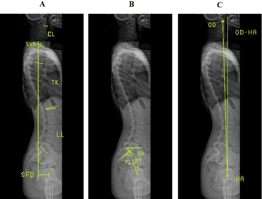
www.nature.com/scientificreports/
SVA/SFD Ratio between the C7 plumb line from the postero-superior corner of the sacrum and SFD
THK Thoracic hypokyphosis
TK Thoracic kyphosis
Adolescent idiopathic scoliosis (AIS) is a three-dimensional deformity of the spine for which the cause has not
yet been i dentified1. Current surgical treatment for AIS involves multi-segmental posterior pedicle screw instru-
mentation and spinal fusion, which can achieve considerable correction in the coronal plane with limited loss of
correction over time2. Most reports on the surgical correction mainly focused on the coronal Cobb angle, with
relatively little emphasis on the changes of sagittal profile. Some studies have demonstrated that an insufficient
surgical correction of hypokyphosis in severe AIS can lead to sagittal malalignment and accelerated degeneration
of the distal unfused vertebral s egments3,4.
Indeed, keeping an ideal standing posture is important to ensure the least amount of energy expenditure to
prevent muscle fatigue and vertebral s train5. Several authors studied the sagittal plane in AIS, and in particular
the relationship between the thoracolumbar spine and the p elvis6. In some cases, cervical spine was i ncluded7.
When considering the changes of the sagittal alignment after surgery, Park et al. followed-up 93 patients for
2 years, for instance, but they limited their analysis to the thoracolumbar s pine4, while other studies included
the cervical spine. Still, the position of the head is often neglected in these analyses. With a mass of 4–5 kg, the
head engages multiple vertebral segments to obtain an economic balance, and the neuromuscular system aims
at keeping the gaze horizontal and the head within a narrow cone above the p elvis8. Including the head position
in the analysis of global sagittal alignment allows to analyze the postoperative changes of alignment in terms of
compensation mechanisms9.
In this study, we aimed to analyze global sagittal alignment in AIS, with a focus on the head position, accord-
ing to thoracic hypokyphosis after posterior spinal fusion, and relative to asymptomatic controls, with the hypoth-
esis that such analysis might improve the understanding of sagittal compensating mechanisms from head to
pelvis during early and later post-operative periods.
Results
Subjects’ characteristics. Twenty-seven patients with AIS (female; mean age: 18.1 ± 4.6yrs, ranging
between 12 and 31 years old) and thirty-six asymptomatic controls (female; mean age: 25.1 ± 2.7yrs) were
recruited (Table 1). Seventeen patients were 18 years old or younger at time of surgery, while 10 were older. Most
patients were presented with Lenke type 1 or 2 curvatures (24 out of 27). Mean Cobb angle at pre-operative
and at immediate post-operative were 65.4 ± 11.9° and 21.5 ± 8.2°, respectively (P < 0.001), indicating a correc-
tion rate of 68 ± 9.9%. Based on Lenke classification10, seven AIS patients had thoracic hypokyphosis (THK;
T5-T12 kyphosis = 1.7 ± 6.6°) and twenty AIS patients had normal thoracic kyphosis (NTK; T5-T12 kypho-
sis = 23.6 ± 7.9°, P < 0.001). Only one THK patient was older than 18 (21 years old, T5-T12 kyphosis of 3.8°);
therefore, no analysis was carried out on the older THK patients’ sub-group. Figure 1 shows the distribution of
T5/T12 kyphosis in NTK and THK groups.
Intraclass correlation coefficient values for the intra- and inter-observer agreement of sagittal parameters,
such as CL, SVA/SFD and pelvic obliquity, were higher than 0.9, indicating reliable measurement methods.
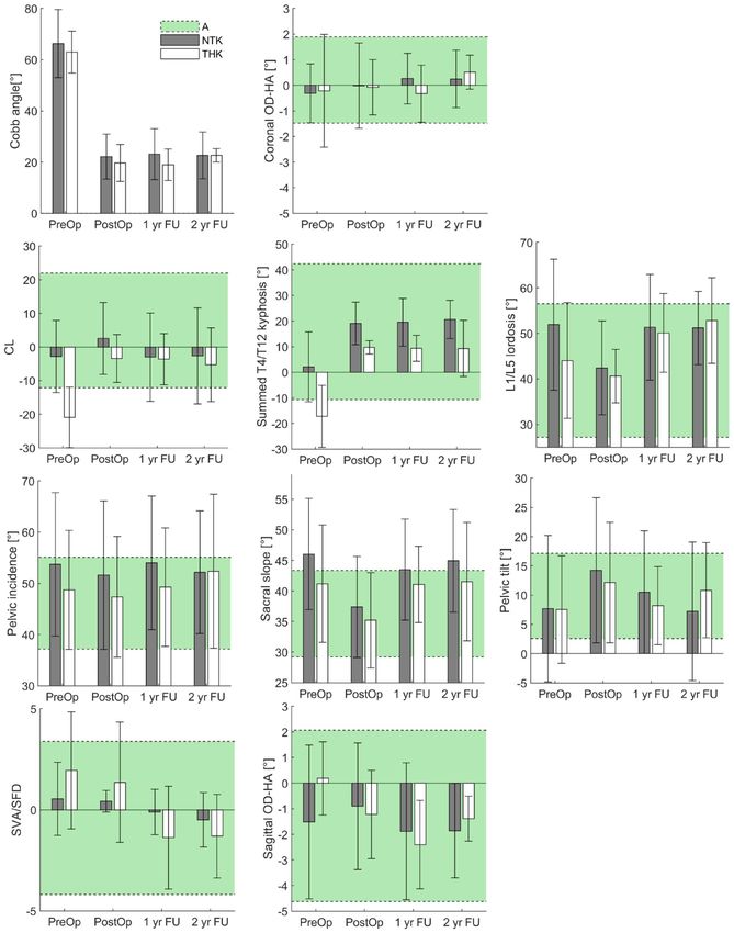
Spinal alignment. Figure 2 shows the main results and group comparisons. In AIS with THK, there was
significant change in CL and TK from pre-operatively to each post-operative stage (P < 0.05), while no signifi-
cant change was found in LL at any stage (P > 0.05) (Table 2). Pre-operatively, THK patients showed significant
differences in CL and TK when compared with the asymptomatic controls (P < 0.05). TK significantly improved
immediately after surgery (P < 0.05).
TK significantly improved post-operatively in NTK patients as well (P < 0.05), with values similar to asympto-
matic subjects (P > 0.05), and remained thus during follow-up. The change of TK was correlated with the change
of LL (R = 0.498, P < 0.05) (Table 3). Although CL also improved at immediate post-operatively in older patients
(age > 18, P = 0.04), there was no change in younger patients (P = 0.4) and all patients underwent a regression of
CL at 1-year follow-up.
Spinopelvic alignment. In AIS with THK, no significant change was found in PI, PT and SS at any stage,
independently of age (P ≥ 0.05). Although significant changes of pelvic obliquity were found at immediate post-
operative, these parameters were normalized at 1-year follow-up (P > 0.05). When compared with asymptomatic
controls, there was significant difference in SS and pelvic obliquity pre-operatively and 1-year follow-up in THK
group (P < 0.05). Also, the changes in LL showed a positive correlation with SS (R = 1.000, P < 0.01) (Table 4).
In NTK group, significant changes were found in PT, SS and pelvic obliquity between pre-operatively and
immediate post-operatively, independently of age (P < 0.05), although the change in PT was at the limit of
significance in young adults (P = 0.05), and all these changes were normalized to pre-operative values at 1-year
follow-up (P > 0.05). PI did not change in average (P > 0.05), but 7 patients (35%) showed changes of 5° or more
in 1-year follow-up. In addition, there were significant differences in PI, SS and pelvic obliquity between AIS
patients pre-operatively and at 1-year follow-up when compared with asymptomatic controls. And similar to
AIS with THK, the increase in PI was correlated to the change in PT (R = 0.711, P < 0.01) (Table 3).
Head and global sagittal alignment. Although there was no significant change in SVA, SFD and coro-
nal OD-HA in AIS with THK, independently of age, there was a significant decrease in the ratio of SVA/SFD at
1-year follow-up (P < 0.05) and a decrease of sagittal OD-HA (P < 0.05). Both SVA/SFD and sagittal OD-HA were
Scientific Reports | (2021) 11:6294 | https://doi.org/10.1038/s41598-021-85782-6 2
Vol:.(1234567890)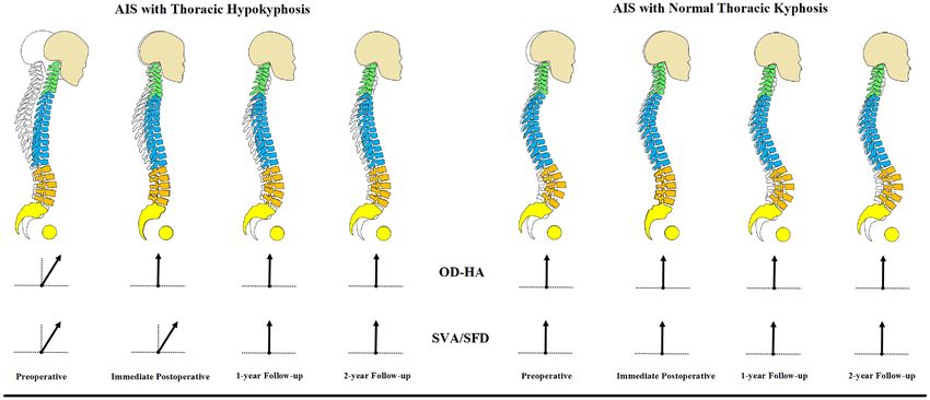
www.nature.com/scientificreports/
AIS Controls
Immediate 1-year 2-year
Included Pre-operation Post-operation Follow-up Follow-up
No. of subject 27 27 27 20 36
Age (years) 18.1 ± 4.6 18.2 ± 4.6 19.5 ± 4.7 21.9 ± 4.9 25.1 ± 2.7
Cobb angle (°) 65.4 ± 11.9 21.5 ± 8.2 22.0 ± 9.0 22.6 ± 8.2 –
Correction rate (%) – 67.7 ± 9.9 67.0 ± 10.5 65.2 ± 10.8 –
Loss of correction (°) – – 0.6 ± 9.7 − 0.3 ± 13.0 –
Normal Thoracic Kyphosis# 20 20 20 16 –
T5-T12 kyphosis (°) 23.6 ± 7.9 – – –
Age (years) 18.7 ± 5.0 18.8 ± 5.1 20.2 ± 5.2 22.5 ± 5.0
Cobb angle (°) 66.3 ± 13.3 22.1 ± 8.8 23.1 ± 9.9 22.6 ± 9.2 –
Correction rate (%) – 67.2 ± 10.5 65.7 ± 11.3 64.7 ± 11.7 –
Loss of correction (%) – – 1.4 ± 10.9 1.3 ± 13.4 –
Thoracic Hypokyphosis# 7 7 7 3 –
T5-T12 kyphosis (°) 1.7 ± 6.6 – – –
Age (years) 16.4 ± 3.1 16.5 ± 3.1 17.8 ± 2.8 17.1 ± 0.3
Cobb angle (°) 62.9 ± 7.6 19.6 ± 6.7 18.9 ± 5.7 22.6 ± 2.1 –
Correction rate (%) – 69.4 ± 7.7 70.3 ± 6.6 67.9 ± 2.6 –
Loss of correction (%) – – − 1.6 ± 4.6 − 9.2 ± 3.6 –
Table 1. Demographic Characteristics are shown for all included adolescent idiopathic scoliosis (AIS) and
controls. Data expressed as mean ± SD. # The values are based on the number of subjects, with the percentage in
parentheses.
Figure 1. Distribution of T5/T12 kyphosis in patients with normal thoracic kyphosis (NTK) and hypokyphosis
(THK).
significantly different between AIS patients at pre-operation and asymptomatic controls (P < 0.05). After surgery,
these parameters improved towards the values of the asymptomatic subjects (Fig. 2).
For AIS patients with NTK, there were significant changes of SVA and SFD immediate post-operatively
(P = 0.01). However, these changes lost significance when looked at by age group (P ≥ 0.05), and they returned
to pre-operative values when follow-up after 1-year and 2-year, when they remained similar to controls at all
time points (P > 0.05). No significant correlation in SVA with these parameters was found, while SFD was cor-
related to the change in PI (R = − 0.738, P < 0.01) and PT (R = − 0.946, P < 0.01). And unlike AIS patients with
Scientific Reports | (2021) 11:6294 | https://doi.org/10.1038/s41598-021-85782-6 3
Vol.:(0123456789)www.nature.com/scientificreports/
Figure 2. Main results comparing asymptomatic subjects (A, green shaded areas representing
average ± 2*standard deviation), normal thoracic kyphosis patients (NTK, mean ± 2SD) and thoracic
hypokyphosis patients (THK).
Scientific Reports | (2021) 11:6294 | https://doi.org/10.1038/s41598-021-85782-6 4
Vol:.(1234567890)www.nature.com/scientificreports/
Immediate 1-year 2-year
Pre-operation Post-operation Follow-up Follow-up
Spinal parameters
NC – – – –
Cobb angle (°) NTK-AIS 66.3 ± 13.3 22.1 ± 8.8* 23.1 ± 9.9* 22.6 ± 9.2*
THK-AIS 62.9 ± 7.6 19.6 ± 6.7* 18.9 ± 5.7* 22.6 ± 2.1*
NC 4.9 ± 8.5 – – –
Cervical lordosis (C2-C7) (°) NTK-AIS − 2.8 ± 10.7# 2.6 ± 10.7 − 3.0 ± 13.1# − 2.7 ± 14.3
THK-AIS − 20.9 ± 8.3# − 3.4 ± 6.6*# − 3.6 ± 7.1*# − 5.3 ± 9.0
NC 15.8 ± 13.3 – – –
Thoracic kyphosis (T4-T12) (°) NTK-AIS 2.2 ± 13.6# 19.1 ± 8.3* 19.5 ± 9.3* 20.7 ± 7.5*
THK-AIS − 17.3 ± 11.2# 9.7 ± 2.4*# 9.4 ± 4.7* 9.3 ± 9.0*
NC 41.8 ± 7.3 – – –
Lumbar lordosis (L1-L5) (°) NTK-AIS 51.9 ± 14.4# 42.4 ± 10.3 51.3 ± 11.6# 51.2 ± 8.1#
THK-AIS 44.0 ± 11.4 40.6 ± 5.2 50.1 ± 7.7 52.8 ± 6.7
Spino-pelvic parameters
NC 46.1 ± 9.0 – – –
Pelvic incidence (PI) (°) NTK-AIS 54.8 ± 12.8# 52.3 ± 13.6# 54.6 ± 12.2# 53.2 ± 12.0#
THK-AIS 50.1 ± 14.4 49.3 ± 14.2 50.0 ± 14.4 55.3 ± 17.6
NC 9.9 ± 7.3 – – –
Pelvic tilt (PT) (°) NTK-AIS 9.6 ± 12.0 16.3 ± 11.7*# 11.5 ± 9.7 9.1 ± 11.3
THK-AIS 5.0 ± 9.4 9.5 ± 11.2 6.9 ± 8.0 9.7 ± 11.3
NC 36.3 ± 7.1 – – –
Sacral slope (SS) (°) NTK-AIS 45.2 ± 9.2# 36.1 ± 7.9* 43.1 ± 7.3# 44.2 ± 8.9#
THK-AIS 45.1 ± 10.3# 39.8 ± 6.7 43.1 ± 7.0# 45.7 ± 8.1
NC 0.8 ± 0.6 – – –
Pelvic obliquity (PO) (°) NTK-AIS 1.8 ± 0.8# 1.3 ± 0.6*# 1.4 ± 0.8# 1.3 ± 0.6*#
#
THK-AIS 1.6 ± 0.4 1.3 ± 0.8* 1.3 ± 0.4 1.3 ± 0.2
Global sagittal alignment parameters
NC − 2.6 ± 16.0 – – –
SVA (mm) NTK-AIS − 0.4 ± 22.5 − 14.4 ± 20.3*# 6.1 ± 22.4 − 6.1 ± 22.8
THK-AIS − 8.0 ± 13.0 − 1.3 ± 17.0 11.5 ± 13.0 − 5.4 ± 15.8
NC − 28.4 ± 15.1 – – –
SFD (mm) NTK-AIS − 21.8 ± 20.8 − 36.4 ± 19.0* − 27.1 ± 18.8 − 18.7 ± 28.2
THK-AIS − 17.0 ± 15.9 − 24.2 ± 20.2 − 23.4 ± 13.9 − 26.3 ± 17.9
NC − 0.4 ± 1.9 – – –
SVA/SFD NTK-AIS 0.5 ± 1.8 0.4 ± 0.5 − 0.1 ± 1.1 − 0.5 ± 1.3
THK-AIS 2.0 ± 2.7# 1.4 ± 2.8 − 1.4 ± 2.4* − 1.3 ± 1.7
NC 0.2 ± 0.8 – – –
Sagittal OD-HA (°) NTK-AIS − 0.3 ± 1.1 0.0 ± 1.7 0.3 ± 1.0 0.2 ± 1.1
THK-AIS − 0.2 ± 2.2 − 0.1 ± 1.1 − 0.3 ± 1.1 0.5 ± 0.7
NC − 1.3 ± 1.6 – – –
Coronal OD-HA (°) NTK-AIS − 1.5 ± 3.0 − 0.9 ± 2.5 − 1.9 ± 2.7 − 1.9 ± 1.8
THK-AIS 0.2 ± 1.3# − 1.2 ± 1.6 − 2.4 ± 1.6* − 1.4 ± 0.9
Table 2. Spinopelvic and global sagittal alignment parameters pre-operatively, immediate post-operatively,
1-year and 2-year follow-up AIS subjects and controls. NC asymptomatic controls; NTK normal thoracic
kyphosis; THK thoracic hypokyphosis; SVA sagittal vertical axis; SFD, sacro-femoral distance; OD-HA,
odontoid-hip axis angle. Data expressed as mean ± SD. *P < 0.05, when compared with AIS at pre-operatively. #
P < 0.05, when compared with NC.
THK, there was no significant correlation with the ratio of SVA/SFD and sagittal parameters being measured.
However, there was correlation found between sagittal OD-HA and the change in SVA (R = − 0.563, P < 0.05) and
SFD (R = 0.522, P < 0.05). OD-HA was also positively correlated with the ratio of SVA/SFD (R = 0.796, P < 0.01).
Discussion and conclusion. In this study, a thorough analysis of global sagittal alignment from head to
pelvis was performed in AIS patients with normal kyphosis and hypokyphosis, before and at different time-
points after surgery. Several important results were highlighted. First, severe scoliosis patients were able to keep
their coronal and sagittal balance (OD-HA within the corridor of normality, Fig. 2), but their sagittal balance
Scientific Reports | (2021) 11:6294 | https://doi.org/10.1038/s41598-021-85782-6 5
Vol.:(0123456789)www.nature.com/scientificreports/
Sag Cor
NTK CA CL TK LL PI PT SS PO SVA SFD SVA/SFD OD-HA OD-HA
CA – N.S N.S N.S N.S N.S N.S .549* − .447* N.S N.S N.S N.S
CL – N.S N.S N.S N.S N.S N.S N.S N.S N.S N.S N.S
TK – 0.498* N.S N.S N.S N.S N.S N.S N.S N.S N.S
LL – N.S N.S .691** N.S N.S N.S N.S N.S N.S
PI – .711** N.S N.S N.S − .738** N.S N.S N.S
PT – N.S N.S N.S − .946** N.S N.S N.S
SS – N.S N.S N.S N.S N.S N.S
PO – N.S N.S N.S N.S N.S
SVA – N.S − .464* − .563* N.S
SFD – .527* .522* N.S
SVA/SFD – .796** N.S
Sag
– N.S
OD-HA
Cor
–
OD-HA
Table 3. Correlation between spinopelvic and global sagittal alignment parameters pre-op AIS with normal
thoracic kyphosis (NTK). Significance level: *P < 0.05, **P < 0.01. NTK normal thoracic kyphosis; CA Cobb
angle; CL cervical lordosis; TK summed thoracic kyphosis; LL lumbar lordosis; PI pelvic incidence; PT pelvic
tilt; SS sacral slope; PO pelvic obliquity; SVA sagittal vertical axis; SFD sarco-femoral distance; Sag OD-HA
sagittal odontoid-hip axis angle; Cor OD-HA coronal odontoid-hip axis angle.
Sag Cor
THK CA CL TK LL PI PT SS PO SVA SFD SVA/SFD OD-HA OD-HA
CA – N.S N.S N.S N.S N.S N.S N.S N.S N.S N.S N.S N.S
CL – N.S N.S N.S N.S N.S N.S − .775* N.S N.S N.S N.S
TK – N.S N.S .857* N.S .929** N.S − .821* N.S N.S N.S
LL – N.S N.S 1.000** N.S N.S N.S N.S − .975** N.S
PI – .775* N.S N.S N.S N.S N.S − .827* N.S
PT – N.S N.S N.S − .964** N.S N.S N.S
SS – N.S N.S N.S N.S N.S N.S
PO – N.S N.S N.S N.S N.S
SVA – N.S N.S N.S N.S
SFD – N.S N.S N.S
SVA/SFD – N.S N.S
Sag
– N.S
OD-HA
Cor
–
OD-HA
Table 4. Correlation between spinopelvic and global sagittal alignment parameters pre-op AIS with thoracic
hypokyphosis (THK). Significance level: *P < 0.05, **P < 0.01. THK thoracic hypokyphosis; CA Cobb angle;
CL cervical lordosis; TK summed thoracic kyphosis; LL lumbar lordosis; PI pelvic incidence; PT pelvic tilt; SS
sacral slope; PO pelvic obliquity; SVA sagittal vertical axis; SFD sarco-femoral distance; Sag OD-HA sagittal
odontoid-hip axis angle; Cor OD-HA, coronal odontoid-hip axis angle.
can improve postoperatively. THK patients can compensate their particularly low kyphosis with a slightly lower
lumbar lordosis, but also with a largely negative cervical lordosis, which allows them to keep the head upon the
pelvis. Still, this compensation appears barely sufficient because their sagittal OD-HA was slightly positive. Sec-
ond, spinopelvic parameters changed between the immediate postop and 1-year post-op. This confirms that sag-
ittal alignment can change for a long time after surgery11, and that the short-term follow-up radiograph shows
the patient while he is still adapting his compensation mechanisms to keep balance after surgery. For instance,
although the position of the head of THK patients was normalized postoperatively (sagittal ODHA, Fig. 2), the
SVA/SFD ratio was still high and became negative at 1-year follow-up, suggesting long-term adaptations of the
balance chain. The third point is that the pelvis appeared to play a minor role in these compensation strategies,
because average PT only changed in the immediate post-op of NTK patients.
The importance of global sagittal alignment, including the entire spine and pelvis, has been well-documented
in the adult s pine12,13. Duval-Beaupere et al. suggested that maintaining a human body in an upright position
should require the least amount of energy to be balanced in terms of muscle fatigue and vertebral strain5. A
disturbance on the sagittal balance can cause p ain14,15 and, it can potentially result from the progression of
Scientific Reports | (2021) 11:6294 | https://doi.org/10.1038/s41598-021-85782-6 6
Vol:.(1234567890)www.nature.com/scientificreports/
Figure 3. The schematic diagram on the evolution of changes in the global sagittal profiles of AIS patients with
and without thoracic hypokyphosis pre-operative and at different time points post-operative. In AIS patients
with thoracic hypokyphosis, the overall balance is compensated with the unusually forward leaning posture
(head forward) in pre-operative stage. This posture is corrected with modification during different stages post-
operatively. Whereas in AIS patients with normal thoracic kyphosis, the overall balance is maintained with head
in a relatively neutral position. There is no significant interval change of the thoracic kyphosis at any time point.
The spinal profiles are superimposed on a non-scoliotic spine. The arrow represents the overall balance of the
corresponding parameters at each time point: ↗, leaning forward; ↑, medial balanced.
spinal curvature into adulthood. Some studies suggested an association between coronal deformity and sagittal
deformity16,17. including an increase of PI in AIS patients3. Despite the differences, these studies utilized a het-
erogeneous AIS group with different curve types. Thus, the current study minimized such concern by recruiting
a homogenous group of AIS subjects with right-sided thoracic curvature only. Based on our findings, significant
different sagittal profile adaptation was found between AIS with and without THK.
In AIS patients with THK, the overall balance was compensated with an unusual forward leaning posture at
pre-operative stage. This was accompanied by a reversed CL, TK and high SS, but normal LL. This posture was
corrected with modification during different stages post-operatively, but not to normal values. This indicated
that THK patients need to undergo substantial change of their sagittal profiles to find a new improved balance,
including a posterior shift of the trunk, whereas a similar maneuver was not necessary for NTK patients. Moreo-
ver, this represents a further confirmation immediate post-op postural configuration is mostly transient, and
it may not truly reflect long-term surgical results (Fig. 3). Part of these changes in sagittal alignment could be
due to behavioral rather than physiological causes, such as increased use of electronics (tablets, smartphones,
etc.) in y outh18,19.
It was previously shown the sagittal alignment in cervical region can be influenced by the corrective spinal
instrumentation20. The spinal fusion at thoracic region could reflect the reciprocal decrease in LL on patients
with pedicle subtraction o steotomy21. Likewise, many studies showed that LL was significantly correlated with
sacral slope12,20. Though PI remained relatively constant, the compensatory mechanism induced a simultaneous
increase in PT and a decrease in SS before and after surgery12,22,23.
Proximal junctional kyphosis is a recent recognized phenomenon in AIS to affect global sagittal alignment
after corrective surgery24. Hence, neglecting the position of the head during alignment assessment might result
in an incomplete evaluation of overall balance pre-operatively for the outcome after corrective spinal surgery.
Our previous studies demonstrated the OD-HA angle has little variation among the population8,9,25. Indeed, this
would remain within a normal range even in surgically-operated AIS patients who have developed proximal
junctional kyphosis13. The more commonly used parameter to evaluate the global sagittal alignment is the SVA/
SFD ratio26. The ratio of SVA/SFD was smaller in both groups during immediate post-operative follow-up, which
indicated that the balance was only partially compensated. On the other hand, OD-HA was only abnormal in
AIS with THK prior to surgical correction. This normalization might suggest that the surgery have helped THK
patients to achieve a compensatory balance immediate after correction, which the ratio of SVA/SFD failed to
indicate. While for the NTK group, the OD-HA remained within normal range at all time-points, suggesting
normal balance (Fig. 3).
The possible explanation of the above observations is that the head plays a vital role in adjustment of balance
during immediate and delayed post-operative periods. AIS patients with instrumental spine need to adopt a new
sagittal profile before the natural physiological balance can be achieved. The normalization of OD-HA soon after
post-op in THK patients suggests that a reasonably compensated balance was achieved. Yet, close monitoring
is still required for these patients as degenerative changes of cervical region and symptomatic pain may occur,
with progression to adulthood17.
Scientific Reports | (2021) 11:6294 | https://doi.org/10.1038/s41598-021-85782-6 7
Vol.:(0123456789)www.nature.com/scientificreports/
Figure 4. Sagittal radiographs of an asymptomatic female control (A) and a female patient with adolescent
idiopathic scoliosis taken pre-operatively (B), and immediate post-operatively (C), at 1-year (D) and 2-year (E)
follow-up.
Although the sample size for this study was relatively small for a generalization of the results, especially in
the THK cohort, it still serves as a reference to the significant changes observed on the spinopelvic parameters,
and as a guideline for future investigations, which should systematically include the analysis of the position of
the head. The mean age of the control group was slightly higher than the AIS group, and the AIS cohort included
patients over 21 years old at the moment of surgery; this is due to the long waiting times for non-life-threatening
surgeries at the at our center—one of the only two the public tertiary hospital designated for AIS surgery. All
AIS patients in this study presented with a Risser sign of 4–5 indicating a cessation of skeletal growth. Thus,
there should be no significant age-effect on the sagittal profile between patients and controls. Also, with only
the position of the head being included in the analysis, the relation with the lower limbs remains unknown.
Hence, incorporating the global sagittal alignment imaging protocol to include the head and lower limbs would
be recommended. In addition, a functional posture balance examination, e.g. gait analysis, would help to better
elucidate these changes before and after surgery.
In conclusion, this study found the changes in global sagittal alignment and mechanism of balance are dif-
ferent in AIS with or without THK. The measurement of OD-HA may be complimentary parameter for global
balance assessment. However, further validation with more expanded studies in using larger cohorts across
multiple centers might help to incorporate this finding as a useful post-operative assessment and follow-up for
AIS patients.
Materials and methods
Subject recruitments. Chinese female adolescents and young adults with AIS, which was confirmed clini-
cally and radiologically, were prospectively recruited at our scoliotic clinics between June 2015 and Feb 2017.
Inclusion criteria were: (1) a major right thoracic curvature with Cobb angle higher than 45°, (2) a planned
surgical treatment by posterior instrumentation with pedicle screw rod construct and fusion with a lower instru-
Scientific Reports | (2021) 11:6294 | https://doi.org/10.1038/s41598-021-85782-6 8
Vol:.(1234567890)www.nature.com/scientificreports/
Figure 5. Illustration of measurements of spinal, pelvic and OD-HA parameters in sagittal radiography,
including (A) CL cervical lordosis; TK thoracic kyphosis; LL lumbar lordosis; SVA sagittal vertical axis; SFD
sacro-femoral distance; (B) PI pelvic incidence; SS sacral slope; PT pelvic tilt; and (C) OD-HA, odontoid-hip
axis angle.
mented vertebra above or at L2, and (3) an absence of pre-operative systemic disorder or neurologic deficit.
Patients with secondary scoliosis of known etiology, such as neuromuscular scoliosis and congenital scoliosis,
were excluded. Asymptomatic girls were recruited from local schools and screened by experienced orthopedic
surgeons to exclude the presence of scoliosis. Other demographic variables, such as the age when the radio-
graphs were made, were also collected.
All study procedures were approved by the institutional review board equivalent ethical committee in our
institution (Joint Chinese University of Hong Kong-New Territories East Cluster Clinical Research Ethics Com-
mittee, reference 2016.722) and conducted in accordance to the Declaration of Helsinki. Written informed
consent was obtained from the subjects or their parents prior to participating in this study.
Radiological evaluation. For each subject, biplanar radiographs (EOS imaging, Paris, France) were
acquired with a standardized radiographic protocol in standing p osition13. Acquisitions were performed pre-
operatively, at immediate post-operatively (1–3 months), 1-year and 2-year follow-up27 (Fig. 4). Patients not in
free-standing position were excluded.
A 3-D patient-specific model was built (SterEOS software, Paris, France), which allowed the automatic com-
putation of Cobb angle and multiple sagittal radiographic parameters, including cervical lordosis (CL), lumbar
lordosis (LL), sagittal vertical axis (SVA), sacro-femoral distance (SFD), as well as the ratio between the C7 plumb
line from the postero-superior corner of the sacrum and SFD (SVA/SFD) (Fig. 5A). Standard spinopelvic param-
eters were also computed: pelvic incidence (PI), pelvic tilt (PT), sacral slope (SS), and pelvic obliquity (Fig. 5B).
In addition, 3-D segmental summed kyphosis was also calculated, using the method described by Newton
et al.28. In brief, local vertebral kyphosis was calculated as the angle between the vectors normal to the two end-
plates, which was projected on the vertebral sagittal plane. The same approach was used to calculate disc kyphosis,
using the adjacent vertebral endplates. Finally, T4-T12 summed kyphosis was obtained by summing the local
kyphosis of all vertebrae and disc between the T4 and T12. Kyphosis was noted as a positive value; lordosis was
recorded as a negative value, and clockwise rotation was defined as positive.
3‑D measurements of odontoid‑hip axis angle. The position of the most superior point of dentiform
apophyse of C2 (OD) was obtained using a validated technique29. The odontoid-hip axis angle (OD-HA) was
Scientific Reports | (2021) 11:6294 | https://doi.org/10.1038/s41598-021-85782-6 9
Vol.:(0123456789)www.nature.com/scientificreports/
defined as the angle between the vertical line crossing the center of the hip axis (HA) and a line between OD and
HA; this angle was computed both in sagittal and coronal views (Fig. 5C).
Statistical analyses. The power analysis on the sample size was performed using G*Power (version 3.1.9.1,
HHU)30: assuming that patients would show a normal OD-HA parameter postoperatively (− 2.3 ± 2.0° according
to Amabile et al.8), and aiming to detect an average change of 1°, a THK cohort of 7 patients yielded a 1-β power
of 0.85. The size of the NTK cohort was the result of including enough patient to attain the minimal cohort of
THK patients. All other statistical analysis was performed with a commercial software (SPSS software version
25.0; IBM SPSS). Normal distribution of the values was confirmed by Shapiro–Wilk normality test for each
series of measurements. For data with normal distribution, analysis of variance with Bonferroni correction was
used for comparison. Comparison between pre- and post-op parameters with non-normal distribution was per-
formed using paired Wilcoxon signed rank test while comparison with healthy control using Mann Whitney U
test. Sub-analyses were performed by age group to compare younger patients (age ≤ 18 years, N = 17) with young
adults (age ≥ 21, N = 10). Correlations between spinopelvic parameters were analyzed using Spearman correla-
tion test. The inter-observer and intra-observer reliability were assessed with an absolute agreement intraclass
correlation coefficient analysis using a two-way random effects model. Agreement was classified as excellent for
an intraclass correlation coefficient of > 0.75. Values of P < 0.05 was considered statistically significant.
Ethics approval and consent to participate. The study procedure was conducted in accordance to
guidelines approved by the institutional clinical research ethics committee (CREC No. 2016.722) and the Decla-
ration of Helsinki. Written informed consent was obtained from all subjects and their parents before participat-
ing in this study.
Consent for publication. All authors have given permission for publication. Consent for publication of
the subject not applicable.
Data availability
The datasets used in this manuscript and all analyzed data from this study are available from the corresponding
author upon reasonable request.
Received: 27 March 2020; Accepted: 4 March 2021
References
1. Cheng, J. C. et al. Adolescent idiopathic scoliosis. Nat. Rev. Dis. Primer 1, 15030 (2015).
2. Xie, J. et al. Change in Cobb angle of each segment of the major curve after posterior vertebral column resection (PVCR): a pre-
liminary discussion of correction mechanisms of PVCR. Eur. Spine J. Off. Publ. Eur. Spine Soc. Eur. Spinal Deform. Soc. Eur. Sect.
Cerv. Spine Res. Soc. 21, 705–710 (2012).
3. Upasani, V. V. et al. Analysis of sagittal alignment in thoracic and thoracolumbar curves in adolescent idiopathic scoliosis: how
do these two curve types differ?. Spine 32, 1355–1359 (2007).
4. Park, S.-J., Lee, C.-S., Lee, K.-J., Lee, J.-W. & Park, J.-S. Analysis of the change patterns of sagittal alignment values after selective
thoracic fusion in lenke 1 adolescent idiopathic scoliosis according to preoperative thoracic kyphosis status. Clin. Spine Surg.
https://doi.org/10.1097/BSD.0000000000000977 (2020).
5. Duval-Beaupère, G., Schmidt, C. & Cosson, P. A Barycentremetric study of the sagittal shape of spine and pelvis: the conditions
required for an economic standing position. Ann. Biomed. Eng. 20, 451–462 (1992).
6. Mac-Thiong, J.-M., Labelle, H., Charlebois, M., Huot, M.-P. & de Guise, J. A. Sagittal plane analysis of the spine and pelvis in
adolescent idiopathic scoliosis according to the coronal curve type. Spine 28, 1404–1409 (2003).
7. Yu, M. et al. Analysis of the cervical spine sagittal alignment in young idiopathic scoliosis: a morphological classification of 120
cases. Eur. Spine J. Off. Publ. Eur. Spine Soc. Eur. Spinal Deform. Soc. Eur. Sect. Cerv. Spine Res. Soc. 22, 2372–2381 (2013).
8. Amabile, C. et al. A new quasi-invariant parameter characterizing the postural alignment of young asymptomatic adults. Eur. Spine
J. Off. Publ. Eur. Spine Soc. Eur. Spinal Deform. Soc. Eur. Sect. Cerv. Spine Res. Soc. 25, 3666–3674 (2016).
9. Alzakri, A. et al. Global sagittal alignment and proximal junctional kyphosis in adolescent idiopathic scoliosis. Spine Deform. 7,
236–244 (2019).
10. Lenke, L. G. et al. Adolescent idiopathic scoliosis: a new classification to determine extent of spinal arthrodesis. J. Bone Joint Surg.
Am. 83-A, 1169–1181 (2001).
11. Pasha, S., Ilharreborde, B. & Baldwin, K. Sagittal spinopelvic alignment after posterior spinal fusion in adolescent idiopathic
scoliosis: a systematic review and meta-analysis. Spine 44, 41–52 (2019).
12. Roussouly, P., Gollogly, S., Berthonnaud, E. & Dimnet, J. Classification of the normal variation in the sagittal alignment of the
human lumbar spine and pelvis in the standing position. Spine 30, 346–353 (2005).
13. Hu, Z. et al. Comparison of clinical and radiologic outcome of three-dimensional correction in lenke 5C curve: uniplanar versus
polyaxial pedicle screws. World Neurosurg. 114, e729–e734 (2018).
14. Jean, L. Influence of age and sagittal balance of the spine on the value of the pelvic incidence. Eur. Spine J. Off. Publ. Eur. Spine Soc.
Eur. Spinal Deform. Soc. Eur. Sect. Cerv. Spine Res. Soc. 23, 1394–1399 (2014).
15. Ilharreborde, B. Sagittal balance and idiopathic scoliosis: does final sagittal alignment influence outcomes, degeneration rate or
failure rate?. Eur. Spine J. Off. Publ. Eur. Spine Soc. Eur. Spinal Deform. Soc. Eur. Sect. Cerv. Spine Res. Soc. 27, 48–58 (2018).
16. Roussouly, P., Labelle, H., Rouissi, J. & Bodin, A. Pre- and post-operative sagittal balance in idiopathic scoliosis: a comparison
over the ages of two cohorts of 132 adolescents and 52 adults. Eur. Spine J. Off. Publ. Eur. Spine Soc. Eur. Spinal Deform. Soc. Eur.
Sect. Cerv. Spine Res. Soc. 22(Suppl 2), S203-215 (2013).
17. Glassman, S. D., Berven, S., Bridwell, K., Horton, W. & Dimar, J. R. Correlation of radiographic parameters and clinical symptoms
in adult scoliosis. Spine 30, 682–688 (2005).
18. Kim, M.-S. Influence of neck pain on cervical movement in the sagittal plane during smartphone use. J. Phys. Ther. Sci. 27, 15–17
(2015).
Scientific Reports | (2021) 11:6294 | https://doi.org/10.1038/s41598-021-85782-6 10
Vol:.(1234567890)www.nature.com/scientificreports/
19. Regiani Bueno, G., Garcia, L. F., Marques Gomes Bertolini, S. M. & Rodrigues Lucena, T. F. The head down generation: musculo-
skeletal symptoms and the use of smartphones among young university students. Telemed. J. E-Health Off. J. Am. Telemed. Assoc.
25, 1049–1056 (2019).
20. Charles, Y. P., Sfeir, G., Matter-Parrat, V., Sauleau, E. A. & Steib, J.-P. Cervical sagittal alignment in idiopathic scoliosis treated by
posterior instrumentation and in situ bending. Spine 40, E419-427 (2015).
21. Klineberg, E. et al. Acute reciprocal changes distant from the site of spinal osteotomies affect global postoperative alignment. Adv.
Orthop. 2011, (2011).
22. La Maida, G. A., Zottarelli, L., Mineo, G. V. & Misaggi, B. Sagittal balance in adolescent idiopathic scoliosis: radiographic study of
spino-pelvic compensation after surgery. Eur. Spine J. Off. Publ. Eur. Spine Soc. Eur. Spinal Deform. Soc. Eur. Sect. Cerv. Spine Res.
Soc. 22(Suppl 6), 859–867 (2013).
23. Clément, J.-L. et al. Relationship between thoracic hypokyphosis, lumbar lordosis and sagittal pelvic parameters in adolescent
idiopathic scoliosis. Eur. Spine J. Off. Publ. Eur. Spine Soc. Eur. Spinal Deform. Soc. Eur. Sect. Cerv. Spine Res. Soc. 22, 2414–2420
(2013).
24. Yan, C., Li, Y. & Yu, Z. Prevalence and consequences of the proximal junctional kyphosis after spinal deformity surgery: a meta-
analysis. Medicine (Baltimore) 95, (2016).
25. Amabile, C., Le Huec, J.-C. & Skalli, W. Invariance of head-pelvis alignment and compensatory mechanisms for asymptomatic
adults older than 49 years. Eur. Spine J. Off. Publ. Eur. Spine Soc. Eur. Spinal Deform. Soc. Eur. Sect. Cerv. Spine Res. Soc. 27, 458–466
(2018).
26. Barrey, C., Roussouly, P., Perrin, G. & Le Huec, J.-C. Sagittal balance disorders in severe degenerative spine. Can we identify the
compensatory mechanisms?. Eur. Spine J. 20, 626–633 (2011).
27. Deschênes, S. et al. Diagnostic imaging of spinal deformities: reducing patients radiation dose with a new slot-scanning X-ray
imager. Spine 35, 989–994 (2010).
28. Newton, P. O. et al. Defining the ‘Three-Dimensional Sagittal Plane’ in thoracic adolescent idiopathic scoliosis. J. Bone Joint Surg.
Am. 97, 1694–1701 (2015).
29. Humbert, L., De Guise, J. A., Aubert, B., Godbout, B. & Skalli, W. 3D reconstruction of the spine from biplanar X-rays using
parametric models based on transversal and longitudinal inferences. Med. Eng. Phys. 31, 681–687 (2009).
30. Faul, F., Erdfelder, E., Buchner, A. & Lang, A.-G. Statistical power analyses using G*Power 3.1: tests for correlation and regression
analyses. Behav. Res. Methods 41, 1149–1160 (2009).
Acknowledgements
The authors would like to thank all the patients who participated in this study and the staff from the Prince of
Wales Hospital, Hong Kong SAR. In addition, the authors are also deeply grateful to have Ms. Min Deng and
Ms. Fiona Wai Ping Yu for their contribution in subject recruitment and data collection.
Author contributions
K.H.Y. handled the conception and design, acquisition of data, analysis and interpretation of data, drafting of
the manuscript, and statistical analysis, G.C.W.M. handled the conception and design, statistical analysis, and
supervision, W.S. handled administrative and material support, and supervision, Z.H. handled the acquisition
of data, V.W.Y.H. handled administrative support, A.L.H.H. handled the acquisition of data, T.P.L. handled the
acquisition of data, B.K.W.N. handled the acquisition of data, J.C.Y.C. handled acquisition of data, obtaining the
funding, administrative and material support, C.V. handled the conception and design, obtaining the funding,
administrative and material support, and supervision, WCWC handled the conception and design, acquisition
and data, obtaining the funding, administrative and material support, and supervision. All authors read and
approved the final manuscript.
Funding
The investigation was fully supported by a grant from the General Research Funding of Hong Kong (Project no.
14206716) (W.C.W.C.), and a funding from the BiomecAM Chair Program on Musculoskeletal Modeling (with
the support of Société Générale, Covea, Yves Cotrel Foundation, ParisTech Foundation and Proteor) (C.V.).
Competing interests
The authors declare no competing interests.
Additional information
Correspondence and requests for materials should be addressed to C.V.
Reprints and permissions information is available at www.nature.com/reprints.
Publisher’s note Springer Nature remains neutral with regard to jurisdictional claims in published maps and
institutional affiliations.
Open Access This article is licensed under a Creative Commons Attribution 4.0 International
License, which permits use, sharing, adaptation, distribution and reproduction in any medium or
format, as long as you give appropriate credit to the original author(s) and the source, provide a link to the
Creative Commons licence, and indicate if changes were made. The images or other third party material in this
article are included in the article’s Creative Commons licence, unless indicated otherwise in a credit line to the
material. If material is not included in the article’s Creative Commons licence and your intended use is not
permitted by statutory regulation or exceeds the permitted use, you will need to obtain permission directly from
the copyright holder. To view a copy of this licence, visit http://creativecommons.org/licenses/by/4.0/.
© The Author(s) 2021
Scientific Reports | (2021) 11:6294 | https://doi.org/10.1038/s41598-021-85782-6 11
Vol.:(0123456789)You can also read