Dermatomyositis-like disease in a Rottweiler
←
→
Page content transcription
If your browser does not render page correctly, please read the page content below
Vet Dermatol 2014; 25: 229–e62                                                                                          DOI: 10.1111/vde.12128

Dermatomyositis-like disease in a Rottweiler
Francesca Bresciani*, Laura Zagnoli*, Federico Fracassi*, Ezio Bianchi†, Carlo Cantile‡, Francesca
Abramo‡ and Marco Pietra*
*Department of Veterinary Medical Sciences, University of Bologna, Via Tolara di Sopra 50, 40064, Ozzano dell’Emilia (BO), Italy
†Department of Veterinary Medical Sciences, University of Parma, Via del Taglio 8, 43126, Parma, Italy
‡Department of Veterinary Sciences, University of Pisa, Viale delle Piagge 2, 56124, Pisa, Italy
Correspondence: Francesca Bresciani, Department of Veterinary Medical Sciences, University of Bologna, Via Tolara di sopra 50, Ozzano dell’Emilia,
Bologna 40064, Italy. E-mail: francesca.bresciani85@gmail.com

     Background – Canine dermatomyositis is a hereditary disease described in collies and Shetland sheep dogs and
     their cross-breeds. A similar disease, called dermatomyositis-like disease, has been described occasionally in
     other breeds but never in the Rottweiler.
     Hypothesis/Objectives – We report on the clinicopathological findings associated with dermatomyositis-like
     disease in a Rottweiler.
     Animal – A 7-month-old female Rottweiler was referred for dermatological abnormalities, regurgitation and
     weakness. Cutaneous abnormalities included alopecia, crusting and scaling on the inner surface of the pinnae,
     the tip of the tail, periorbital and perilabial skin. The dog also presented onychogryphosis and onychalgia.
     Methods – Complete blood count, serum biochemistry panel, thoracic radiographs, electromyography, nerve-
     conduction studies and skin and muscle biopsies were performed.
     Results – Megaoesophagus, pneumonia, ischaemic dermatopathy and generalized myositis were documented.
     The final diagnosis was dermatomyositis-like disease.
     Conclusions and clinical importance – This is the first report of dermatomyositis-like disease in a Rottweiler.

                                                                             esophagus may develop and can be complicated by
Introduction
                                                                             subsequent aspiration pneumonia.1,3,4,14
Canine familial dermatomyositis is an uncommon                                  The primary antigenic target in dermatomyositis is
hereditary disease of collies, Shetland sheep dogs,                          the endothelium of the capillaries.15 Skin and muscle
Beauceron shepherds, Belgian Tervurens and Portu-                            lesions are considered to be a consequence of an is-
guese water dogs.1–7 A similar disease, called derma-                        chaemic vasculopathy, characterized by swollen endo-
tomyositis-like disease, has been described in the                           thelial cells, vacuolization, capillary necrosis, perivascular
Pembroke Welsh corgi, Lakeland terrier, chow chow,                           inflammation and ischaemia.8,11,15,16 Electromyographic
Jack Russell terrier, German shepherd dog, Kuvasz and                        abnormalities include fibrillation potential, positive sharp
in mongrels.7–12                                                             waves and bizarre high-frequency discharges.17,18 In
   The disease is characterized by skin lesions and gen-                     some cases, serum creatinine kinase concentration is
eralized myositis, with early onset between 7 weeks                          mildly increased and a nonregenerative anaemia may
and 6 months of age.2 Multifocal alopecia, with demar-                       develop. We describe a case with clinical presentation,
cated patches of erythema, scaling, ulceration and                           skin histopathology and neuromuscular abnormalities typ-
crusting accompanied by changes in pigmentation, are                         ical of dermatomyositis in a breed other than the ones
common. Lesions occur primarily around the eyes and                          described to date.
lips, on the inner surface of the pinnae, bridge of
the nose, tip of the tail, distal extremities and bony
                                                                             Case report
prominences.1–3
   Myositis may develop after dermatological lesions and                     A 7-month-old female Rottweiler dog was referred for
is generally more severe in collies than in Shetland sheep                   weight loss, regurgitation, asthenia and skin lesions
dogs.4,13 Symmetrical atrophy of temporal muscles is fre-                    noticed 3 months before presentation. The dog was vacci-
quently the first muscle abnormality observed.1 Muscular                     nated 4 months prior to presentation for parvovirus,
lesions tend to coincide with the skin lesions and are                       canine distemper virus, adenovirus, parainfluenza virus
more severe in the head and distal extremities.4 Megao-                      and leptospirosis. The dog had a poor body condition score
                                                                             (three of nine), dyspnoea, cyanotic mucous membranes
                                                                             and no gross evidence of atrophy of body muscles.
                                                                             Dermatological examination showed multifocal alopecia
                                                                             involving perilabial and periocular regions, the inner
Accepted 19 February 2014
                                                                             surface of the pinnae and the tip of the tail. The alopecic
Sources of Funding: This study was self-funded.
                                                                             skin had ulcers, crusts and scales. The dog’s claws
Conflict of Interest: No conflicts of interest have been declared.

© 2014 ESVD and ACVD, Veterinary Dermatology, 25, 229–e62.                                                                                    229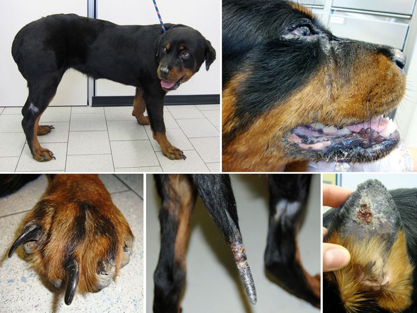
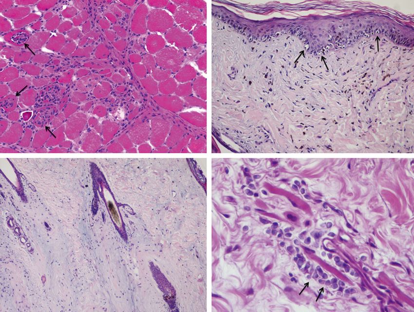
Bresciani et al.

(a)                                                                            (b)

(c)                                                 (d)                                                           (e)

Figure 1. (a) A 7-month-old Rottweiler with dermatomyositis. Note the periocular lesion and onychogryphosis. (b) Crusted periocular and perilabial
skin lesions, with partial hair loss and hypopigmentation. (c) Dog’s claws elongated and distorted (onychogryphosis; nails IV and V), with spontane-
ous avulsion (nails II and III). (d, e) The alopecic, crusted and ulcerated skin lesions at the tip of the tail and the pinna.

appeared elongated and distorted (onychogryphosis) and                       complex repetitive discharges in all muscles evaluated
were painful (onychalgia), with spontaneous avulsion (Fig-                   (Figure S2). The distal appendicular, coccygeal and head
ure 1).                                                                      muscles were the most severely affected. Motor function
   Laboratory findings showed mild nonregenerative anae-                     of peripheral nerves, evaluated by means of motor nerve-
mia [haematocrit 32%, reference range (RR) 37–55%],                          conduction and F-wave studies, and sensory function of
mild hypoalbuminaemia (2.19 g/dL, RR 2.80–3.70 g/dL)                         peripheral nerves, evaluated by means of sensory nerve-
and moderate increases in serum creatinine kinase                            conduction studies and cord dorsum potentials, were
(378 U/L, RR 50–290 U/L), serum phosphorus (6.6 mg/                          within normal limits. The EMG findings were indicative of
dL, RR 2.6–4.9 mg/dL) and serum C-reactive protein                           the presence of spontaneously firing hypersensitive myo-
concentrations (8.98 mg/dL, RR 0.0–0.5 mg/dL). Urinary                       fibres, as a result of destabilization of the sarcolemmal
specific gravity was adequate (1.044), with a normal                         membrane.19 These findings, in conjunction with normal
urinary protein-to-creatinine ratio. Serology tests for                      peripheral nerve function, were suggestive of a general-
canine distemper virus and leishmaniosis were negative,                      ized myopathy, with variable degrees of involvement of
as was an antinuclear antibody test.                                         different muscle groups.
   Chest radiographs showed a diffuse megaoesopha-                              Skin biopsies collected from the tip of the ear, tail and
gus, with severe aspiration pneumonia (Figure S1).                           perilabial skin had mild hyperplastic changes and
Fungal culture of nail and periungual tissues was neg-                       scattered basal apoptotic and vacuolated keratinocytes
ative for dermatophytes.                                                     (Figure 2b, arrows). In the dermis, a sparse interstitial
   Due to the clinical suspicion of dermatomyositis, elec-                   infiltrate of mononuclear cells, represented by small lym-
tromyography (EMG) was performed on the epaxial,                             phocytes, histiocytes and plasma cells, was seen. Some
appendicular and head muscles of the right side of the                       of the small dermal vessels had thickening of the wall,
body by inserting the recording needle to different depths                   with lack of endothelial cells. Hair follicles were atrophic,
and in different places for each muscle evaluated. The                       with increased prominence of the connective tissue of
EMG revealed diffuse, mild to moderate (graded as + or                       the external root sheath (Figure 2c). Mononuclear cells
++; range: 0–++++)19 abnormal spontaneous activity con-                      were seen in the deep dermis, associated with a few
sisting of fibrillation potentials, positive sharp waves or                  muscle fibres (Figure 2d, arrows).
230                                                                            © 2014 ESVD and ACVD, Veterinary Dermatology, 25, 229–e62.
Dermatomyositis-like disease

(a)                                                                       (b)

(c)                                                                       (d)

Figure 2. Histopathological examination of skin and muscle biopsies. (a) Temporal muscle biopsy with lymphocyte and macrophage infiltrates in
the perimysial and muscle tissue, associated with variation in myofibre size and phagocytosis of muscle fibres (arrows) (haematoxylin and eosin,
9200). (b) Scattered keratinocytes of the epidermal basal layer, with hydropic degeneration (arrows) (haematoxylin and eosin, 9200). (c) Atrophic
follicles, with small sebaceous glands and dermal mucin (haematoxylin and eosin, 9100). (d) Plasma cells around a few muscle fibres in the deep
dermis (arrows) (haematoxylin and eosin, 9600).

   Muscle biopsies were collected from the middle third                     Italy)]. After resolution of aspiration pneumonia, predni-
of the lateral head of the triceps brachii muscle and the                   sone [1 mg/kg p.o. twice daily (Deltacortene; Bruno
temporal muscle. The triceps brachii muscle had no histo-                   Farmaceutici, Rome, Italy)] was added. The dog’s condi-
pathological alterations, while in the perilabial muscle                    tion remained unchanged after 3 months of treatment.
(captured with the perilabial skin biopsy) perimysial                       Unfortunately, the dog was lost to long-term follow-up,
lymphocytic infiltration was found.                                         and no information about the parents or littermates of this
   Histologically, the temporal muscle showed mild varia-                   dog was provided by the owner.
tion in myofibre size, with rounded atrophic myofibres at
the periphery of the muscle bundles adjacent to the fas-
                                                                            Discussion
cia. In these areas, mild multifocal mononuclear inflam-
matory infiltrates within perimysial and perivascular areas                 Canine dermatomyositis is an idiopathic disease reported
were also observed (Figure 2a). All muscle biopsies were                    in dogs and humans. In dogs, it has been widely described
collected from the left side of the body.                                   in Shetland sheep dogs and collies, in which onset is
   Clinical signs, laboratory test results, chest radiogra-                 typically noted within the first few months of life.1–4 It may
phy, electromyography and histopathological examination                     also occur sporadically in other breeds, where the term
of skin and muscle biopsies were indicative of dermato-                     ‘dermatomyositis-like disease’ is used.5–12 In humans,
myositis-like disease associated with megaoesophagus                        dermatomyositis is an inflammatory muscular, cutaneous
and aspiration pneumonia. The dog was initially treated                     and sometimes vascular connective tissue disease20,21
with intravenous (i.v.) fluids, oxygen insufflation,                        affecting children (juvenile dermatomyositis) and adults
ampicillin–sulbactam [15 mg/kg i.v. and then per os (p.o.)                  (associated with malignancy in one-third of cases).20–22
twice daily (Unasyn, Pzifer Italia, Latina, Italy)], marboflox-             Canine familial dermatomyositis resembles type II juve-
acin [2 mg/kg i.v. and then p.o. once daily (Marbocyl FD                    nile dermatomyositis.2 In humans, neither the cause nor
1%; Vetoquinol S.A., Lure, France)] and pentoxifylline                      the pathogenesis of this disease is completely known.
[15 mg/kg p.o. twice daily (Trental, Sanofi-Aventis, Milan,                 Genetic factors associated with autoimmune conditions,
© 2014 ESVD and ACVD, Veterinary Dermatology, 25, 229–e62.                                                                                   231
Bresciani et al.
as well as environmental factors, such as viral, drug and 9. White SD, Shelton GD, Sisson A et al. Dermatomyositis in an
vaccine triggers, have been implicated. The pathogene- adult Pembroke Welsh Corgi. J Am Anim Hosp Assoc 1992; 28:
398–401.
sis in dogs is unclear; the role of factors such as
10. Evans J, Levesque D, Shelton GD. Canine inflammatory myopa-
viruses has not been elucidated,17 and in collies and thies: a clinicopathologic review of 200 cases. J Vet Intern Med
Shetland sheep dogs it is assumed to be a familial 2004; 18: 679–691.
disease.3,4,23 11. Gross TL, Ihrke PJ, Walder EJ et al. Interface diseases of
The case of canine dermatomyositis presented in the dermal–epidermal junction. In: Skin Diseases of the Dog
this report had similar features to those described in and the Cat. 2nd edition. Oxford: Blackwell Science, 2005;
collies and Shetland sheep dogs (skin and muscular 49–52.
12. Miller WH, Griffin CE, Campbell KL. Congenital and hereditary
lesions and megaoesophagus); however, this case
defects. In: Muller and Kirk’s Small Animal Dermatology. 7th edi-
also had nail lesions similar to those reported in tion. Missouri: Elsevier, 2012; 573–617.
humans, which are thought to be a consequence of 13. Rees CA, Boothe D. Therapeutic response to pentoxifylline and
microangiopathy.24 Unfortunately, nail tissue was not its active metabolites in dogs with familial canine dermatomyosi-
collected for histopathology to confirm vascular com- tis. Vet Ther 2003; 4: 234–241.
promise. In other tissues, subtle vasculopathy was 14. Hargis AM, Mundell AC. Familial canine dermatomyositis.
recognized together with the effects of vascular dam- Compend Contin Educ Vet 1992; 14: 855–863.
15. Dalakas MC, Hohlfeld R. Polymyositis and dermatomyositis.
age to the skin, such as hydropic degeneration and
Lancet 2003; 362: 971–982.
apoptosis of basal keratinocytes, focally hypercellular 16. Jackson HA, Olivry T. Ulcerative dermatosis of the Shetland
dermis and atrophic follicles; all of these alterations sheepdog and rough collie dog may represent a novel vesicular
were grouped under the term ‘ischaemic dermatopa- variant of cutaneous lupus erythematosus. Vet Dermatol 2001;
thy’. This histological entity embraces the following 12: 19–27.
five clinical subtypes: (i) canine familial dermatomyosi- 17. Haupt KH, Prieur DJ, Hargis AM et al. Familial canine dermato-
myositis: clinicopathologic, immunologic and serologic studies.
tis; (ii) juvenile-onset ischaemic dermatopathy (similar
Am J Vet Res 1985; 46; 1870–1875.
to canine familial dermatomyositis except for the 18. Haupt KH, Prieur DJ, Moore MP et al. Familial canine dermato-
breed predilection); (iii) focal post-rabies vaccination myositis: clinical, electrodiagnostic and genetic studies. Am
reaction; (iv) generalized vaccination-induced ischaemic J Vet Res 1986; 46;1861–1869.
dermatopathy; and (v) adult-onset (nonvaccine-induced) 19. Kimura J. Types of electromyographic abnormalities. In: Kimura
generalized ischaemic dermatopathy.11 The present J, ed. Electrodiagnosis in Diseases of Nerve and Muscle: Princi-
case is representative of group (ii). The pattern of ples and Practice. 3rd edition. New York: Oxford University
Press, 2002; 339–369.
muscular involvement as diagnosed by EMG, where
20. Femina AN, Vleugels RA, Callen JP. Cutaneous dermatomyosi-
the distal appendicular, coccygeal and head muscles tis: an updated review of treatment options and internal associa-
were most severely affected, was also similar to the tion. Am J Clin Dermatol 2013; 14: 291–313.
distribution of muscular lesions reported in collies and 21. Gowdie PJ, Allen RC, Kornberg AJ et al. Clinical features and
Shetland sheep dogs.2,10,25,26 disease course of patients with juvenile dermatomyositis. Int
In conclusion, the diagnosis was made based on clinical J Rheum Dis 2013; 16: 561–567.
signs, type and distribution of electromyographic and his- 22. Travassos AR, Borges-Costa J, Filipe P et al. Malignancy
associated with dermatomyositis – a retrospective single-cen-
tological findings, and exclusion of other causes. This is
ter study with 33 patients. Acta Reumatol Port 2013; 38:
the first report regarding a dermatomyositis-like condition 92–97.
in a Rottweiler. 23. Clark LA, Credille KM, Murphy EK et al. Linkage of dermatomyo-
sitis in the Shetland Sheepdog to chromosome 35. Vet Dermatol
2005; 16: 392–394.
References 24. Koler RA, Montemarano A. Dermatomyositis Am Fam Physician
1. Hargis AM, Haupt KH, Hegreberg GA et al. Familial canine 2001; 64: 1565–1572.
dermatomyositis. Initial characterization of the cutaneous and 25. Hargis AM, Prieur DJ, Haupt KH et al. Prospective study of
muscular lesions. Am J Pathol 1984; 116: 234–244. familial canine dermatomyositis. Correlation of severity of
2. Hargis AM, Haupt KH, Prieur DJ et al. Animal model of human dermatomyositis and circulating immune complex levels. Am
disease. Dermatomyositis. Familial canine dermatomyositis. Am J Pathol 1986; 123: 465–479.
J Pathol 1985; 120: 323–325. 26. Podell M. Inflammatory myopathies. Vet Clin North Am Small
3. Ferguson EA, Cerundolo R, Lloyd DH et al. Dermatomyositis in Anim Pract 2002; 32: 147–167.
five Shetland sheepdogs in the United Kingdom. Vet Rec 2000;
146: 214–217.
4. Wahl IM, Clark LA, Skalli O et al. Analysis of gene transcript pro- Supporting Information
filing and immunobiology in Shetland sheepdogs with dermato-
Additional Supporting Information may be found in the
myositis. Vet Dermatol 2008; 19: 52–58.
online version of this article.
5. Campbell KL, Lowe AD, Lichtensteiger CA. Dermatomyositis in
three Portuguese water dog littermates. Vet Dermatol 2008; 19 Figure S1. Left lateral (a) and ventrodorsal view of the
(Suppl. 1): 69 (abstract). thorax (b).
6. Bensignor E. A propros d’une observation de dermatomyosite Figure S2. (a) Electromyographic spontaneous activity of
chez un beauceron. Rec Me d Ve t 1997; 173: 125–129. the palmar interossei muscles of the right forelimb, con-
7. Gross T, Ihrke PJ, Walder EJ et al. Vascular disease of the der- sisting of fibrillation potentials and positive sharp waves.
mis. In: Skin Diseases of the Dog and Cat. 2nd edition. Oxford:
(b) Electromyographic spontaneous activity of the right or-
Blackwell Science, 2005; 247–250.
8. Yoon JS, Minami T, Takizawa Y et al. Two dogs with juve-
bicularis oris muscle, consisting of complex repetitive dis-
nile-onset skin diseases with involvement of extremities. J Vet charges (20 ms/division; 50 lV/division).
Med Sci 2010; 72: 1513–1516.
232 © 2014 ESVD and ACVD, Veterinary Dermatology, 25, 229–e62.Dermatomyositis-like disease
Resume
Contexte – La dermatomyosite canine est une maladie he re
ditaire de crite chez les colleys, shetlands et
croises. Une maladie identique, appele e dermatomyosite-like, a e te
decrite occasionnellement dans d’au-
tres races mais jamais chez le Rottweiler.
Hypothe ses/Objectifs – Nous de crivons les donne es clinicopathologiques associe es
a une dermatomyo-
site-like chez un Rottweiler.
Sujet – Une femelle Rottweiler de 7 mois a e te
re
fe
re
e pour le sions cutane es, re
gurgitation et faiblesse.
Les anomalies cutane es consistaient en de l’alopecie, des crou ^tes et des squames sur la face interne des
mite
pavillons, l’extre de la queue, autour des yeux et des le vres. Le chien pre sentait e
galement de l’ony-
chogryphose et de l’onychalgie.
Me thodes – Une nume ration formule, une biochimie, des radiographies thoraciques, une e lectromyogra-
phie, une e tude de la conduction nerveuse, et des biopsies cutane es et musculaires ont e te
re
alise
es.
Resultats – Un me gaœsophage une pneumonie, une dermatopathie ische mique et une myosite
ge ne
ralise
e ont e
te
mis en evidence. Le diagnostic final e tait une dermatomyosite-like.
Conclusions et importance clinique – Ceci est le premier cas de crit de dermatomyosite-like chez un
Rottweiler.
Resumen
Introduccio n – la dermatomiositis canina es una enfermedad hereditaria que se describe en Collies y per-
ros pastores de Shetland y sus cruces. Una enfermedad similar, denominada enfermedad parecida a der-
matomiositis, ha sido descrita ocasionalmente en otras razas, pero nunca en la raza Rottweiler.
Hipo tesis/Objetivos – describimos los hallazgos clınico-patolo gicos asociados con la enfermedad parecida
a dermatomiositis en un Rottweiler.
Animales – una hembra de siete meses de edad de raza de raza Rottweiler fue referida por la presencia de
anormalidades dermatolo gicas, regurgitacio n y debilidad. Las anomalıas cut aneas incluıan alopecia, co-
stras, descamacio n de la superficie interna de las orejas, la punta de la cola, zona periorbital, y zona perilabi-
al. El perro tambie n presento onicogriposis y onicalgia.
Me todos – se realizo analisis hematologico completo, y un panel de bioquımica se rica, radiografıas tor
aci-
cas, electromiografıa, estudios de conduccio n nerviosa, y biopsias de piel y musculo.
Resultados – se documentaron megaeso fago, pneumonıa, dermatopatıa isque mica, miositis generalizada.
El diagno stico final fue de enfermedad parecida a la dermatomiositis.
Conclusiones e importancia clınica – e ste es el primer caso publicado de enfermedad parecida a derma-
tomiositis en un Rottweiler.
Zusammenfassung
Hintergrund – Die Dermatomyositis des Hundes ist eine heredit€ are Erkrankung, die bei Collies, Shelties
und ihren Mischungen beschrieben ist. Eine € ahnliche Erkrankung, die Dermatomyositis-€ahnliche Krankheit
bezeichnet wird, wurde schon gelegentlich bei anderen Rassen beschrieben, allerdings nicht beim Rott-
weiler.
Hypothese/Ziele – Wir beschreiben die klinisch-pathologischen Befunde, die mit einer Dermatomyositis-
€ahnlichen Krankheit beim Rottweiler einhergingen.
Tiere – Eine sieben Monate alte Rottweilerhu €ndin wurde aufgrund dermatologischer Ver€ anderungen, re-
gurgitieren und Schw€ache u €berwiesen. Die Hautver€ anderungen bestanden aus Alopezie, Krusten-und
Schuppenbildung an der inneren Oberfl€ache der Pinnae, der Schwanzspitze, rund um die Augen und rund
um die Lippen. Der Hund zeigte auch Onychogryphose und Onychalgie.
Methoden – Blutbild, Serum Biochemie, Brustro €ntgen, Elektromyographie, Nervenkonduktionsstudien
und Haut und Muskelbiopsien wurden durchgefu €hrt.
Ergebnisse – Megao €sophagus, Pneumonie, isch€ amische Dermatopathie und eine generalisierte Myositis
konnten dokumentiert werden. Die endgu €ltige Diagnose war Dermatomyositis-€ ahnliche Krankheit.
Schlußfolgerungen und klinische Bedeutung – Es handelt sich hierbei um den ersten Bericht einer Der-
matomyositis-€ahnlichen Krankheit beim Rottweiler.
© 2014 ESVD and ACVD, Veterinary Dermatology, 25, 229–e62. e61Bresciani et al. e62 © 2014 ESVD and ACVD, Veterinary Dermatology, 25, 229–e62.
You can also read