Mycobacterium avium subspecies hominissuis disseminated infection in a Basset Hound dog
←
→
Page content transcription
If your browser does not render page correctly, please read the page content below
418616 XXXXXX10.1177/1040638711418616Cam
pora et al.Mycobacterium avium subspecies hominissuis disseminated infection
Journal of Veterinary Diagnostic Investigation
Mycobacterium avium subspecies 23(5) 1083–1087
© 2011 The Author(s)
Reprints and permission:
hominissuis disseminated infection sagepub.com/journalsPermissions.nav
DOI: 10.1177/1040638711418616
http://jvdi.sagepub.com
in a Basset Hound dog
Luca Campora,1 Michele Corazza, Cristina Zullino, Valentina V. Ebani,
Francesca Abramo
Abstract. In the current report, a case in Italy of disseminated Mycobacterium avium subsp. hominissuis infection in a dog
from an American lineage of Basset Hounds is described. A 2-year-old intact female Basset Hound presented with persistent
lymphadenopathy, lameness, and a history characterized by coccidiosis, bacterial gastroenteritis, and alopecia. Lymphadenitis,
with macrophages containing a few intracytoplasmic, negative staining, Ziehl–Neelsen-positive bacilli, was detected by
a popliteal fine-needle aspirate leading to the diagnosis of mycobacteriosis. Ultrasound and X-ray examinations revealed
visceral and mediastinal lymphadenopathy. Because of the extent of the disease, the dog was humanely euthanized. Significant
gross abnormalities, such as enlargement of the cranial mediastinal lymph nodes with encapsulated areas of caseous necrosis
and generalized lymphadenopathy, were observed at necropsy. Granulomatous lesions were histopathologically detected in
the liver and spleen. Ziehl–Neelsen-positive bacilli were observed in all examined lymph node, liver, spleen, lung, and bone
marrow smears. Lymph nodes and liver were collected in order to pursue speciation by bacterial culture and molecular
biology; multiplex polymerase chain reaction results classified the pathogen as M. avium subsp. hominissuis. Although an
immune system deficiency was not investigated, anamnesis suggests that the dog was immunocompromised. Furthermore,
the dog came from an American stock of Basset Hound, and for some of this breed, a predisposition to this infection has been
hypothesized.
Key words: Basset Hound; dogs; Mycobacterium avium subspecies hominissuis.
A 2-year-old intact female Basset Hound dog was presented authors in another Basset Hound dog, and Basset Hounds are
for evaluation of persistent lymphadenopathy and lameness. known to be predisposed to Mycobacterium avium intracel-
From its birth by Caesarian section, which gave rise to 4 lulare complex (MAIC) infection,10 further investigation was
empty amniotic sacs, the history of the dog had been charac- recommended.
terized by episodes of coccidiosis and severe bacterial gas- The dog was thus referred to the Department of Veterinary
troenteritis responsive to anticoccidial and antibiotic therapy. Clinics of the University of Pisa. On physical examination,
At 4 months of age, the dog spent a short period in a breeding the dog was bright, alert, and responsive, with a body condition
house in northern Italy, and soon after its return, the dog score of 3 out of 5, and temperature of 38.9°C. The mucous
showed periodic lameness, without a detectable neurologic membranes were pink, and the respiratory and heart rates
defect, and diffuse alopecia. A systemic form of lupus was sus- were within normal limits. The main abnormal findings were
pected, and an anti-nuclear antibody test was positive. Con- generalized lymphadenopathy, with increased consistency of
tinuous treatments with prednisone (almost 6 cycles) were all palpable lymph nodes, and splenomegaly. Abnormal hema-
necessary to prevent recurrence of lameness. tological findings were limited to a mildly microcytic, hypo-
At 2 years of age, a popliteal fine-needle aspirate was per- chromic anemia, and hypoalbuminemia.
formed and sent to the Department of Animal Pathology of Thoracic radiographs revealed a 3 cm in diameter, medi-
the University of Pisa (Pisa, Italy) for cytopathological eval- astinal mass compatible with an enlarged cranial mediastinal
uation. One of the 2 obtained slides was stained with a lymph node. On abdominal ultrasonography, the spleen
Romanowsky stain, and low cellularity was noted with appeared enlarged, with scattered, small, round, hypoechoic
prevalent macrophages and small lymphocytes. A few rod-
shaped, nonstaining organisms were seen within the cyto- From the Departments of Animal Pathology (Campora, Ebani,
plasm of 1 macrophage (Fig. 1A). A Ziehl–Neelsen (ZN) Abramo) and Veterinary Clinics (Corazza), University of Pisa, Italy; and
stain was performed on the remaining unstained slide and veterinarian in private practice (Zullino), Montecatini, Pistoia, Italy.
revealed a few acid-fast bacilli morphologically consistent 1
Corresponding Author: Luca Campora, Department of Animal
with Mycobacterium species (Fig. 1B). Since mycobacterial Pathology, University of Pisa, viale delle Piagge 2, 56124 Pisa, Italy.
infection had been previously diagnosed by one of the campora@vet.unipi.it
Downloaded from vdi.sagepub.com by guest on November 2, 20151084 Campora et al.
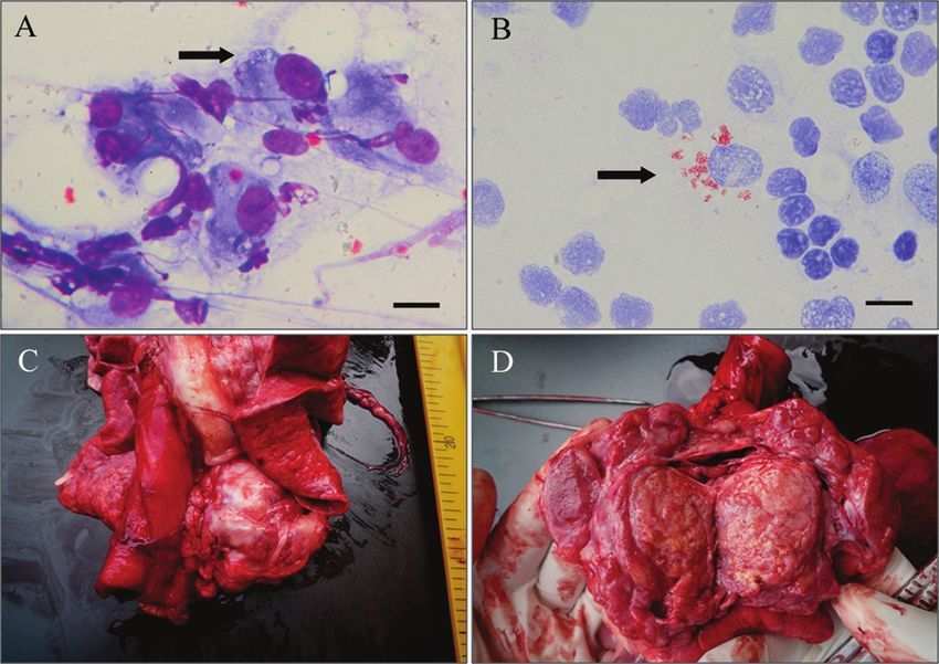
Figure 1. Basset Hound dog. Popliteal lymph node fine-needle aspirate (A, B) and postmortem view (C, D). A, macrophages with
a few nonstaining, rod-shaped organisms (arrow). Romanowsky stain. Bar = 10 µm. B, macrophages containing numerous acid-fast,
intracytoplasmic bacilli morphologically consistent with Mycobacterium sp. (arrow). Ziehl–Nielsen. Bar = 10 µm. C, grossly enlarged
mediastinal lymph node. D, cut surface of the mediastinal lymph node with encapsulated areas of caseous necrosis.
areas. The liver was mildly enlarged, but had normal echo- mediastinal, mesenteric, and submandibular lymph nodes,
genicity, and the abdominal lymph nodes were enlarged and lungs, heart, liver, spleen, stomach, intestine, kidney, brain,
hypoechoic, with rounded edges. and bone marrow were sampled for histologic evaluation.
New fine-needle aspirates were obtained from prescapu- Microscopically, all examined lymph nodes showed mas-
lar lymph nodes. Some of these samples were stained with a sive infiltration of epithelioid macrophages, leading to com-
Romanowsky stain and ZN, while others were sent to the plete loss of the normal lymphoid tissue architecture.
Microbiology Laboratory of the Department of Animal Furthermore, in the cranial mediastinal lymph node, multiple
Pathology of the University of Pisa for bacterial culture. The foci of necrosis were observed (Fig. 2). The lungs were
cytological findings were characterized by a granulomatous markedly congested. The liver showed sinusoidal ectasia and
lymphadenitis with a predominance of large macrophages congestion, and portal fibrosis with mixed infiltration by
and hypersegmented neutrophils. Within the macrophage macrophages, lymphocytes, and neutrophils. Bile ducts were
cytoplasm and free in the background, nonstaining negative hyperplastic with evidence of cholestasis. Multiple granulo-
images of bacterial rods were present, which stained brilliant matous foci composed of epithelioid macrophages were ran-
pink with ZN. Morphological changes thus confirmed the domly distributed throughout the parenchyma (Fig. 3). The
presence of mycobacterial infection characterized by high splenic red pulp was diffusely infiltrated by epithelioid
numbers of bacteria within the cytoplasm. macrophages; occasional megakaryocytes, extramedullary
Because of the extent of the disease, the owners opted not hematopoietic foci, and hemosiderosis were also present.
to pursue treatment, and the dog was humanely euthanized. The bone marrow was partially replaced by epithelioid mac-
At necropsy, the significant gross abnormalities were limited rophages with residual aggregates of erythropoietic cells
to enlargement of the cranial mediastinal lymph nodes (Fig. (Fig. 4). No alterations were present in samples examined
1C), with encapsulated areas of caseous necrosis (Fig. 1D) from other tissues. Ziehl–Neelsen stain was performed on all
and enlargement of all peripheral and visceral lymph nodes, samples obtained. Numerous epithelioid macrophages con-
with loss of corticomedullary distinction. No gross altera- taining acid-fast, intracytoplasmic, bacterial rods were seen
tions of the joints that could be considered a source of the in all lymph nodes, liver, spleen, bone marrow, and, to a lesser
lameness were seen. Representative portions of cranial extent, in the lungs.
Downloaded from vdi.sagepub.com by guest on November 2, 2015Mycobacterium avium subspecies hominissuis disseminated infection 1085
Figure 2. Histology; Basset Hound dog. Mediastinal lymph node
with massive infiltration of epithelioid macrophages around an area Figure 4. Histology; Basset Hound dog. Bone marrow is partially
of caseous necrosis. Hematoxylin and eosin. Bar = 100 µm. replaced by macrophages with residual aggregates of erythropoietic
cells. Hematoxylin and eosin. Bar = 50 µm.
used in the PCR protocol to amplify a 577-bp fragment, whereas
the primers IS1245-f (5′-GAGTTGACCGCGTTCATCG-3′)
and IS1245-r (5′-CGTCGAGGAAGACATACGG-3′) were
employed to amplify a 385-bp fragment. The fragments cor-
respond to the IS901 and IS1245 genes, respectively, which
are both present in M. avium subsp. avium and M. avium
subsp. silvaticum, whereas only the IS1245 gene is present in
M. avium subsp hominissuis.17 The PCR amplification was
performed in 50 µl total volume containing 200 µM of
deoxynucleoside triphosphates, 0.5 µM of each primer, 1.25
U of Taq polymerase,b 5 µl of 10× PCR buffer,b and 2 µl of
extracted DNA. The PCR amplifications were performed in
an automated thermal cyclerc for 40 cycles. Each cycle con-
sisted of a denaturation phase (95°C for 1 min), an annealing
Figure 3. Histology; Basset Hound dog. Granulomatous hepatic phase (58°C for 30 sec), and an extension phase (72°C for 1
lesion with necrosis and epithelioid macrophages. Hematoxylin and min). An initial denaturation of 5 min at 95°C and a final
eosin. Bar = 50 µm. extension of 5 min at 72°C were performed. The PCR prod-
ucts were analyzed by electrophoresis on a 1.5% agarose gel
at 100 V for 45 min; the gel was stained with ethidium bro-
Lymph node aspirates collected antemortem, and lymph mide and observed under ultraviolet light. All isolates
node and liver samples taken at necropsy were cultured in showed the 385-bp band, but not the 577-bp band. On the
Dubos medium after decontamination with a 1% hexadecyl- basis of these results, the isolates were identified as M. avium
pyridinium chloride solution. For each sample, a single tube subsp. hominissuis.
was incubated at 37°C while another tube was incubated at In dogs, infections due to M. avium are sporadically
43°C in order to differentiate between M. avium and other reported, and in most of the cases, the subspecies was not
Mycobacterium species. Colonies that grew at 43°C after 10 days identified.* Mycobacterium avium subsp. hominissuis is gener-
were submitted for ZN staining. The acid-fast isolates were ally isolated from pigs and human beings,14,15 and cases of infec-
identified as M. avium by DNA probe hybridizationa performed tion have been reported in a horse,13 pet parrots,21 and 2 dogs.9
following the manufacturer’s instructions. DNA samples Mycobacterium avium intracellulare complex bacilli are
were extracted from the isolates using a commercial kitb ubiquitous in the environment and can remain viable for over
according to manufacturer’s recommendations. The samples 2 years.3,22 In dogs, infection with M. avium subsp. hominis-
were successively used in a multiplex polymerase chain reac- suis occurs because of direct contact with infected animals or
tion (PCR) assay in order to identify the M. avium subspecies.
The primers IS901-f (5′-GGATTGCTAACCACGTGGTG-
*
3′) and IS901-r (5′-GCGAGTTGCTTGATGAGCG-3′) were References 1,2,4–7,9,10,12,16,18–20.
Downloaded from vdi.sagepub.com by guest on November 2, 20151086 Campora et al.
from the environment. It has also been documented that dogs Certain breeds of dog, such as the Basset Hound and
can become infected by their owners, but the opposite situa- Miniature Schnauzer, appear to be predisposed to this dis-
tion, namely the dog as a source of infection for immuno- ease10; the basis of this breed predisposition is unclear. Only
compromised human beings, is the higher risk.13 The source in a few cases has the efficiency of the immune status been
of infection was not determined in the current case; however, investigated. Other authors4 have suggested that Basset
the dog lived outdoors for a long time in northern Italy in a Hounds might be predisposed to a cell-mediated immunode-
rural area, and during that period, it was exposed to caged pet ficiency against intracellular pathogens due to a defect in
birds and pigeons, and had access to a pond, which was fre- either T cells or intracellular killing ability of macrophages.
quented by migrating waterfowl. No known exposure to In another case,18 a concurrent infection with 3 types of patho-
chicken or swine carcasses, or human beings known to have gens, one of which was M. avium, was reported in a Great
tuberculosis in that area was suspected. Pyrenees dog, suggesting that an immunodeficient state also
Mycobacterium avium subsp. hominissuis can invade the existed for this dog. These examples support the hypothesis
body through the skin, or the respiratory or gastrointestinal tracts. that an immunocompromised state might predispose to the
Normally, the infected subject develops an early granuloma- sporadic form of the disease. In the case herein described,
tous lesion at the site of entry and in the regional lymph node the breed is one previously reported to be predisposed to the
(complete primary complex) or only in the regional lymph infection, and severity and massive dissemination of the
node (incomplete primary complex), with or without secondary lesions suggest that glucocorticoids might have promoted
lymphohematogenous dissemination.8 Mycobacterium avium spread of the infection.
intracellulare complex infections in dogs are frequently From the study that described the 5 cases of MAIC infec-
associated with the disseminated form of disease, with mul- tion in Basset Hounds,4 pedigrees were available and studied
tiple organ system involvement.18,22 In the present case, the only in 3 dogs; from these, it emerged that 1 male was the sire
extensive mediastinal and tracheobronchial lymph node of one of the affected dogs and that one of his sons was the
involvement suggests a respiratory route of infection, with sire of the other 2 dogs. For the cases of MAIC described in
formation of an incomplete primary complex and secondary mixed breeds,2,12 whether the dogs were genetically related to
lymphohematogenous dissemination. Basset Hounds was not presented. The dog described in the
Diagnosis is a challenge since clinical signs may be current report was born from an American lineage of Basset
vague. Cytology is very helpful in the early diagnostic work- Hounds (both mother and father). Direct documentation does
up, allowing for rapid detection of the rod-shaped bacteria; not exist on whether the parents were direct descendants of
however, these can be few in number and easily overlooked. the 5 Basset Hounds described in 1988.4
In the current case, cytology revealed the infectious nature of Since human beings are relatively resistant to infection
the disease, with further methods used to better characterize with MAIC, unless they are immunocompromised, and zoo-
the involved pathogen. Cytology and histopathology are notic transmission is no more likely than environmental
indeed useful to detect the rod-shaped, acid-fast bacilli, but acquisition, treatment of affected dogs might be considered.
do not help in differentiating between zoonotic and nonzoo- However, in most of the reported cases, dogs were eutha-
notic mycobacterial species. The lepromatous type of MAIC nized for the progressive and often rapid worsening of clini-
infection, characterized by florid, monomorphic, epithelioid cal signs without any attempt at specific therapy.1,5-7,9,12,18
macrophage responses with numerous intracytoplasmic bac- Only in a few cases has specific antimycobacterial therapy
teria, is cytologically and histologically different from the been initiated. One affected dog was treated with isoniazid
pleocellular granulomatous response characteristic of infec- for 10 months; however, the subject was euthanized because
tion due to Mycobacterium tuberculosis or Mycobacterium of continuing deterioration.20 Three of the 5 Basset Hounds
bovis. This lepromatous type of infection includes Langhan with MAIC disseminated infection described in the United
type, multinucleated giant cells, and few intracytoplasmic States4 were specifically treated for periods lasting from 7
bacilli. However, definitive diagnosis relies on the use of days to 1 month, and in all 3 cases, a lack of response and
supplementary tools such as bacterial culture and molecular progressive course of disease prompted the decision for
genetic techniques such as PCR. Mycobacterial culture euthanasia. Antimycobacterial therapy was also attempted in
requires a long period of time (4–12 weeks) until a definitive a Schnauzer dog whose condition seemed to initially
diagnosis is provided. improve, but the dog rapidly deteriorated.16Mycobacterium
The molecular techniques designed to identify the various avium intracellulare complex organisms are resistant to com-
subspecies of M. avium are a relatively recent development, mon anti-tuberculosis drugs; however, fluoroquinolones and
and in many published cases, were not used, and therefore new quinolones have been shown to be active in vitro against
the subspecies was not identified. In the present case, multi- many mycobacterial species.11 Unfortunately, since clinical
plex PCR allowed the identification of the pathogen as M. signs tend to be vague and often lead to misdiagnosis if rod-
avium subsp. hominissuis, and this is, to the authors’ knowl- shaped organisms are not detected in cytological specimens,
edge, the third case described worldwide in the dog and the diagnosis is not usually achieved in an early, more treatable
first in Italy. stage of the disease. In the current case, therapy was not
Downloaded from vdi.sagepub.com by guest on November 2, 2015Mycobacterium avium subspecies hominissuis disseminated infection 1087
attempted, mostly because of dissemination of the disease Greene CE, 3rd ed., pp. 313–321. WB Saunders Elsevier,
with multi-organ involvement. St. Louis, MO.
In conclusion, the authors report a case of disseminated 9. Haist V, Seehusen F, Moser I, et al.: 2008, Mycobacterium
mycobacteriosis in Italy in a Basset Hound of American lin- avium subsp. hominissuis infection in 2 pet dogs, Germany.
eage. The introduction of molecular biology to the diagnos- Emerg Infect Dis 14:988–989.
tic plan allowed for the identification of the pathogen as M. 10. Horn B, Forshaw D, Cousins D, Irwin PJ: 2000, Disseminated
avium subsp. hominissuis, and to the authors’ knowledge, Mycobacterium avium infection in a dog with chronic diar-
systemic MAIC infection due to this subspecies is rarely rhoea. Aust Vet J 78:320–325.
described and has not been reported in Italy. 11. Jacobs MR: 1999, Activity of quinolones against mycobacte-
ria. Drugs 58(Suppl 2):19–22.
Sources and manufacturers 12. Kim DY, Cho DY, Newton JC, et al.: 1994, Granulomatous
a. INNO-LiPA Mycobacteria v2, Innogenetics NV, Ghent, myelitis due to Mycobacterium avium in a dog. Vet Pathol
Belgium. 31:491–493.
b. DNeasy®, Qiagen GmBH, Hilden, Germany. 13. Kriz P, Jahn P, Bezdekova B, et al.: 2010, Mycobacterium
c. Gene-Amp PCR System 2700, Perkin-Elmer Inc., Waltham, MA. avium subsp. hominissuis infection in horses. Emerg Infect Dis
16:1328–1329.
Declaration of conflicting interests 14. Matlova L, Dvorska L, Ayele WY, et al.: 2005, Distribution
The author(s) declared no potential conflicts of interest with of Mycobacterium avium complex isolates in tissue samples of
respect to the research, authorship, and/or publication of this pigs fed peat naturally contaminated with mycobacteria as a
article. supplement. J Clin Microbiol 43:1261–1268.
15. Mijs W, de Haas P, Rossau R, et al.: 2002, Molecular evidence
Funding to support a proposal to reserve the designation Mycobacterium
The author(s) received no financial support for the research, avium subsp. avium for bird-type isolates and “M. avium subsp.
authorship, and/or publication of this article. hominissuis” for the human/porcine type of M. avium. Int J Syst
Evol Microbiol 52:1505–1518.
References 16. Miller MA, Greene CE, Brix AE: 1995, Disseminated Myco-
1. Bauer N, Burkhardt S, Kirsch A, et al.: 2002, Lymphadenop- bacterium avium-intracellulare complex infection in a minia-
athy and diarrhea in a Miniature Schnauzer. Vet Clin Pathol ture schnauzer. J Am Anim Hosp Assoc 31:213–216.
31:61–64. 17. Moravkova M, Hlozek P, Beran V, et al.: 2008, Strategy for
2. Beaumont PR, Jezyk PF, Haskins ME: 1981, Mycobacterium the detection and differentiation of Mycobacterium avium spe-
avium infection in a dog. J Small Anim Pract 22:91–97. cies in isolates and heavily infected tissue. Res Vet Sci 85:
3. Biet F, Boschiroli ML, Thorel MF, Guilloteau LA: 2005, 257–264.
Zoonotic aspects of Mycobacterium bovis and Mycobacterium 18. Naughton JF, Mealey KL, Wardrop KJ, et al.: 2005, Systemic
avium-intracellulare complex (MAC). Vet Res 36:411–436. Mycobacterium avium infection in a dog diagnosed by poly-
4. Carpenter JL, Myers AM, Conner MW, et al.: 1988, Tuber- merase chain reaction analysis of buffy coat. J Am Anim Hosp
culosis in five basset hounds. J Am Vet Med Assoc 192: Assoc 41:128–132.
1563–1568. 19. O’Toole D, Tharp S, Thomsen BV, et al.: 2005, Fatal mycobac-
5. Eggers JS, Parker GA, Braaf HA, Mense MG: 1997, Dis- teriosis with hepatosplenomegaly in a young dog due to Myco-
seminated Mycobacterium avium infection in three miniature bacterium avium. J Vet Diagn Invest 17:200–204.
schnauzer litter mates. J Vet Diagn Invest 9:424–427. 20. Shackelford CC, Reed WM: 1989, Disseminated Mycobacte-
6. Friend SC, Russell EG, Hartley WJ, Everist P: 1979, Infection rium avium infection in a dog. J Vet Diagn Invest 1:273–275.
of a dog with Mycobacterium avium serotype II. Vet Pathol 21. Shitaye EJ, Grymova V, Grym M, et al.: 2009, Mycobacte-
16:381–384. rium avium subsp. hominissuis infection in a pet parrot. Emerg
7. Gow AG, Gow DJ: 2008, Disseminated Mycobacterium avium Infect Dis 15:617–619.
complex infection in a dog. Vet Rec 162:594–595. 22. Thorel MF, Huchzermeyer H, Weiss R, Fontaine JJ: 1997,
8. Greene CE, Gunn-Moore DA: 2006, Mycobacterial infections. Mycobacterium avium infections in animals. Literature review.
In: Infectious diseases of the dog and cat–revised reprint, ed. Vet Res 28:439–447.
Downloaded from vdi.sagepub.com by guest on November 2, 2015You can also read