New Sonographic Classification of Adenomyosis: Do Type and Degree of Adenomyosis Correlate to Severity of Symptoms?
←
→
Page content transcription
If your browser does not render page correctly, please read the page content below
Original Article
New Sonographic Classification of Adenomyosis: Do Type and Degree
of Adenomyosis Correlate to Severity of Symptoms?
Caterina Exacoustos, MD, PhD, Giulia Morosetti, MD, Francesca Conway, MD,
Sara Camilli, MD, Francesco Giuseppe Martire, MD, Lucia Lazzeri, MD, PhD,
Emilio Piccione, MD, and Errico Zupi, MD
From the Department of Biomedicine and Prevention Obstetrics and Gynecological Clinic, University of Rome “Tor Vergata” (Drs. Exacoustos, Morosetti,
Conway, Camilli, Giuseppe Martire, Piccione, and Zupi), Rome, and Department of Molecular and Developmental Medicine, Obstetrics and Gynecological
Clinic, University of Siena (Dr. Lazzeri), Siena, Italy.

ABSTRACT Study Objective: To correlate the type and degree of adenomyosis, scored through a new system based on the features of
         transvaginal sonography, to patients’ symptoms and fertility.
         Design: This is a multicenter, observational, prospective study.
         Setting: Two endometriosis tertiary referral centers (University of Rome “Tor Vergata” and University of Siena).
         Patients: A total of 108 patients with ultrasonographic signs of adenomyosis.
         Interventions: A new ultrasonographic scoring system designed to assess the severity and the extent of uterine adenomyo-
         sis was used to stage the disease in correlation with the clinical symptoms. Menstrual uterine bleeding was assessed by a pic-
         torial blood loss analysis chart, painful symptoms were evaluated using a visual analog scale, and infertility factors were
         considered.
         Measurements and Main Results: A total of 108 patients with ultrasonographic signs of adenomyosis (mean age § stan-
         dard deviation, 37.7 § 7.7 years) were classified according to the proposed scoring system. Women with ultrasound diagno-
         sis of diffuse adenomyosis were older (p = .04) and had heavier menstrual bleeding (p = .04) than women with focal disease;
         however, no statistically significant differences were found regarding the presence and severity of dyspareunia and dysme-
         norrhea. Higher values of menstrual bleeding were found for severe diffuse adenomyosis, with the highest values being
         found in those with adenomyomas. In patients trying to conceive, the presence of ultrasound findings of focal disease was
         associated with a higher percentage of infertility than in those with diffuse disease, and the focal involvement of the junc-
         tional zone showed a higher percentage of at least 1 miscarriage than in those with diffuse adenomyosis.
         Conclusion: The ultrasonographic evaluation of the type and extension of adenomyosis in the myometrium seems to
         be important in correlation to the severity of symptoms and infertility. Journal of Minimally Invasive Gynecology (2020)
         27, 1308−1315. © 2019 AAGL. All rights reserved.
Keywords:        Adenomyosis; Transvaginal ultrasound; Classification; Heavy menstrual bleeding; Pain

    Adenomyosis is a frequent benign gynecologic disease,                     from focal to diffuse adenomyosis. It can also present as a
defined by the presence of endometrial glands and stroma                      nodular lesion forming an adenomyoma [1].
within the myometrium, associated with the hypertrophy of                        The real prevalence of adenomyosis is difficult to accu-
the smooth muscle. Adenomyosis may involve different                          rately assess owing to the requirement of histologic confir-
sites of myometrium or most of the uterine wall, ranging                      mation after a clinical diagnosis, which is based on the
                                                                              patient’s symptoms and results of imaging studies. Its prev-
                                                                              alence has been reported to range from 5% to 70% in hys-
The authors declare that they have no conflict of interest.                   terectomy specimens [2−6]. However, the study of
Corresponding author: Lucia Lazzeri, MD, PhD, Department of Molecular         prevalence through histologic diagnosis presents a large
and Developmental Medicine, Obstetrics and Gynecological Clinic, Uni-         selection bias because of the advanced age of and clinical
versity of Siena, Viale Bracci 16, 53100, Siena, Italy.
                                                                              symptoms observed in patients undergoing hysterectomy.
E-mail: lucialazzeri79@gmail.com
                                                                              Moreover, a study highlighted the variability in the histo-
Submitted June 11, 2019, Revised September 26, 2019, Accepted for publica-    logic diagnosis of adenomyosis from center to center and
tion September 26, 2019.                                                      among pathologists [7].
Available at www.sciencedirect.com and www.jmig.org

1553-4650/$ — see front matter © 2019 AAGL. All rights reserved.
https://doi.org/10.1016/j.jmig.2019.09.788
Exacoustos et al. New Sonographic Classification of Adenomyosis 1309
Certainly, it becomes more difficult to ascertain preva- All patients underwent a detailed ultrasound evaluation
lence when various diagnostic criteria are used during of the uterus in which type and extension of adenomyosis
imaging assessment in patients, with or without symptoms, were classified according to a previously published study
not undergoing surgery. [22].
Transvaginal sonography (TVS) has recently been used Furthermore, demographic data and detailed medical
for the noninvasive diagnosis of adenomyosis and to study history were recorded before the TVS scan, and the pres-
its prevalence [1,8−10]. When TVS is performed by dedi- ence of painful symptoms (including dysmenorrhea, dys-
cated sonographers, it shows high accuracy in detecting this pareunia, dyschezia, and dysuria) of heavy menstrual
pathology [11−13]. bleeding and/or infertility was evaluated.
Adenomyosis seems to be associated with endometriosis
Historic Information
[14,15]. Surely, both conditions share pathogenesis and
symptoms such as dysmenorrhea, heavy menstrual bleeding, A complete medical, surgical, and obstetric history
infertility, dyspareunia, and chronic pelvic pain [14,16−18]. including women’s age, body mass index (kg/m2), age at
Some authors suggest that the severity of symptoms and menarche, gravidity and parity (number of all previous
the clinical features correlate with the extent and depth of pregnancies: spontaneous pregnancy loss and/or live
adenomyosis [18−21]. So far, however, the only classifica- births), and the mode of delivery were recorded. Infertility
tion proposed for the extension of the disease is based on was defined as no pregnancy after 12 months of unprotected
the histologic findings after surgery and not on imaging. intercourse.
Because adenomyosis often results in poorly defined Patients were also asked about any medication they were
lesions, possibly disseminated in different parts of the myo- taking including the use of analgesics for the treatment of
metrium, it is difficult to express its severity in quantitative painful periods. The presence of the following painful
terms. symptoms was noted: dysmenorrhea, dyspareunia, dysuria,
Through TVS, it is possible to assess the characteristics dyschezia, and chronic pelvic pain. Symptom intensity was
of adenomyosis. Therefore, recently we have proposed a evaluated through the visual analog scale (VAS) system,
scoring system [22] that grades the type of adenomyosis using a 10-cm line with the extreme points 0 and 10 corre-
and its extension inside the uterus. sponding to “no pain” and “maximum pain,” respectively.
To the best of our knowledge, there are no prospective Severe symptoms were considered if VAS score was equal
studies using noninvasive techniques to assess the link to or more than 5. Furthermore, the presence of heavy men-
between type and degree of adenomyosis and the severity strual bleeding was investigated. Women were asked about
of clinical symptoms. the frequency and duration of menstrual periods and about
The aim of this study was to correlate the type and any episodes of intermenstrual bleeding. To obtain an
degree of adenomyosis to symptoms and fertility through a objective evaluation regarding the amount of menstrual
new scoring system [22] based on the features of TVS. loss, a pictorial blood loss analysis chart (PBAC) was used.
The PBAC provides a score that depends on the number of
tampons or sanitary towels used during the menstrual cycle
Materials and Methods
and on the degree to which each item is soiled. The PBAC
This is a multicenter, prospective, observational study score has been shown to have a high specificity and sensi-
conducted in 2 tertiary referral university hospitals (“Tor tivity when used as a diagnostic test for objective menstrual
Vergata” University Hospital, Department of Biomedicine bleeding [23], and a PBAC score of more than 100 is con-
and Prevention; “Santa Maria alle Scotte” University Hos- sistent with heavy menstrual bleeding.
pital, Department of Molecular and Developmental Medi-
cine) between January 2017 and March 2018. The study
Ultrasound Examination
was approved by the local research ethics committee.
A total group of 148 women aged between 29 and All transvaginal ultrasound examinations were per-
46 years old referred to our center for pelvic pain were formed by 2 experienced sonographers (C.E. and L.L.)
included in the study and divided in 2 groups based on the using a 4- to 9-MHz probe with a three-dimensional (3D)
presence of sonographic signs of adenomyosis, according capability (Voluson E6; GE Medical Systems, Zipf, Aus-
to previous studies [1,13,22]. Inclusion criteria were pre- tria). The 2 sonographers were blinded to the patient’s clini-
menopausal status, availability to perform transvaginal cal symptoms, and the clinical examination was performed
ultrasound, and no hormonal therapy, whereas exclusion after the ultrasound. Briefly, a conventional two-dimen-
criteria were an ongoing pregnancy, a gynecologic malig- sional (2D) ultrasound with gray scale and power Doppler
nant disease, and the presence of more than 3 uterine myo- for assessment of the pelvis was performed. First, we care-
mas. A total of 108 women with ultrasound diagnosis of fully evaluated the uterus, the myometrium, and the endo-
adenomyosis were finally enrolled in the study, and 40 metrium. The myometrium was systematically examined
patients without any sonographic signs of myometrial for the presence of any abnormalities. A 2D examination
pathology were excluded from the analysis. was followed by the acquisition of 3D volume of the uterus1310 Journal of Minimally Invasive Gynecology. Vol 27, No 6, September/October 2020
with and without power Doppler. Thereafter, the scan The ultrasound detection of each type of adenomyotic
examined the adnexa, the pouch of Douglas, and the other lesion in the external myometrium and in the JZ was classi-
pelvic organs (bladder, rectum, rectosigmoid junction, ure- fied in 4 grades according to the parameters published in a
ters) and sites (posterior, lateral and anterior parametria, previous study [22]. Through the use of uterine drawings, the
rectovaginal septum, vesicouterine pouch, uterosacral liga- previously published adenomyosis extension score (using the
ments), looking for features of endometriosis according to a same criteria) was simplified to help clarify it and make it
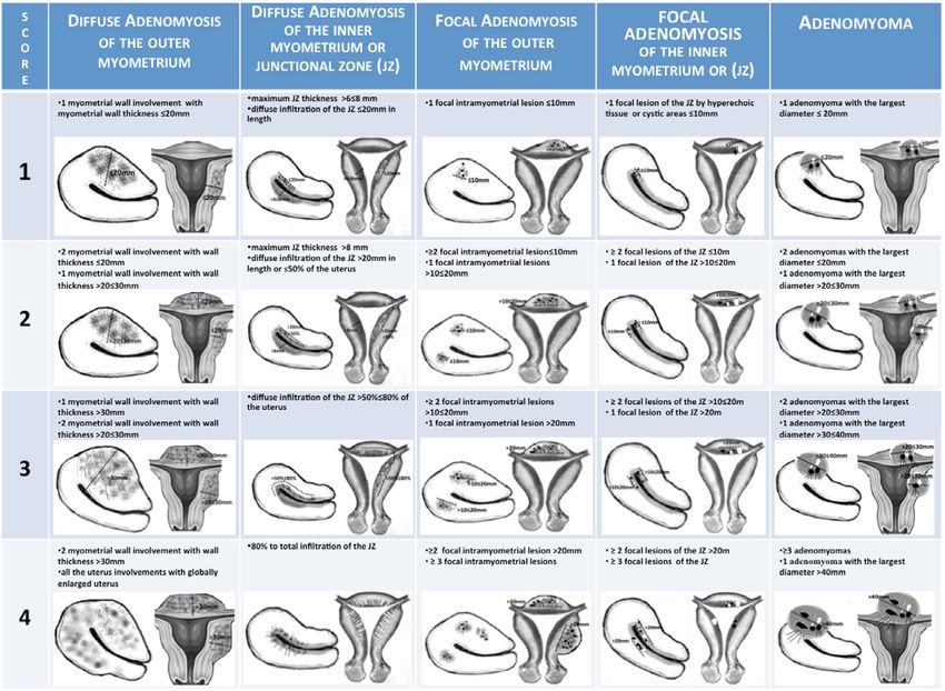
previous ultrasound mapping system [24]. Women were easier to apply to all sonographers as shown in Fig. 1.
considered affected by endometriosis if an ovarian endome- Briefly, as previously described [22], we considered 4
trioma or deep endometriotic nodules were detected at the degrees of extension for each type of disease considered: dif-
ultrasound evaluation. Adhesions of the anterior and poste- fuse and focal adenomyosis (of inner and outer myometrium)
rior compartments were suspected by the presence or and adenomyoma. For diffuse adenomyosis of the outer
absence of the “sliding sign” and the pain induced during myometrium, degree is assigned according to the thickness
the examination was recorded, carefully mapping all pain- of the uterine wall (> or 40) and number of adeno-
Asymmetrically enlarged uterus (1 uterine wall thicker myomas. A score from 1 to 4 was attributed to each degree
than others) unrelated to leiomyoma of disease considered. Then, the ultrasound extent of the dis-
Round cystic area within the myometrium surrounded ease was calculated through the sum of the scores obtained
by a hyperechoic halo and classified in 3 groups: mild (ranged, 1−3), moderate (4−6),
Inhomogeneous, irregular myometrial echotexture in an and severe (≥7) adenomyosis.
indistinctly defined myometrial area with decreased or Patients’ characteristics, severity of symptoms, and uter-
increased echogenicity; hyperechogenic islands; and ine menstrual bleeding were correlated to the type of adeno-
subendometrial lines and buds myosis and score. Finally, the extent of adenomyosis, which
Myometrial hypoechoic linear striations seen as a radi- was classified as mild, moderate, or severe, was correlated
ating pattern of thin acoustic shadows not arising from to the severity of symptoms, uterine menstrual bleeding,
echogenic foci or leiomyoma (fan-shaped shadowing) presence of pelvic endometriosis (ovarian and deep infiltrat-
Indistinct, fuzzy endometrial-myometrial border (ill- ing), and infertility.
defined endometrial stripe)
Presence of diffuse minimal vascularity seen as diffuse
Statistical Analysis
spread of small vessels within the myometrium
Question mark sign [9], defined as when the corpus Statistical analysis was undertaken using the Statistical
uterus is flexed backward, the fundus of uteri is facing Package for the Social Sciences (SPSS v.15.0; SPSS, Inc.,
the posterior pelvic compartment, and the cervix is Chicago, IL). All continuous variables were expressed in
directed frontally toward the urinary bladder terms of mean § standard deviation, whereas categorical
variables were expressed in terms of frequency and percent-
All these ultrasound features have been previously age.
described, and there is a wide consensus that they are reliable A PBAC score of ≥100 was estimated to be consistent
morphologic markers of adenomyosis [1,11−13,22,26−29]. with heavy menstrual bleeding. Severe symptoms were con-
The type of adenomyosis was divided into focal, diffuse, sidered if VAS score was equal to or more than 5.
or adenomyomas according to the TVS features described Prevalence of symptoms and percentage related to the
in a previous study [22]. Focal adenomyosis is classified single type and score of adenomyosis were calculated. Prev-
when typical ultrasonographic adenomyotic signs are cir- alence of pelvic endometriotic lesions at TVS evaluation
cumscribed in aggregates and surrounded by normal myo- was evaluated.
metrium, whereas diffuse adenomyosis is classified when Two analyses were performed using predetermined com-
typical alterations at TVS spread throughout the myome- binations of predictor variables: (1) correlation between sin-
trium [30−33]. Adenomyomas are a subgroup of focal gle ultrasound types of adenomyosis and their extension
adenomyosis surrounded by hypertrophic myometrium. inside the myometrium and patients’ symptoms, infertility,
In our analysis, we considered focal and diffuse adeno- miscarriage, and age; and (2) correlation between the total
myosis of the outer and inner myometrium junctional zone adenomyosis score and the patients’ symptoms, infertility,
(JZ) separately. miscarriage, and age.Exacoustos et al. New Sonographic Classification of Adenomyosis 1311 Fig. 1 Ultrasound score system used to classify the severity of adenomyosis [20]. The characteristics between adenomyosis groups were Different ultrasound types of adenomyosis are coex- compared using chi-square tests for categorical variables istent: diffuse adenomyosis of the outer myometrium and independent samples t tests or Mann-Whitney U tests and of the JZ are associated in 51% of cases (55/108). as appropriate for continuous data. Fisher exact test was Focal adenomyosis of the JZ occurs together with focal used to compare the prevalence, and p
1312                                                                   Journal of Minimally Invasive Gynecology. Vol 27, No 6, September/October 2020

  Table 1                                                                     Discussion

  Patients’ characteristics and the symptoms of the study population
                                                                                  The aim of this study was to find a correlation between
  (N = 108)                                                                   the TVS evaluation of type and severity of adenomyosis
                                                                              and painful symptoms, amount of uterine bleeding, and
  Patient demographics and characteristics            Study group             infertility.
  Age, yrs                                            37.7 § 7.7
                                                                                  The correlation between amount of histopathology fea-
  BMI                                                 22.4 § 4.3              tures and clinical manifestations has been clarified in previ-
  Gravidity                                           0.43 § 0.8              ous studies performed on uterine specimens obtained from
  Parity                                              0.21 § 0.5              hysterectomies. These studies showed no increase in the
  Amount of menstrual bleeding with PBAC              248.3 § 201.8           number of adenomyotic foci in women with heavy men-
  Heavy menstrual bleeding, PBAC ≥ 100                91 (84.2)
  Dysmenorrhea VAS score                              6.0 § 3.6
                                                                              strual bleeding but confirmed direct correlation of foci num-
  Severe dysmenorrhea, VAS score ≥ 5                  78 (72.2)               ber with severe dysmenorrhea [5,34,35]. Bird et al [5]
  Dyspareunia VAS score                               3.2 § 3.7               reported that dysmenorrhea was present in 4.3% of women
  Severe dyspareunia, VAS score ≥ 5                   43 (39.8)               whose uterus had histologically defined grade I penetration,
  Infertility (of 70 women trying to conceive)        39 (55.7)               and in 42.4% and 83.3% of women with grade II and grade
  Miscarriage (of 70 women trying to conceive)        24 (34.3)
  Endometrioma                                        17 (15.7)
                                                                              III penetration, respectively. Furthermore, Levgur et al [35]
  Deep endometriosis                                  43 (39.8)               found that heavy menstrual bleeding was also related to the
                                                                              depth of the adenomyotic foci within the myometrium.
  BMI = body mass index; PBAC = pictorial blood loss analysis chart;
  VAS = visual analog scale.
                                                                                  On the basis of these findings, there was the belief of a
  Values are given in mean § standard deviation or number (%).                direct correlation between the extent of histopathologic fea-
                                                                              tures and clinical manifestations with the consequent
                                                                              hypothesis of a causal relationship between the number and
                                                                              depth of the adenomyotic foci and specific symptoms.
    During the analysis of the single ultrasonographic type                       However, several authors did not show significant differ-
of adenomyosis according to the score, which reflects the                     ences in the prevalence of adenomyosis among women
extension of the disease inside the uterus, we only observed                  with or without a history of heavy menstrual bleeding [4,36
a difference in age and PBAC mean score between scores 1                      −38]. Unfortunately, all these studies showed a great bias,
and 4 for diffuse adenomyosis of the outer myometrium                         because they were conducted on the uteri of patients who
and between score 1 and 3 for focal disease. Patients with                    were scheduled for hysterectomy for severe symptoms,
low score for diffuse and focal adenomyosis showed a                          mostly of older age, and had no desire for pregnancy.
younger age. Mean dysmenorrhea VAS score was signifi-                             The TVS diagnosis of adenomyosis through specific fea-
cantly higher in patients with a score of 4 for diffuse adeno-                tures showed an accuracy up to 91% [22]. TVS is a highly
myosis than in those with a score of 1 for the same type and                  tolerable examination, and owing to its reduced invasive-
a score of 4 for adenomyoma.                                                  ness, it could be performed in all patients, including youn-
    In patients trying to conceive, the presence of ultrasound                ger patients with fewer symptoms and a desire for
findings of focal disease was associated with a higher per-                   pregnancy.
centage of infertility than in those with diffuse disease                         Therefore, TVS gave us the ability to evaluate the real
(Table 3). In addition, women with focal disease affecting                    impact of adenomyosis on specific symptoms. Previous
the JZ showed a higher rate of at least 1 miscarriage than                    studies have reported a correlation between the ultrasound
did women affected by diffuse adenomyosis. Regarding the                      features of adenomyosis and specific symptoms including
association between adenomyosis and endometriosis,                            infertility [18,20,21]; however, no correlation was ever
women with moderate and severe adenomyosis showed a                           demonstrated between the severity of symptoms and the
statistically significant association with endometriosis com-                 type and extent of adenomyosis within the uterus.
pared with those with mild adenomyosis.                                           We found that ultrasound features of diffuse adenomyo-
    Table 4 reports patients’ age, menstrual bleeding, pain                   sis were more frequent in older women with heavy men-
symptoms, contemporary presence of and infertility based                      strual bleeding compared with those with focal disease. We
on the 3 degrees of uterine involvement by adenomyosis.                       also observed a higher percentage of infertility and miscar-
The sum of the single score of each type of adenomyosis                       riage in focal adenomyosis of the outer myometrium and
(i.e., adenomyosis total score) determined these 3 catego-                    the JZ, respectively. These findings could lead us to believe
ries: mild, moderate, and severe. There was a statistically                   that different types and depth of adenomyosis (in terms of
significant difference between severe and mild disease                        localization in the outer or inner myometrium) have an
regarding age, but not for any other features. With regard to                 impact on symptoms and fertility.
miscarriage, there was a tendency, although not significant,                      We also demonstrated that severe diffuse adenomyosis is
toward a higher percentage in patients with severe adeno-                     correlated to severe dysmenorrhea and heavy menstrual
myosis than in those with mild adenomyosis.                                   bleeding. However, we were not able to find any other
Exacoustos et al. New Sonographic Classification of Adenomyosis 1313
Table 2
Correlation of adenomyosis classified in different forms (diffuse, focal, and adenomyoma), location inside the myometrium (outer and JZ), and exten-
sion inside the uterus scored in 4 points according to symptoms
Adenomyosis (N = 108) Age, yrs Menstrual bleeding Dysmenorrhea Dyspareunia
PBAC VAS score VAS score
Diffuse outer myometrium (n = 60) 38.8 § 7.2* 279.2 § 233* 5.6 § 3.8 3.0 § 3.7
Score 1 (n = 16) 34.8 § 7.3y 200.7 § 128.2y 5.3 § 2.3y 5.3 § 2.3
Score 2 (n = 19) 38.7 § 7.5 190.6 § 94.5 4.4 § 4.2 2.3 § 3.6
Score 3 (n = 9) 42.1 § 5.2 283.7 § 140.8 5.9 § 3.0 3.0 § 4.1
Score 4 (n = 16) 41.1 § 6.4y 427.2 § 338.2y 7.5 § 3.1y,z 3.0 § 3.6
Diffuse inner myometrium (JZ), (n = 91) 38.7 § 7.2 249.5 § 193.5 5.9 § 3.7 3.1 § 3.8
Score 1 (n = 19) 37.5 § 7.3 208.5 § 115.1 5.6 § 3.5 2.8 § 3.8
Score 2 (n = 21) 37.7 § 8.9 233.0 § 160.9 6.4 § 3.5 4 § 3.6
Score 3 (n = 16) 40.4 § 5.9 248.0 § 176.9 6.4 § 3.4 2.9 § 3.6
Score 4 (n = 35) 39.1 § 6.7 282.4 § 246.7 5.7 § 4.1 2.9 § 4.0
Focal outer myometrium (n = 42) 35.5 § 7.5*,x 194.7 § 119.5* 6.8 § 2.9x 3.5 § 3.8
Score 1 (n = 6) 31.3 § 6.8k 157.7 § 82.7 5.5 § 4.4 3.3 § 3.8
Score 2 (n = 23) 34.2 § 7.9 185.5 § 133.2 6.3 § 2.9 3.9 § 3.8
Score 3 (n = 12) 38.8 § 4.6k 232.7 § 109.9 8.4 § 1.5 2.3 § 3.9
Score 4 (n = 1) 50 256 9 7
Focal inner myometrium (JZ) (n = 30) 35.2 § 7.1*,x 175.4 § 98.5* 5.9 § 3.6 3.6 § 3.9
Score 1 (n = 15) 33 § 4.8 150.5 § 68.2 6.6 § 3.0 5.0 § 3.9
Score 2 (n = 12) 34 § 4.5 239.9 § 115.1 4.7 § 3.7 2.0 § 3.7
Score 3 (n = 3) 37.0 § 4.3 156.7 § 83.3 6.7 § 5.8 2.3 § 2.5
Score 4 (n = 0)
Adenomyoma (n = 21) 40.8 § 7.6x 243.3 § 163.7 4.5 § 4.1x 2.2 § 3.3
Score 1 (n = 7) 39.8 § 4.8{ 170.7 § 74.2 4.4 § 4.1 2.7 § 3.9
Score 2 (n = 4) 37.5 § 8.8 239.7 § 143.8 6.7 § 3.7 1.5 § 1.7
Score 3 (n = 6) 40.5 § 10.9 339.7 § 235.0 5.5 § 4.4 2.5 § 3.9
Score 4 (n = 4) 46.0 § 2.9{ 229.2 § 153.9 1.0 § 2.0z 1.7 § 3.5
JZ = junctional zone; PBAC = pictorial blood loss analysis chart; VAS = visual analog scale.
Values are mean § standard deviation.
* p1314 Journal of Minimally Invasive Gynecology. Vol 27, No 6, September/October 2020 Table 3 Correlation of infertility and at least 1 miscarriage in the past 2 years with adenomyosis of different types (diffuse, focal, and adenomyoma), locations inside the myometrium (outer and JZ), and extension into the uterus scored in 4 points according to our scheme Type of adenomyosis in women who try to conceive (Total n = 70) Infertility, n (%) Miscarriage, n (%) Diffuse outer myometrium (n = 42) 22 (52)* 15 (36)* Score 1 (n = 10) 8 (80) 5 (50) Score 2 (n = 12) 5 (42) 3 (42) Score 3 (n = 6) 2 (33) 2 (33) Score 4 (n = 12) 7 (50) 5 (36) Diffuse inner myometrium (JZ), (n = 62) 33 (53) 23 (37) Score 1 (n = 10) 7 (70) 3 (30) Score 2 (n = 13) 7 (54) 5 (38) Score 3 (n = 13) 4 (31) 3 (23) Score 4 (n = 26) 15 (58) 12 (46) Focal outer myometrium (n = 22) 18 (82)* 9 (43) Score 1 (n = 2) 2 (100) 1 (100) Score 2 (n = 9) 8 (89) 3 (33) Score 3 (n = 10) 7 (70) 5 (50) Score 4 (n = 1) 1 (100) 0 (0) Focal inner myometrium (JZ), (n = 16) 10 (62) 11 (69)* Score 1 (n = 4) 3 (75) 2 (50) Score 2 (n = 9) 5 (56) 6 (67) Score 3 (n = 3) 2 (67) 3 (100) Score 4 (n = 0) Adenomyoma (n = 19) 7 (37) 4 (21) Score 1 (n = 7) 4 (57) 0 Score 2 (n = 4) 2 (50) 1 (25) Score 3 (n = 4) 0 1(25) Score 4 (n = 4) 1 (25) 2 (50) JZ = junctional zone. * p
Exacoustos et al.    New Sonographic Classification of Adenomyosis                                                                                  1315

evaluating the effectiveness of medical and surgical man-                          a systematic review and meta-analysis. Hum Reprod. 2014;29:964–
agement, as well as the possible relationship between                              977.
adenomyosis and infertility.                                                 19.   Bergeron C, Amant F, Ferenczy A. Pathology and physiopathology
                                                                                   of adenomyosis. Best Pract Res Clin Obstet Gynaecol. 2006;20:511–
                                                                                   521.
References                                                                   20.   Tomassetti C, Meuleman C, Timmerman D, D’Hooghe T. Adenomyo-
                                                                                   sis and subfertility: evidence of association and causation. Semin
 1. Van den Bosch T, Dueholm M, Leone FP, et al. Terms, definitions and            Reprod Med. 2013;31:101–108.
    measurements to describe sonographic features of myometrium and          21.   Naftalin J, Hoo W, Nunes N, Holland T, Mavrelos D, Jurkovic D.
    uterine masses: a consensus opinion from the Morphological Uterus              Association between ultrasound features of adenomyosis and severity
    Sonographic Assessment (MUSA) group. Ultrasound Obstet Gynecol.                of menstrual pain. Ultrasound Obstet Gynecol. 2016;47:779–783.
    2015;46:284–298.                                                         22.   Lazzeri L, Morosetti G, Centini G, et al. A sonographic classification
 2. Parazzini F, Bertulessi C, Pasini A, et al. Determinants of short term         of adenomyosis: interobserver reproducibility in the evaluation of type
    recurrence rate of endometriosis. Eur J Obstet Gynecol Reprod Biol.            and degree of the myometrial involvement. Fertil Steril. 2018;110.
    2005;121:216–219.                                                              1154−1161.e3.
 3. Vercellini P, Parazzini F, Oldani S, Panazza S, Bramante T, Cro-         23.   Higham JM, O’Brien PM, Shaw RW. Assessment of menstrual blood
    signani PG. Adenomyosis at hysterectomy: a study on frequency dis-             loss using a pictorial chart. Br J Obstet Gynaecol. 1990;97:734–739.
    tribution and patient characteristics. Hum Reprod. 1995;10:1160–         24.   Exacoustos C, Malzoni M, Di Giovanni A, et al. Ultrasound mapping
    1162.                                                                          system for the surgical management of deep infiltrating endometriosis.
 4. Bergholt T, Eriksen L, Berendt N, Jacobsen M, Hertz JB. Prevalence             Fertil Steril. 2014;102. 143−150.e2.
    and risk factors of adenomyosis at hysterectomy. Hum Reprod.             25.   Guerriero S, Ajossa S, Gerada M, Virgilio B, Angioni S, Melis GB.
    2001;16:2418–2421.                                                             Diagnostic value of transvaginal ‘tenderness-guided’ ultrasonography
 5. Bird CC, McElin TW, Manalo-Estrella P. The elusive adenomyosis of              for the prediction of location of deep endometriosis. Hum Reprod.
    the uterus—revisited. Am J Obstet Gynecol. 1972;112:583–593.                   2008;23:2452–2457.
 6. Bromley B, Shipp TD, Benacerraf B. Adenomyosis: sonographic find-        26.   Bazot M, Daraı̈ E, Rouger J, Detchev R, Cortez A, Uzan S. Limita-
    ings and diagnostic accuracy. J Ultrasound Med. 2000;19:529–534.               tions of transvaginal sonography for the diagnosis of adenomyosis,
    quiz 535−536.                                                                  with histopathological correlation. Ultrasound Obstet Gynecol.
 7. Seidman JD, Kjerulff KH. Pathologic findings from the Maryland                 2002;20:605–611.
    Women’s Health Study: practice patterns in the diagnosis of adeno-       27.   Kepkep K, Tuncay YA, G€oyn€umer G, Tutal E. Transvaginal sonogra-
    myosis. Int J Gynecol Pathol. 1996;15:217–221.                                 phy in the diagnosis of adenomyosis: which findings are most accu-
 8. Naftalin J, Hoo W, Pateman K, Mavrelos D, Holland T, Jurkovic D.               rate. Ultrasound Obstet Gynecol. 2007;30:341–345.
    How common is adenomyosis? A prospective study of prevalence             28.   Reinhold C, McCarthy S, Bret PM, et al. Diffuse adenomyosis: com-
    using transvaginal ultrasound in a gynaecology clinic. Hum Reprod.             parison of endovaginal US and MR imaging with histopathologic cor-
    2012;27:3432–3439.                                                             relation. Radiology. 1996;199:151–158.
 9. Di Donato N, Montanari G, Benfenati A, et al. Prevalence of adeno-       29.   Naftalin J, Hoo W, Pateman K, Mavrelos D, Holland T, Jurkovic D.
    myosis in women undergoing surgery for endometriosis. Eur J Obstet             How common is adenomyosis? A prospective study of prevalence
    Gynecol Reprod Biol. 2014;181:289–293.                                         using transvaginal ultrasound in a gynaecology clinic. Hum Reprod.
10. Shwayder J, Sakhel K. Imaging for uterine myomas and adenomyosis.              2012;27:3432–3439.
    J Minim Invasive Gynecol. 2014;21:362–376.                               30.   Siegler AM, Adenomyosis Camilien L. J Reprod Med. 1994;39:841–
11. Bazot M, Cortez A, Darai E, et al. Ultrasonography compared with               853.
    magnetic resonance imaging for the diagnosis of adenomyosis: corre-      31.   Ferenczy A. Pathophysiology of adenomyosis. Hum Reprod Update.
    lation with histopathology. Hum Reprod. 2001;16:2427–2433.                     1998;4:312–322.
12. Dueholm M, Lundorf E. Transvaginal ultrasound or MRI for diagnosis       32.   Haines M, Taylor CW. Haines and Taylor Obstetrical and Gynaeco-
    of adenomyosis. Curr Opin Obstet Gynecol. 2007;19:505–512.                     logical Pathology (Claud W, Fox H (Harold), Wells M (Michael).
13. Exacoustos C, Manganaro L, Zupi E. Imaging for the evaluation of               New York: Churchill Livingstone; 1995.
    endometriosis and adenomyosis. Best Pract Res Clin Obstet Gynaecol.      33.   Van den Bosch T, de Bruijn AM, de Leeuw RA, et al. Sonographic
    2014;28:655–681.                                                               classification and reporting system for diagnosing adenomyosis. Ultra-
14. Lazzeri L, Di Giovanni A, Exacoustos C, et al. Preoperative and post-          sound Obstet Gynecol. 2019;53:576–582.
    operative clinical and transvaginal ultrasound findings of adenomyosis   34.   Nishida M. Relationship between the onset of dysmenorrhea and histo-
    in patients with deep infiltrating endometriosis. Reprod Sci.                  logic findings in adenomyosis. Am J Obstet Gynecol. 1991;165:229–231.
    2014;21:1027–1033.                                                       35.   Levgur M, Abadi MA, Tucker A. Adenomyosis: symptoms, histology,
15. Leyendecker G, Bilgicyildirim A, Inacker M, et al. Adenomyosis and             and pregnancy terminations. Obstet Gynecol. 2000;95:688–691.
    endometriosis. Re-visiting their association and further insights into   36.   Parazzini F, Vercellini P, Panazza S, Chatenoud L, Oldani S, Crosignani
    the mechanisms of auto-traumatisation. An MRI study. Arch Gynecol              PG. Risk factors for adenomyosis. Hum Reprod. 1997;12:1275–1279.
    Obstet. 2015;291:917–932.                                                37.   Weiss G, Maseelall P, Schott LL, Brockwell SE, Schocken M, John-
16. Leyendecker G, Wildt L, Mall G. The pathophysiology of endometri-              ston JM. Adenomyosis a variant, not a disease? Evidence from hyster-
    osis and adenomyosis: tissue injury and repair. Arch Gynecol Obstet.           ectomized menopausal women in the Study of Women’s Health
    2009;280:529–538.                                                              Across the Nation (SWAN). Fertil Steril. 2009;91:201–206.
17. Alabiso G, Alio L, Arena S, et al. Adenomyosis: what the patient         38.   Sakhel K, Abuhamad A. Sonography of adenomyosis. J Ultrasound
    needs. J Minim Invasive Gynecol. 2016;23:476–488.                              Med. 2012;31:805–808.
18. Vercellini P, Consonni D, Dridi D, Bracco B, Frattaruolo MP,             39.   Naftalin J, Hoo W, Pateman K, Mavrelos D, Foo X, Jurkovic D. Is adeno-
    Somigliana E. Uterine adenomyosis and in vitro fertilization outcome:          myosis associated with menorrhagia. Hum Reprod. 2014;29:473–479.
You can also read