A Practical, Clinical User-Friendly Format for Breast Ultrasound Report
←
→
Page content transcription
If your browser does not render page correctly, please read the page content below
Alipour et al. Breast Ultrasound Reports

Original Article                                                                                                Eur J Breast Health 2021; 17(2): 165-172
                                                                                                                     DOI: 10.4274/ejbh.galenos.2021.6344

A Practical, Clinical User-Friendly Format for Breast
Ultrasound Report
    Sadaf Alipour1,2, Bita Eslami1, Mahboubeh Abedi1,3, Nasrin Ahmadinejad4,5, Ali Arabkheradmand6,
    Arvin Aryan4, Khadijeh Bakhtavar7, Leila Bayani1,3, Ahmad Elahi1,8, Masoumeh Gity1,4, Maryam Rahmani4,
    Nahid Sedighi4, Adel Yazdankhahkenari9, Ramesh Omranipour1,10
1
  Breast Disease Research Center, Tehran University of Medical Sciences, Tehran, Iran
2
  Department of Surgery, Arash Women’s Hospital, Tehran University of Medical Sciences, Tehran, Iran
3
  Department of Radiology, Arash Women’s Hospital, Tehran University of Medical Sciences, Tehran, Iran
4
  Department of Radiology, Advanced Diagnostic and Interventional Radiology Research Center (ADIR), Tehran University of Medical Sciences
(TUMS), Tehran, Iran
5
  Medical Imaging Center, Cancer Research Institute, Imam Khomeini Hospital, Tehran, Iran
6
  Department of Surgery, Cancer Institute, Tehran University of Medical Sciences, Tehran, Iran
7
  Department of Radiology, Sina Hospital, Tehran University of Medical Sciences, Tehran, Iran
8
  Division of Breast Surgical Oncology, Department of Surgery, Alborz University of Medical Sciences, Karaj, Iran
9
  Trauma and Surgery Research Center, Sina Hospital, Tehran University of Medical Sciences, Tehran, Iran
10
   Department of Surgical Oncology, Cancer Institute, Tehran University of Medical Sciences, Tehran, Iran

    ABSTRACT
    Objective: Breast ultrasound (BUS) is often performed as an adjunct to mammography in breast cancer screening or for evaluating breast lesions. Our aim
    was to design a practical and user-friendly format for BUS that could include the details of the Breast Imaging Reporting and Data System.
    Materials and Methods: As a team of radiologists and surgeons trained in the management of breast diseases, we gathered and carried out the project
    in four phases-literature search and collection of present report formats, summarizing key points and preparing the first draft, seeking expert opinion and
    preparing the final format, and pilot testing-followed by a survey was answered by the research team’s radiologists and surgeons.
    Results: It produced a list of items to be stated in the BUS report, the final BUS report format, and the pilot format guide. Then, the radiologists used
    the format in three active ultrasound units in university-affiliated centers, and reports were referred to the surgeons. At the end of the project, the survey
    showed a high degree of ease of use, clarity, conciseness, comprehensiveness, and well-classified structure of the report format; but radiologists believed that
    the new organization took more time.
    Conclusion: We propose our design as a user-friendly and practical format for BUS reports. It should be used for a longer time and by various ultrasound
    centers in order to ascertain its benefits.
    Keywords: Breast, ultrasonography, breast diseases

    Cite this article as: Alipour S, Eslami B, Abedi M, Ahmadinejad N, Arabkheradmand A, Aryan A, Bakhtavar K, Bayani L, Elahi A, Gity M, Rahmani M,
    Sedighi N, Yazdankhahkenari A, Omranipour R. A Practical, Clinical User-Friendly Format for Breast Ultrasound Report.
    Eur J Breast Health 2021; 17(2): 165-172

    Key Points
    •    Breast ultrasound is one of the most frequently used modality in breast screening.
    •    BUS can detect and define lesions and assist both in diagnosis and treatment planning of breast disorders.
    •    An applied format for BUS report that could be user-friendly for breast care practitioners was designed and tested.

Corresponding Author:                                                                                                                             Received: 16.12.2020
Ramesh Omranipour; omranipour@tums.ac.ir                                                                                                          Accepted: 01.02.2021   165

©Copyright   2021 by the the Turkish Federation of Breast Diseases Societies / European Journal of Breast Health published by Galenos Publishing House.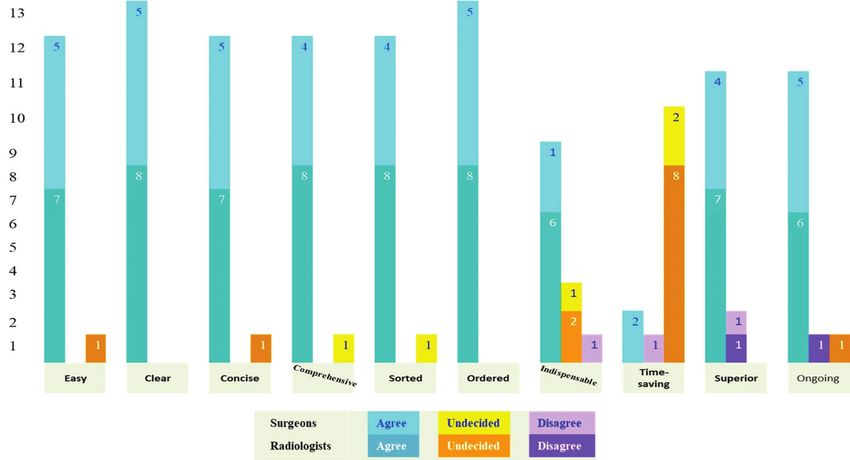
Eur J Breast Health 2021; 17(2): 165-172
Introduction assessed and ethically approved by its Ethics Committee (ethics code:
IR.TUMS.VCR.REC.1 397.846).
Breast cancer is the most prevalent female cancer and the first cause of
death from cancer in women worldwide (1, 2). Breast cancer screening We formed a team including radiologists and surgeons who are expert
is achieved by clinical breast examination (CBE) and mammography, in the management of breast diseases. All radiologists were dedicated
but under numerous circumstances, breast ultrasound (BUS) is used to gynecological imaging or breast imaging and have practiced in
as a complementary modality (3). Breast complaints are also among the radiology departments of Tehran University of Medical Sciences
the most common causes for women to attend surgery and gynecology (Tehran), which ranks first in education and research among medical
clinics (4). In addition to breast examination, many cases need to be universities of the country. In Iran, as in many other countries,
further examined by imaging, and BUS is one of the most frequently patients undergo ultrasound in different centers on the basis of which
used modalities in this regard. Also, in many referral centers, breast they can book an appointment. In addition, radiologists generally
surgeons regularly use ultrasound imaging as an adjunct to clinical mention all the lesions they detect in the BUS in their reports,
examinations. although unfortunately most clinicians do not mention an individual
target lesion to be assessed when they are requesting the BUS. In our
BUS can detect and define lesions and assist both in diagnosis and country, surgeons are responsible for the clinical management of breast
treatment planning of breast disorders, especially in discrimination diseases, and surgical oncologists and breast surgical oncologists are
of solid and cystic masses, which is beyond the diagnostic field of trained and entitled for subspecialized practice over the subject. All
mammography, and in detection of hidden masses in dense breast the surgeons of our research team were surgical oncologists or breast
mammogram. However, in many situations, lesions detected by BUS surgical oncologists and practiced as full-time or part-time professors
undergo serial ultrasound to follow the probable changes of that at our university. We performed the study in the following four phases.
specific lesion [usually Breast Imaging Reporting and Data System
(BIRADS) III Category: a probably benign lesions], in order to Phase 1: Literature search and collection of present report formats
discriminate benign and suspicious ones. This phase consisted of two stages that were accomplished by one of the
surgeons and a research expert. In the first stage, an extended search of
How to define findings in BUS and which features to note in the
the English literature from 1990 to the present time was performed for
report have been described in the ultrasound lexicon and the Breast
similar works and different viewpoints about the ultrasound BIRADS
Imaging Reporting and Data System (5).
lexicon. The rationale for beginning the search from the 1990s
A precise BUS report denoting all details is certainly helpful, but two was because the first version of BIRADS was issued in 1993 by the
key problems arise. First, which of these details would help the in- American College of Radiology (ACR) (6). The keywords consisted of
charge physicians in medical decision-making and would affect the BUS report, BIRADS ultrasound, BUS interpretation, breast imaging
management plan? Second, how should the arrangement of the report report, and breast mass radiologist assessment. All articles containing
be in order to make it practical and easy to use? In other words, the user relevant data or viewpoints were gathered. Also, chapters or paragraphs
of a BUS report is the physician that is managing the breast disorder, about the subject in referral radiology or breast books were investigated
who is usually a breast or general surgeon, a gynecologist, or perhaps a in this stage.
family practitioner general physician. In the second stage, we collected BUS reports from high-volume and
The format of the report, the arrangement of the details, and the low-volume radiology centers in Tehran, the capital of Iran, and from
number of significant or nonsignificant details affect the practicality several centers in large or small cities around the country. In order to
and usefulness of the report. A wisely organized report, with the details provide a basis for detection of defects of the reports and compare
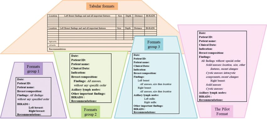
applied in an orderly manner, would save the clinicians’ time and help them, we outlined them grossly as four types of formats, which are
them figure out the clinically significant points aptly. summarized in Figure 1.
Phase 2: Summarizing key points and preparing the first draft
In addition, all BUS must be compared with the previous ones in
order to identify changes in previous lesions or new findings. At Two of the surgeons completed this phase in three steps. In the first step,
present, various ultrasound units use different formats, although the key points in the BUS report were extracted from the ultrasound
many observe the key problems of the BIRADS system and BIRADS lexicon and the few related returned articles in our search.
lexicon. These various styles may make the comparison between In the second step, all collected reports were reviewed and evaluated
ultrasound reports very time-consuming and sometimes ambiguous. regarding precision, clarity, and ease to use of the arrangement, as well
A comparison would be simpler if all BUS reports were arranged as ambiguous, vague, or complicated definitions or organization of the
systematically and uniformly and especially if they were all arranged items in the second step.
in the same form.
In the third step, the items to be defined in a BUS report were designated
As a team of radiologists and clinicians whose main field of interest according to the BIRADS lexicon; and the most appropriate order for
is the management of breast diseases, we have carried out a study to the reporting of those items was argued, in an effort to describe an
design the applied format for BUS report, which would yield the above order which could provide the highest clarity. Several designs were
advantages. prepared as drafts to be discussed.
Phase 3: Seeking expert opinion and preparing the final format
Materials and Methods
The third phase consisted of an expert panel, attended by two breast
This project was supported by the Vice-Chancellor in Research surgeons, three surgical oncologists, eight breast and gynecologic
166 Affairs of Tehran University University of Medical Sciences and was radiologists, and a research expert and then several virtual meetingsAlipour et al. Breast Ultrasound Reports
Figure 1. General gross classification of the frameworks of existing breast ultrasound report and comparison with the approved pilot format
(all frameworks are shortened to fit in the figure)
ID: Identification; BIRADS: Breast Imaging Reporting and Data System
in the era of coronavirus disease-2019 (COVID-19) through a virtual Following the establishment of the program in the three units,
group including all the named experts as members. The drafted designs radiologists’ reports were printed according to the proposed format,
for BUS report were introduced and debated during the meetings. One and the patients brought them to their surgeons according to their
design was designated as most user-friendly, and further modifications schedules.
were proposed. After several revisions, a final framework was defined
A brief survey was designed to investigate the impression of the
and approved as a BUS report pilot format.
specialists about the new format, and the responses were rated on a
Phase 4: Pilot testing 5-point Likert scale: strongly disagree, disagree, undecided, agree, and
strongly agree. The survey contained 11 questions, as seen in Table 4.
The last phase consisted of pilot testing of the approved format. This
After 5.5 months project execution, all surgeons and radiologists filled
was supposed to be uniformly held in ultrasound units of university
the survey. The results of the survey for each group and for all experts
hospitals for 4 months, so that a comparison of two subsequent
are demonstrated in Figure 1 and Figure 2. The average number of
results could take place in some cases that underwent two BUS in
BUS performed by each radiologist and seen by each surgeon per
a 3-month interval. Due to COVID-19 conditions and the delay in
month is approximately 200 cases in non-COVID-19 conditions.
many schedules including holding of most screening programs, the
number of monthly BUS dropped largely; however, three major units
remained active, although with a small number of patients. These were
Discussion and Conclusion
the centers where the radiologists and surgeons of the research team We performed a study to design and test an applied format for BUS
were practicing. Therefore, the pilot was held in these three units for report that could be user-friendly for breast care practitioners. After
around 5.5 months. After this time, a survey was carried out to assess gathering the existing formats and assessing them, we defined a
the format from the point of view of the research team’s radiologists framework and its user guide through several panels and tested it in
and surgeons. three high-volume BUS units, with favorable outcomes.
The sensitivity of mammography in detecting suspicious lesions is
Results variable and is overall lower in dense breast tissue (3, 7, 8). Adding
The first product of the panel was the list of items to be stated in the BUS to mammography increases the sensitivity for detection of breast
BUS report based on the ultrasound BIRADS lexicon, as demonstrated cancer in women with high mammographic breast tissue density (9).
in Table 1. Berg et al. (10) performed a multicenter study involving 2809 women
at high risk for breast cancer to find out if the inclusion of BUS to
The second product was the final BUS report format, which was mammography may have an effect in the diagnostic yield of the latter
proposed as a straightforward, user-friendly framework for reporting during breast cancer screening. They showed a diagnostic yield of 7.6
BUS. Since the format could only demonstrate the basic structure for versus 11.8 per 1,000 women screened for mammography alone and
writing the report, a guideline (the pilot format guide) was also written the combination of the two modalities, respectively (2). Gharekhanloo
to explain how and where to describe the items in the framework. et al. (11) also confirmed the additional sensitivity provided by adding
Table 2 illustrates the BUS report pilot format, and Table 3 shows the BUS to mammography for the detection of breast cancer in their study
pilot format guide. on 300 cases. The additional advantage of BUS in mammographic 167Eur J Breast Health 2021; 17(2): 165-172
Table 1. Items to be stated in the BUS report as approved in the expert panel
General items
History Previous breast medical and surgical history or previous biopsy results
Family history Of breast cancer
Indication For performing the BUS
Homogenous background echotexture-fat/homogenous background echotexture-fibroglandular/
Breast composition
heterogenous background echotexture
Findings
Mass Described as below
Tissue distortion Described as below
Retraction Described as below
Calcification Described as below
Location (axillary, in breast), significance, cortical thickness, hilum changes, extracapsular
Lymphadenopathies
invasion, matted nodes
Skin changes Edema, thickness, retraction
Nipple changes Retraction
Postoperative findings
Descriptions for any breast finding
Side Left/right
Location On a clock face
Distance (mm) From the nipple
Depth (mm) From the skin
Comparison Comparison with previous ultrasound examination
Descriptions for masses
Size (mm) The largest dimension or the three dimensions
Type Cystic, solid
Echopattern Anechoic, hypoechoic, isoechoic, hyperechoic, heteroechoic
Shape Round, oval, irregular
Lobulations Microlobulations, macrolobulations; number
Margins Circumscribed, indistinct, angular, spiculated
Orientation Horizontal, vertical
Posterior features None, enhancement, shadowing, combined pattern
Vascularity Absent, internal, vessels in rim
Elasticity Soft, intermediate, hard
Intracystic details Septations, masses
Postoperative findings
Significant recent change in findings
Correlation with mammographic, MRI, or clinical findings
BIRADS
Recommendations of the sonographer
BUS: Breast ultrasound; BIRADS: Breast Imaging Reporting and Data System; MRI: Magnetic resonance imaging
breast cancer screening in women at high risk of breast cancer has been regarding their shape, size, or other features; a part of the follow-up is
maintained by the American College of Radiology Imaging Network performed by serial BUS. Hence, overall, BUS plays a significant role
via a multicenter trial (12). in the approach to the breast.
In addition to screening purposes, BUS assists in the evaluation of In 2003, the ACR released a BIRADS lexicon for ultrasound that
breast symptoms and signs, including lumps or nipple discharge. intended to standardize BUS reports and simplify comparisons with
168 In many instances, lesions that appear benign need to be followed previous imaging (5). This has yielded a kind of international sharedAlipour et al. Breast Ultrasound Reports
Figure 2. Results of survey among radiologists and surgeons
language between radiologists. Abdullah et al. (13) evaluated the
Table 2. Framework of approved pilot format
concordance of definitions of five sonographists about ultrasound
characteristics of 267 benign and malignant breast lumps based
- Date: on the BIRADS lexicon for BUS. They detected an overall “good”
- Patient ID: level of agreement but a fair one for evaluating lesion borders. The
- Patient name: concordance of their description was lower for smaller lumps as well
- Clinical data: as malignant cases. Their overall conclusion was in favor of a good
interobserver agreement. This was confirmed by the studies of Lazarus
- Indication:
et al. (14) and Costantini et al. (15) on 91 and 178 breast lesions,
- Breast composition: respectively.
- Findings:
While BUS is performed by radiologists, the clinician has to decide
♣ Left breast
on the suitable approach to a breast lesion based on the findings of
Solid masses
breast exam and breast imaging. Consequently, the BIRADS system
and the lexicon also aim to ease the communication between the
Cystic masses sonographer and the clinician. As a creditable product should be
produced by the cooperation of stakeholders with diverse viewpoints
♣ Right breast:
from different aspects, several medical organizations have cooperated
in the production of the BIRADS lexicon, including associations of
Solid masses
surgeons, who could be seen as the end-users of the lexicon product,
or the BUS report (16). Items that should be mentioned in a report,
Cystic masses: descriptors for every item, and the gross order of the report are
explained in the BIRADS lexicon for BUS. However, the order of the
- Axillary lymph nodes: details, the scope and number of details, and the visual method for
emphasizing on more important findings can also be outlined, giving
Left axilla:
rise to user-friendly reports that could easily be compared. This is
what our team aimed for, by delineating an orderly structure for the
Right axilla: BUS report, where details appear in accord with the BIRADS lexicon,
and the usual classifications of breast lesions. In our proposed format,
- Other important findings: sorting the lesions by type allows users to selectively pick up the parts
they are concerned about or first pay attention to components that
are more important to them. By writing the clockface location of each
- BIRADS:
lesion first, the users localize the lesion in their mind and match it
Left breast: with the CBE or other imaging modalities. The size of each lesion
Right breast: immediately follows, because size change is almost the most important
- Recommendations: alteration that can affect the significance of a finding. Then, the other
location coordinates including depth from the skin and distance from
ID: Identification; BIRADS: Breast Imaging Reporting and Data System
the nipple depicted as near zone, mid zone, and far zone are described 169Eur J Breast Health 2021; 17(2): 165-172
Table 3. Guide for approved pilot format
- Date:
- Patient ID:
- Patient name:
- Clinical data (age, history, family history)
- Indication (cause for requesting ultrasound)
- Tissue composition (according to ACR format: homogenous background echotexture-fat/homogenous background echotexture-
fibroglandular/heterogenous background echotexture)
Findings:
For suspicious lesions: please write in BOLD + mention ZONE (near zone, mid zone, far zone) and DEPTH (anterior zone, mid zone,
posterior zone) + write the BIRADS of that specific lesion
For new lesions or lesions with recent changes, please write in BOLD
For lesions in location of clinician interest, please write in BOLD
For any suspicious finding in the breast other than masses, like tissue distortion or retraction, please write it next to the mass or in
the relevant location among masses
If typical, please write the probable diagnosis of the mass (probably fibroadenoma, fat necrosis, hamartoma, intramammary lymph
node…)
For any significant finding, if correlated with mammographic, MRI, or clinical findings, please mention it, with BIRADS
Please follow this order:
♣ Left breast:
Solid masses
In order of clock hours, first retroareolar, then 1 to 12
In each line, please first write the location (…. O’clock) and the size, then if needed the zone (NZ, MZ, FZ) and the depth (….mm from
skin), then the features of the mass as needed (irregular margin, orientation, posterior features, vascularity, elasticity, …)
Cystic masses
In order of clock hours, first retroareolar, then 1 to 12
Please only mention BIRADS 3 and 4 cysts, those in region of relevant findings in other imagings, and those in region of clinician
interest as requested in their order.
Multiple cystic lesions may be defined in a row.
In each line, please first write the location (…. O’clock), then if needed the zone (NZ, MZ, or FZ) and the depth (…mm from skin),
then the features as needed (intracystic mass, septations …).
♣ Right breast:
As above
- Axillary lymph nodes:
For normal or reactive lymph nodes please only mention nonsignificance, and do not mention size and other features
Please mention when lymph nodes are relevant to a clinical or other imaging finding
For suspicious nodes, please mention features as needed (cortical thickness, hilum changes, extracapsular invasion, matted nodes,
etc.), the BIRADS of that specific lymph node and the recommendation (short-term follow-up, tissue diagnosis…)
- Other important findings (skin changes, duct changes, seroma, etc.)
- BIRADS
Recommendations (follow-up/further imaging/suggestion of tissue diagnosis, etc. for breast or axillary lesions)
Please do not mention type of surgical management
ID: Identification; ACR: American College of Radiology; BIRADS: Breast Imaging Reporting and Data System
in order to ascertain the site of the lesion and a correct comparison believed that the organization of the report took more time. This could
with the previous BUS. be permanently true or may be temporary due to the novelty of the
structure, which might take time to get used to by the radiologists
After using the format, the survey showed a high level of agreement and their assistants who are preparing the report. Two of the surgeons
of the team members with ease of use, clarity, conciseness, thought the format was time-consuming, and one could not decide
170 comprehensiveness, and well-classified structure. However, radiologists about the time; these were considering the time for preparing theAlipour et al. Breast Ultrasound Reports
Table 4. Survey questions
Number Abbreviation* Question
1 Easy The format is easy to use
2 Clear The definitions are clear
3 Concise The format is concise and useful
4 Comprehensive The format contains all key elements
5 Sorted The format contains necessary classifications
6 Ordered The arrangement is appropriate for comparison of two BUSRs
7 Indispensable The present details cannot be deleted
8 Time-saving Using this format takes less time
9 Superior The format increases the quality of the BUSR
10 Ongoing I am eager to use the format in all my BUSRs
11** --------- I will take part in similar studies
*Abbreviations demonstrating the subject of each question in calculations and in the figures, ** Question number 11 is not depicted in the figure because it
had no direct relation with the format assessment.
BUSR: Breast ultrasound report
report; the two other surgeons agreed that in comparison with the M.R., N.S., A.Y., R.O., Al.A.; Supervision: S.A., B.E., M.A., N.A., A.A., K.B.,
BUS reports they were receiving before the study, this one took much L.B., A.E., M.G., M.R., N.S., A.Y., R.O.; Materials: S.A., B.E., M.A., N.A.,
less time to read, understand, and specially compare with the previous A.A., K.B., L.B., A.E., M.G., M.R., N.S., A.Y., R.O., Al.A.; Data Collection
report. The indispensability of all details and whether the report could or Processing: S.A., B.E., M.A., N.A., A.A., K.B., L.B., A.E., M.G., M.R.,
N.S., A.Y., R.O., Al.A.; Analysis or Interpretation: S.A., B.E., M.A., N.A.,
be further shortened were also questionable for some of the members.
A.A., K.B., L.B., A.E., M.G., M.R., N.S., A.Y., R.O., Al.A.; Literature Search:
Our study had some limitations. First and foremost, the COVID-19 S.A., B.E., M.A., N.A., A.A., K.B., L.B., A.E., M.G., M.R., N.S., A.Y., R.O.,
Al.A.; Writing: S.A., B.E., M.A., N.A., A.A., K.B., L.B., A.E., M.G., M.R.,
situation disrupted the usual flow of patients and BUS. In addition,
N.S., A.Y., R.O., Al.A.
the users of the format were the same as the designers. Therefore, the
study should also be performed by other users in other centers provide Conflict of Interest: No conflict of interest was declared by the authors.
a more valid assessment of the proposed format.
Financial Disclosure: This study was supported by grant in aid of Tehran
In conclusion, we propose our format as a user-friendly format for University of medical Sciences (no: # 97-03-218-40362).
BUS reports, which may be used and introduced as an adjunct to
the BIRADS ultrasound lexicon. The format should be applied for a References
longer time in university hospitals in order to find out if the apparent
time-consuming nature for radiologists would be solved by routine 1. Maajani K, Jalali A, Alipour S, Khodadost M, Tohidinik HR, Yazdani
use. Also, the format should be tested in other centers in order to K. The global and regional survival rate of women with breast cancer: a
systematic review and meta-analysis. Clin Breast Cancer 2019; 19: 165-
ascertain its positive features.
177. (PMID: 30952546) [CrossRef ]
2. Bray F, Ferlay J, Soerjomataram I, Siegel RL, Torre LA, Jemal A. Global
cancer statistics 2018: GLOBOCAN estimates of incidence and mortality
Acknowledgements
worldwide for 36 cancers in 185 countries. CA Cancer J Clin 2018; 68:
We would like to acknowledge Dr. Mehrnoush Hadadi (Dezfool, Iran), Dr. 394-424. (PMID: 30207593) [CrossRef ]
Mehdi Ghassemi (Andimeshk, Iran) and Mrs. Marzieh Orooji (Tehran, Iran)
3. Posso M, Louro J, Sánchez M, Román M, Vidal C, Sala M, et al.
for their kind collaboration in providing and collecting BUS reports from
Mammographic breast density: how it affects performance indicators
various centers and cities.
in screening programmes? Eur J Radiol. 2019;110: 81-87. (PMID:
30599878) [CrossRef ]
Ethics Committee Approval: This project was supported by the Vice- 4. Dawson C, Armstrong MW, Michaels J, Faber RG. Breast disease and the
Chancellor in Research Affairs of Tehran University University of Medical general surgeon. II. Effect of audit on the referral of patients with breast
Sciences and was assessed and ethically approved by its Ethics Committee problems. Ann Royal Coll Surg Engl 1993; 75: 83. (PMID: 8476191)
(Ethics code: IR.TUMS.VCR.REC.1 397.846). [CrossRef ]
5. Mendelson E, Böhm-Vélez M, Berg W, et al. ACR BI-RADS ultrasound.
Informed Consent: Informed consent was obtained. In ACR BI-RADS atlas, breast imaging reporting and data system. 5th ed.
2013:1-173. Philadelphia: Clinical Research Center: 2013.
Peer-review: Externally peer-reviewed.
6. Burnside ES, Sickles EA, Bassett LW, Rubin DL, Lee CH, Ikeda DM,
Authorship Contributions et al. The ACR BI-RADS® experience: learning from history. J Am Coll
Conception: S.A., B.E., M.A., N.A., A.A., K.B., L.B., A.E., M.G., M.R., N.S., Radiol 2009; 6: 851-860. (PMID: 19945040) [CrossRef ]
A.Y., R.O. Al.A.; Design: S.A., B.E., M.A., N.A., A.A., K.B., L.B., A.E., M.G., 171Eur J Breast Health 2021; 17(2): 165-172
7. Lynge E, Vejborg I, Andersen Z, von Euler-Chelpin M, Napolitano G. 12. The American College of Radiology: ACR practice parameter for the
Mammographic density and screening sensitivity, breast cancer incidence performance of whole-breast ultrasound for screening and staging. Last
and associated risk factors in danish breast cancer screening. J Clin Med Accessed Date: 24.07.2020. Available from: https://www.acr.org/-/media/
2019; 8: 2021. (PMID: 31752353) [CrossRef ] ACR/Files/Practice-Parameters/USWholeBreast.pdf [CrossRef ]
8. von Euler-Chelpin M, Lillholm M, Vejborg I, Nielsen M, Lynge E. 13. Abdullah N, Mesurolle B, El-Khoury M, Kao E. Breast imaging reporting
Sensitivity of screening mammography by density and texture: a cohort and data system lexicon for US: interobserver agreement for assessment
study from a population-based screening program in Denmark. Breast of breast masses. Radiology 2009; 252: 665-672. (PMID: 19567644)
Cancer Res 2019; 21: 111. (PMID: 31623646) [CrossRef ] [CrossRef ]
9. Hollingsworth AB. Redefining the sensitivity of screening mammography: 14. Lazarus E, Mainiero MB, Schepps B, Koelliker SL, Livingston LS. BI-
a review. Am J Surg 2019; 218: 411-418. (PMID: 30739738) [CrossRef ] RADS lexicon for US and mammography: interobserver variability
10. Berg WA, Zhang Z, Lehrer D, Jong RA, Pisano ED, Barr RG, et and positive predictive value. Radiology. 2006; 239: 385-391. (PMID:
al. Detection of Breast Cancer with Addition of Annual Screening 16569780) [CrossRef ]
Ultrasound or a Single Screening MRI to Mammography in Women with 15. Costantini M, Belli P, Lombardi R, Franceschini G, Mulè A, Bonomo
Elevated Breast Cancer Risk. JAMA 2021; 307: 1394-1404. (PMID: L. Characterization of solid breast masses: use of the sonographic breast
22474203) [CrossRef ] imaging reporting and data system lexicon. J Ultrasound Med 2006; 25:
11. Gharekhanloo F, Torabian S, Kamrani S. Survey of the role of combined 649-659. (PMID: 16632790) [CrossRef ]
screening method with ultrasonography in the diagnosis of breast cancer. 16. Levy L, Suissa M, Chiche JF, Teman G, Martin B. BIRADS
Avicenna J Clin Med 2011; 17: 57-60. [CrossRef ] ultrasonography. Eur J Radiol 2007; 61: 202-211. (PMID: 17215097)
[CrossRef ]
172You can also read