Clinical and imaging findings of walled-off pancreatic necrosis misdiagnosed as an intra-abdominal neoplasia in a Schnauzer dog: A case report
←
→
Page content transcription
If your browser does not render page correctly, please read the page content below
Case Report                                                           Veterinarni Medicina, 66, 2021 (01): 32–39

                                                                       https://doi.org/10.17221/123/2020-VETMED

Clinical and imaging findings of walled-off pancreatic
necrosis misdiagnosed as an intra-abdominal
neoplasia in a Schnauzer dog: A case report
Jayon Kim1, Jaeeun Ko1, Hakyoung Yoon1,3, Hyoju Kim2,
Jeongyeon Hwang2, Kidong Eom1, Jaehwan Kim1*
1
  Department of Veterinary Medical Imaging, College of Veterinary Medicine,
  Konkuk University, Gwangjin-gu, Seoul, Republic of Korea
2
  Helix Animal Medical Center, Seocho-gu, Seoul, Republic of Korea
3
  Department of Veterinary Medical Imaging, College of Veterinary Medicine,
  Jeonbuk National University, Iksan-si, Jeollabuk-do, Republic of Korea
*Corresponding author: jaehwan@konkuk.ac.kr

Citation: Kim J, Ko J, Yoon H, Kim H, Hwang J, Eom K, Kim J (2021): Clinical and imaging findings of walled-off pancreatic
necrosis misdiagnosed as an intra-abdominal neoplasia in a Schnauzer dog: A case report. Vet Med-Czech 66, 32–39.

Abstract: A 10-year-old Schnauzer presented with a 1-month history of vomiting, anorexia, and abdominal pain,
and a recently detected intra-abdominal mass. The round, soft-tissue opacity masses identified on the radiography
in the left mid-abdomen were confirmed as multifocal, cystic masses via ultrasonography. The necrotic masses
mimicked an intra-abdominal neoplasia on the initial imaging examinations. The computed tomography (CT)
clearly showed encapsulated masses with a necrotic fluid arising from the left limb of the pancreas and extending
to the peripancreatic, paracolic, and perigastric regions. Based on the multimodal imaging, surgical exploration,
and histopathology, the mass was diagnosed as a walled-off pancreatic necrosis (WOPN). CT is an effective diag-
nostic modality for diagnosing acute pancreatitis in WOPN.

Keywords: acute pancreatitis; computed tomography; necrotic debris; necrotizing pancreatitis; peripancreatic
fluid collections

   Pancreatitis is a common gastrointestinal disor-           (Thoeni 2012; Zaheer et al. 2013). Since the dis-
der in dogs; it is classified into acute and chronic          ease has high morbidity and mortality associated
pancreatitis. Although acute pancreatitis is not              with systemic inflammatory response syndrome,
associated with permanent parenchymal changes,                imaging modalities, primarily computed tomog-
it can induce various local and systemic compli-              raphy (CT), play an essential role in the diagno-
cations that have a clinical importance in dogs               sis and assessment of the peripancreatic fluid
(Mansfield 2012). Acute pancreatitis is subdivided            collections (Trout et al. 2010; Bharwani et al. 2011;
into interstitial oedematous pancreatitis and necro-          Baudin et al. 2012). Based on the revised Atlanta
tising pancreatitis (NP); both conditions can lead            classification system (Thoeni 2012), peripancreatic
to various complications in the pancreatic paren-             fluid collections are classified into four different
chyma and the adjacent organs.                                types: acute peripancreatic fluid collection, pan-
   In humans, NP is a severe form of acute pancre-            creatic pseudocyst, acute necrotic collection, and
atitis and is defined as the necrosis of the pan-             walled-off pancreatic necrosis (WOPN). Among
creatic parenchyma with peripancreatic tissues                them, pseudocyst and WOPN occur more than

32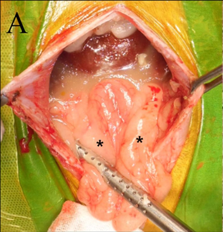
Case Report                                                          Veterinarni Medicina, 66, 2021 (01): 32–39

                                                                      https://doi.org/10.17221/123/2020-VETMED

4 weeks after the onset of pancreatitis and have              (3 300; reference range, 388−1 007 IU/l), lipase
clinical relevance due to the mass-like lesions that          (1 991; reference range, 0−1 800 IU/l), C-reactive
are formed and often misdiagnosed as abdominal                protein (CRP) (130; reference range, 0−20 mg/l), and
masses. There have been several reports on WOPN,              canine pancreatic lipase (cPL) (881; reference range,
which is a known NP complication in humans; how-              0−200 ng/ml) levels.
ever, to our knowledge, there has been only one                 Routine radiographs were obtained (Figure 1)
report on WOPN in veterinary medicine (Hwang                  (Titan 2000M; Comed Medical Systems, Seoul,
et al. 2018). The aim of this case report was to de-          Republic of Korea). In the lateral radiograph, a de-
scribe the clinical and imaging findings of WOPN              creased serosal detail of the entire abdominal cav-
as an NP complication in dogs to establish the basis          ity with a moderate mass effect was detected. The
of a diagnosis in the future.                                 hazy, round masses were compressing and dislo-
                                                              cating the adjacent small intestines caudoventrally.
                                                              On the ventrodorsal projection, heterogeneous
Case description                                              soft-tissue opacity masses were noted on the left-
                                                              side of the abdomen, dislocating the small intes-
   A 10-year-old, spayed, female, Schnauzer weigh-            tines to the contralateral side.
ing 7 kg, presented with a 1-month history of vom-              An ultrasonographic examination was performed
iting, anorexia, and abdominal pain, and a recently           (Figure 2) (Aplio 500; Toshiba Medical System,
detected intra-abdominal mass on an ultrasono-                Tokyo, Japan). Enlarged, an oedematous hypoecho-
graphic examination. The dog was diagnosed with               ic pancreatic parenchyma with irregular septations
acute pancreatitis and treated at the first visited           was visible, specifically in the left pancreatic limb.
animal hospital. However, the owner recently rec-             Multifocal, round, hypoechoic cystic masses were
ognised an abdominal distension, thus raising                 noted in the left mid-abdomen, adjacent to the de-
a concern.                                                    scending colon, greater curvature of the stomach,
   On the physical examination, only mild hyper-              and spleen. A small amount of peritoneal effu-
thermia (39.0 °C) and an abdominal distension were            sion and increased mesenteric and omental fat
noted. The complete blood counts and serum bio-               echogenicity were noted. There were no remarkable
chemistry profile revealed leukocytosis (45; refer-           findings in the descending duodenum and biliary
ence range, 6−17 × 10 9/l) and increased amylase              tracts.

     (A)                                                            (B)

Figure 1. Lateral (A) and ventrodorsal (B) radiographs of the abdominal cavity
At the left-mid abdomen, soft-tissue opacity round intra-abdominal masses are visible (asterisks). The masses deviate
the small intestines towards the opposite site. Around the mass, remarkably decreased serosal details are found (arrow-
heads). Concurrently, round radiopaque materials at the region of the urinary bladder consistent with calculi are visible

33
Case Report                                                          Veterinarni Medicina, 66, 2021 (01): 32–39

                                                                      https://doi.org/10.17221/123/2020-VETMED

       Figure 2

     (A)                                                           (B)

Figure 2. Abdominal ultrasonographic findings at the left limb of the pancreas (A) and the intra-abdominal mass (B)
The left limb of the pancreas (P) shows an irregularly increased echogenicity with a multiple hypoechoic septum. The mul-
tifocal, round, hypoechoic masses with multiple cysts are visible. Note that the hypoechoic, round fluid collection is
visible at the peripancreatic area (asterisk)

  Computed tomography (Brivo CT385; GE Health-                4.1 cm) regions with well-defined margins. Around
care, Waukesha, WI, USA) was used to evaluate the             the spleen, extensive fat stranding with loculated
intra-abdominal mass and pancreas. Anaesthesia                fluid collections were noted; this was consistent
was induced with propofol (6 mg/kg, Provive 1%;               with acute necrotic fluid collections. The fluid col-
Myungmoon Pharmaceutical Co., Seoul, Republic                 lections compressed the adjacent organs includ-
of Korea). Following the endotracheal intubation,             ing the left kidney, spleen, and descending colon.
the anaesthesia was maintained with 2% isoflurane             A regional lymphadenopathy involving the gastric,
(Forane solution; Choongwae Pharma Corporation,               hepatic, and pancreaticoduodenal lymph nodes,
Seoul, Republic of Korea). With the dog in the ven-           was noted. The dog was tentatively diagnosed with
tral recumbency position, a CT was performed                  NP with extensive peripancreatic necrosis and dif-
in the helical mode using soft tissue algorithms with         fuse peritonitis based on the clinical and imaging
the following scan parameters: 100 kVp; 200 mAs;              findings.
slice thickness 0.125 mm, without gantry tilting.               A peritoneal lavage and exploratory laparotomy
To obtain post-contrast images, a contrast medi-              were performed to relieve the abdominal pain and
um (Omnihexol 300; Korea United Pharmaceutical,               peritonitis (Figure 4A). The surgery was performed
Seoul, Republic of Korea) at a dose of 600 mg                 in a routine manner with a ventral midline inci-
iodine/kg was rapidly injected by hand into the left          sion. At the level of the left upper abdomen and
cephalic vein, and one post-contrast scan was per-            retroperitoneum, a severe adhesion formation,
formed 60 s after the injection.                              fat necrosis, and peritonitis were noted. Most
  On the post-contrast transverse plane image (Fig-           of the small intestine was deviated to the right
ure 3), the left limb of the pancreatic parenchy-             side, and a fat necrosis and a palpable mass sur-
ma was mildly enlarged and showed oedema-                     rounded by fat were mainly identified on the left
tous changes with peripancreatic fat stranding.               side. The right limb of the pancreas was identified
At the tip of the pancreas, an irregular heteroge-            as having a normal appearance, but identification
neous enhancement of the pancreatic parenchyma,               of the left limb was difficult due to the severe adhe-
consistent with necrosis, was noted. Encapsulated             sion. The mass was excised using a blunt dissection
hypoechoic non-enhancing soft-tissue attenuat-                to free it from the adhesion regions. Furthermore,
ing masses (15−20 Hounsfield units) were located              350 ml of a cloudy and viscous peritoneal effusion
in the peripancreatic (5.3 × 5.8 × 2.6 cm), paracolic         was suctioned. After flushing the abdominal cav-
(3.2 × 6.5 × 12.8 cm), and perigastric (5.2 × 3.5 ×           ity several times with warm saline, a closed active

34
Case Report Veterinarni Medicina, 66, 2021 (01): 32–39
https://doi.org/10.17221/123/2020-VETMED
Figure 3. Post-contrast com-
(A) (B) puted tomography findings at
the level of the pancreas (A),
descending colon (B and C),
and kidney (D)
The left limb of the pancreas shows
an oedematous, heterogeneously
enhanced parenchyma (open
arrowheads, 80−100 Hounsfield
units). Sharply demarcated, mul-
tifocal hypoattenuating masses
(C) (D) (15−20 Hounsfield units) are vis-
ible at the peripancreatic region
(asterisks), paracolic space (dag-
gers), and perigastric area (double
dagger). There is also fat stranding
and a fluid attenuating lesion in
the left-mid abdomen consistent
with acute necrotic fluid collec-
tions (white arrowheads). A and B,
transverse plane; C and D, dorsal
plane
Jackson-Pratt drain was installed. The abdominal macrophages without any infectious agents. The
wall, subcutaneous tissues, and cutaneous tissues aerobic and anaerobic cultures of the peritoneal
were closed in a routine manner. effusion were negative. The histopathologic re-
The cytologic analysis of the peritoneal effu- sults (Figure 4B) revealed that the adipose tis-
sion revealed only a few red blood cells and foamy sue was multifocal in the coalescing areas of the
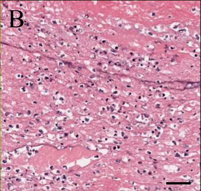
(A) (B)
Figure 4. Intraoperative (A) and histopathological (B) findings in the dog
On the exploratory laparotomy, extensive adhesion formation and fat necrosis in the left upper abdomen and retroperitoneal
cavity can be observed. The encapsulated masses surrounded by the intra-abdominal fat (asterisks) compress the adjacent
small intestines. The histopathologic result shows necrotic adipocytes with saponification, multifocal to the coalescing
area of the necrosis with numerous visible degenerate neutrophils, macrophage, fibrin, cell debris, and basophilic granular
materials. Bar = 5 μm
35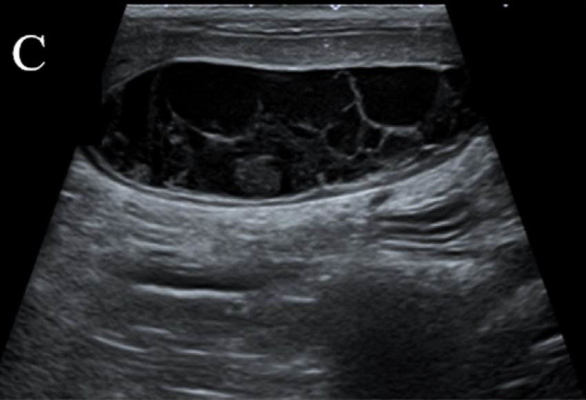
Case Report                                                        Veterinarni Medicina, 66, 2021 (01): 32–39

                                                                    https://doi.org/10.17221/123/2020-VETMED

necrosis with numerous degenerative and viable              DISCUSSION AND CONCLUSION
neutrophils, macrophages, fibrin, cell debris, and
basophilic granular materials. The mass showed                WOPN is a long-term sequel of acute pancrea-
inflammation and necrosis in the fat tissue with-           titis that occurs at least 4 weeks after the onset
out evidence of a neoplasm; the affected adipo-             of NP (Thoeni 2012). In humans, NP accounts for
cytes were necrotic with saponification consistent          20−30% of acute pancreatitis cases (Stamatakos
with WOPN.                                                  et al. 2010; Hughey et al. 2017) and can develop
  After surgery, the dog recovered uneventfully             into a life-threatening disease if adequate treat-
with postoperative antibiotics administered for             ment is delayed (Mier et al. 1997; Thoeni 2012).
9 days (cefazolin 22 mg/kg, i.v., every 12 h and            Therefore, the clinical and imaging characteristics
metronidazole 10 mg/kg, i.v., every 12 h). At the           of WOPN are well-established, and the Atlanta clas-
follow-up visit 5 weeks after the surgery, the hae-         sification has outlined clear criteria for the clinical
matological profile had returned to its reference           assessment and management of acute pancreatitis
ranges. No intra-abdominal mass or a mass ef-               (Thoeni 2012). Acute pancreatitis is also a com-
fect was visible on the radiographs. Additionally,          mon disease in veterinary medicine and although
the hypoechoic masses were distinctly decreased             the prevalence of acute pancreatitis in the canine
in size and were only identified in the peripan-            population is not well-known, the mortality rate
creatic area on the abdominal ultrasonography               in dogs was reported to be 27−58% in some recent
(Figure 5). Based on the clinical and multimodal            studies (Mansfield 2012; French et al. 2019). There
imaging findings, the dog was finally diagnosed             have been several reports of acute pancreatitis and
with NP and WOPN caused by extensive sterile                pancreatic abscesses which are known complica-
peritonitis.                                                tions of NP; however, WOPN is not well-established

 (A)                                                            (B)

 (C)

Figure 5. Radiographs (A and B) and abdominal ultrasonographic images (C) for the dog at the 5 week-follow-up
after surgery
There are no remarkable masses and normal serosal details can be observed throughout the abdominal cavity. The ultra-
sonography revealed that the hypoechoic masses distinctly decreased in size showing more cystic characteristics

36
Case Report Veterinarni Medicina, 66, 2021 (01): 32–39
https://doi.org/10.17221/123/2020-VETMED
in veterinary medicine. This case report is intend- ing an imaging-based assessment, especially CT.
ed to provide a basis for the diagnosis of WOPN, However, there is no classification system based
which may be misdiagnosed as an intra-abdominal on the clinical and radiological characteristics
mass in small animals based on the clinical and im- in veterinary medicine. Therefore, WOPN is still
aging findings. being reported as a pancreatic abscess in veterinary
WOPN consists of an encapsulated collection medicine, as formerly defined in human medicine.
of pancreatic and/or peripancreatic necrosis and NP requires close monitoring because the neces-
a thickened non-epithelised wall between the ne- sity and timing of the surgery differ depending
crosis and the adjacent tissue (Thoeni 2012). There on the NP classification. Moreover, the prognosis
can be single or multiple necrotic fluid collections is poor in NP cases; therefore, an accurate diag-
(Hughey et al. 2017). Low-attenuated fluid col- nosis according to the classification is necessary.
lections replace the pancreatic necrosis area and Furthermore, reporting on WOPN might have been
can be observed to extend into the peripancre- underestimated because an abdominal CT is not
atic space with well-defined walls on CT imaging routinely performed in cases of acute pancreati-
in humans (Takahashi et al. 2008; Thoeni 2012; tis in veterinary medicine. General anaesthesia
Zaheer et al. 2013). Similar to previous reports must be used to conduct the CT examinations,
in humans, acute necrotic fluid collections with and the use of anaesthesia can lead to hypoperfu-
well-defined walls were identified in the left mid- sion, exacerbating the pancreatic inflammatory
abdomen, adjacent to the descending colon, greater response. Additionally, a CT examination is not
curvature of the stomach, and spleen in the pre- routinely performed because the imaging criteria
sent case. Pancreatic necrotic debris without any for WOPN have not been well-established in vet-
enhancement is another common characterisation erinary medicine. Finally, the owner’s inevitable
of WOPN (Cunha et al. 2014). Necrotic debris medical expense and the smaller number of hos-
is not always identified using CT, but it may be pitals with the ability to perform CT examina-
analysed using a narrower window (Morgan et al. tions may also be additional reasons that WOPN
1997). In WOPN, pancreatic necrotic debris is is underestimated in veterinary medicine. More
identified because the pancreatic necrosis replaces cases might have been reported if CT was rou-
part of the pancreatic parenchyma (Thoeni 2012; tinely used in the diagnosis of WOPN in veteri-
Cunha et al. 2014). However, in the only previ- nary medicine. Therefore, the present case report
ously reported case in veterinary medicine (Hwang suggests the possibility of using the same WOPN
et al. 2018) known to us, ill-defined borders with classification in veterinary medicine as that used
heterogeneous peripancreatic fluid collections, in human medicine. This recommendation can be
which were confirmed in other NPs, were identi- considered in the future, and lessons learnt from
fied. Moreover, the identification of necrotic debris this case could assist in driving new research ini-
using CT was not described in the previously re- tiatives.
ported case. In the present case, the necrotic debris Differentiating WOPN from pancreatic pseudo-
with well-defined walls was identified using the CT cysts is clinically important since pseudocysts are
and the findings were similar to those from previ- long term sequelae of interstitial oedematous pan-
ous reports in humans. creatitis; thus, their treatment and prognosis differ
In human medicine, the 2012 revision of the At- from those of WOPN. In humans, the use of CT
lanta classification of acute pancreatitis outlined is relatively accurate in differentiating between
improvements to the clinical assessment and man- WOPN and a pseudocyst. On the CT, the WOPN
agement of acute pancreatitis and the terminol- characteristics differ from those of pseudocysts
ogy used for peripancreatic fluid collections, and and include a larger size, extension into the para-
pancreatic and/or peripancreatic necrosis (Thoeni colic space, an irregular wall definition, the pres-
2012). Terms such as acute peripancreatic fluid ence of fat attenuation debris in the pancreatic
collections, a pancreatic pseudocyst, acute ne- fluid collections, a pancreatic parenchymal de-
crotic collections, and WOPN were revised based formity or discontinuity, and the absence of dila-
on the classification of the peripancreatic fluid col- tion of the main pancreatic duct (Takahashi et al.
lections. The revision of the Atlanta classification 2008). Differentiating WOPN from pseudocysts
focuses on the morphologic criteria obtained us- is also important in veterinary medicine because
37Case Report Veterinarni Medicina, 66, 2021 (01): 32–39
https://doi.org/10.17221/123/2020-VETMED
pseudocysts are frequently reported (Jerram et al. 2004). The CT severity index and the modified CT
2004; Park et al. 2018), and WOPN misdiagnosed severity index, which evaluate the degree of necrosis
as a pseudocyst can lead to an inaccurate treatment of the pancreatic parenchyma, are used for the ear-
and a poor prognosis. In the present case, the CT ly detection of the disease severity. In previous
characteristics described above, with the exception reports, the morbidity and mortality rates were
of the pancreatic parenchymal deformity or discon- higher when the extent of the pancreatic necrosis
tinuity, were identified and used to differentiate identified based on the modified CT severity index
the WOPN from a pseudocyst. Further studies are was greater than 30% (Balthazar 2002). Applying
needed to determine if the CT diagnostic criteria this criterion to the present case suggests a good
in veterinary medicine differ significantly from prognosis because the necrosis in the pancreatic
those applied in human medicine. parenchyma was not severe; additionally, the pa-
WOPN can often be difficult to distinguish from tient showed good recovery. Therefore, the CT
an intra-abdominal neoplasia, especially a pancre- imaging characteristics and the prognostic criteria
atic neoplasia, as the presenting clinical and ra- for WOPN did not significantly differ from those
diographic features are often similar. In particular, of humans in the present case. More research is
in veterinary medicine, there have been few reports needed to confirm the criteria for the prognostic
regarding WOPN; therefore, residual fluid collec- evaluation based on CT in veterinary medicine.
tions with necrotic debris can be misdiagnosed In conclusion, WOPN should be considered as
as an intra-abdominal neoplasia. Residual fluid a differential diagnosis when multifocal, round, and
collections can also appear as cystic pancreatic poorly vascularised cystic masses are identified
neoplasms; this is more common if the masses are on the radiography and ultrasonography, especially
multifocal or extensive. Likewise, there have been in dogs with clinically suspected acute pancreatitis.
cases in which NP, which is thought to be WOPN, WOPN can be misdiagnosed as an intra-abdominal
mimicked pancreatic cancer in humans (Thurnher neoplasia if the clinical and radiographic features
et al. 2001). Since NP is characterised by a high are similar; therefore, an additional CT examina-
incidence of local complications and a high mor- tion is recommended for an accurate diagnosis.
tality rate (Munsell and Buscaglia 2010; Cunha A contrast-enhanced CT examination is a useful
et al. 2014), the accurate diagnosis and proper and tool for the diagnosis, prognostic evaluation, and
timely treatment are very important. In the present treatment planning in dogs with WOPN.
case, the radiographs and ultrasonographic images
showed radiologically necrotic masses in the left
mid-abdomen. These necrotic masses were consid- Conflict of interest
ered as an intra-abdominal neoplasia based on the
initial imaging examinations. However, following The authors declare no conflict of interest.
the CT examinations, the masses were diagnosed
as WOPN, thus ruling out the initial diagnosis
of an intra-abdominal neoplasia. Therefore, WOPN REFERENCES
should be considered as a differential diagnosis
when multifocal, round, poorly vascularised cystic Balthazar EJ. Acute pancreatitis: Assessment of severity with
masses are identified on the radiography and ul- clinical and CT evaluation. Radiology. 2002 Jun;223(3):
trasonography. Additionally, a CT examination is 603-13.
recommended for the accurate diagnosis. Baudin G, Chassang M, Gelsi E, Novellas S, Bernardin G,
A contrast-enhanced CT is considered to be Hebuterne X, Chevallier P. CT-guided percutaneous cath-
the gold standard imaging modality for diagnosing eter drainage of acute infectious necrotizing pancreatitis:
acute pancreatitis in human medicine (Trout et al. Assessment of effectiveness and safety. AJR Am J Roent-
2010; Bharwani et al. 2011). It plays a critical role genol. 2012 Jul;199(1):192-9.
in not only diagnosing the morphological complica- Bharwani N, Patel S, Prabhudesai S, Fotheringham T,
tions and monitoring treatment (Baudin et al. 2012), Power N. Acute pancreatitis: The role of imaging in diag-
but also in excluding the possibility of neoplasms. nosis and management. Clin Radiol. 2011 Feb;66(2):164-75.
Moreover, CT has a clinical value as a prognos- Cunha EF, Rocha Mde S, Pereira FP, Blasbalg R, Baroni RH.
tic tool for NP in human medicine (Mortele et al. Walled-off pancreatic necrosis and other current con-
38Case Report Veterinarni Medicina, 66, 2021 (01): 32–39
https://doi.org/10.17221/123/2020-VETMED
cepts in the radiological assessment of acute pancreatitis. uating acute pancreatitis: Improved correlation with pa-
Radiol Bras. 2014 May-Jun;47(3):165-75. tient outcome. AJR Am J Roentgenol. 2004 Nov;183(5):
French JM, Twedt DC, Rao S, Marolf AJ. Computed tomo- 1261-5.
graphic angiography and ultrasonography in the diagno- Munsell MA, Buscaglia JM. Acute pancreatitis. J Hosp Med.
sis and evaluation of acute pancreatitis in dogs. J Vet 2010 Apr;5(4):241-50.
Intern Med. 2019 Jan;33(1):79-88. Park J, Lee M, Lee H, Jeong SM. Treatment of pancreatic
Hughey M, Taffel M, Zeman RK, Patel S, Hill MC. The di- pseudocyst with omentalization in a dog. Korean J Vet
agnostic challenge of the sequelae of acute pancreatitis on Res. 2018;58(3):163-5.
CT imaging: A pictorial essay. Abdom Radiol. 2017 Apr; Stamatakos M, Stefanaki C, Kontzoglou K, Stergiopoulos S,
42(4):1199-209. Giannopoulos G, Safioleas M. Walled-off pancreatic ne-
Hwang TS, Park SJ, Lee JH, Jung DI, Lee HC. Walled-off crosis. World J Gastroenterol. 2010 Apr 14;16(14):1707-12.
pancreatic necrosis in a dog. J Vet Clin. 2018 Aug;35(4): Takahashi N, Papachristou GI, Schmit GD, Chahal P, LeRoy
146-9. AJ, Sarr MG, Vege SS, Mandrekar JN, Baron TH. CT find-
Jerram RM, Warman CG, Davies ES, Robson MC, Walker ings of walled-off pancreatic necrosis (WOPN): Differ-
AM. Successful treatment of a pancreatic pseudocyst entiation from pseudocyst and prediction of outcome
by omentalisation in a dog. N Z Vet J. 2004 Aug;52(4): after endoscopic therapy. Eur Radiol. 2008 Nov;18(11):
197-201. 2522-9.
Mansfield C. Acute pancreatitis in dogs: Advances in un- Thoeni RF. The revised atlanta classification of acute pan-
derstanding, diagnostics, and treatment. Top Companion creatitis: Its importance for the radiologist and its effect
Anim Med. 2012 Aug;27(3):123-32. on treatment. Radiology. 2012 Mar;262(3):715-64.
Mier J, Leon EL, Castillo A, Robledo F, Blanco R. Early ver- Thurnher MM, Schima W, Turetschek K, Thurnher SA, Fug-
sus late necrosectomy in severe necrotizing pancreatitis. ger R, Oberhuber G. Peripancreatic fat necrosis mimick-
Am J Surg. 1997 Feb;173(2):71-5. ing, pancreatic cancer. Eur Radiol. 2001 May;11(6):922-5.
Morgan DE, Baron TH, Smith JK, Robbin ML, Kenney PJ. Trout AT, Elsayes KM, Ellis JH, Francis IR. Imaging of acute
Pancreatic fluid collections prior to intervention: Evalu- pancreatitis: Prognostic value of computed tomographic
ation with MR imaging compared with CT and US. Ra- findings. J Comput Assist Tomogr. 2010 Jul;34(4):485-95.
diology. 1997 Jun;203(3):773-8. Zaheer A, Singh VK, Qureshi RO, Fishman EK. The revised
Mortele KJ, Wiesner W, Intriere L, Shankar S, Zou KH, Atlanta classification for acute pancreatitis: Updates in
Kalantari BN, Perez A, vanSonnenberg E, Ros PR, Banks imaging terminology and guidelines. Abdom Imaging.
PA, Silverman SG. A modified CT severity index for eval- 2013 Feb;38(1):125-36.
Received: June 1, 2020
Accepted: October 26, 2020
39You can also read