Case Report: Surgical Correction of a Cystic Duct Stump Leakage Following Cholecystectomy Using an Autologous Rectus Sheath Graft in a Dog - Frontiers
←
→
Page content transcription
If your browser does not render page correctly, please read the page content below
CASE REPORT
                                                                                                                                           published: 01 February 2021
                                                                                                                                       doi: 10.3389/fvets.2021.584975

                                             Case Report: Surgical Correction of a
                                             Cystic Duct Stump Leakage
                                             Following Cholecystectomy Using an
                                             Autologous Rectus Sheath Graft in a
                                             Dog
                                             Hyun-Jung Han 1 , Kyu-Cahng Kim 2 and Hun-Young Yoon 2*
                                             1
                                              Department of Veterinary Emergency and Critical Care, Konkuk Veterinary Medical Teaching Hospital, Konkuk University,
                                             Seoul, South Korea, 2 Department of Veterinary Surgery, College of Veterinary Medicine, Konkuk University, Seoul, South
                                             Korea

                                             A 2.7 kg, 13-year-old, castrated male Yorkshire Terrier was presented with bile peritonitis
                                             after cholecystectomy. Exploratory coeliotomy to identify and correct bile leakage
                        Edited by:
                 Seong Mok Jeong,            revealed that the transected end of the cystic duct was open with no in-situ ligatures
        Chungnam National University,        or vascular clips. The residual cystic duct stump was too short to ligate or seal directly.
                       South Korea
                                             An autologous rectus sheath graft, harvested from the internal leaf of the rectus sheath,
                       Reviewed by:
             Floryne Ottilie Buishand,
                                             was used to patch the cystic duct stump. The graft was secured over the open duct using
             University of Edinburgh,        several simple interrupted sutures and covered with an omentalization. The clinical signs
                      United Kingdom         resolved after surgery, except for a transient increase in hepatobiliary enzyme levels and
                   Young-Sam Kwon,
       Kyungpook National University,        intrahepatic bile duct dilatation. The enzyme levels returned to near normal on day 25 after
                           South Korea       surgery. No intrahepatic bile duct dilatation was detected on day 55 after surgery. The
                      Philipp Mayhew,
        University of California, Davis,
                                             owner was contacted for 3 years post-operatively and reported that the dog remained
                        United States        healthy without any long-term complications. Grafting using autologous rectus sheath
                  *Correspondence:           can be used to treat cystic duct stump leakage that cannot be managed with direct
                     Hun-Young Yoon          closure using traditional modalities due to spatial constraints.
                  yoonh@konkuk.ac.kr
                                             Keywords: autologous rectus sheath, bile peritonitis, cholecystectomy, cystic duct stump leakage, graft, yorkshire
                    Specialty section:       terrier
         This article was submitted to
                Veterinary Surgery and
                        Anesthesiology,      BACKGROUND
                a section of the journal
        Frontiers in Veterinary Science      Post-operative cystic duct stump leakage (CDSL) resulting in bile peritonitis is a reported
             Received: 19 July 2020          complication of open and laparoscopic cholecystectomy in humans and animals (1–4). CDSL
         Accepted: 07 January 2021           accounts for 3–8.7% of complications associated with cholecystectomy in dogs (2, 5). Most CDSL
        Published: 01 February 2021          in veterinary medicine is iatrogenic and due to technical failures, such as dislodgement of an
                              Citation:      improperly sized or improperly placed surgical clip (6, 7). For surgical treatment of CDSL in dogs,
    Han H-J, Kim K-C and Yoon H-Y            direct closure of the remaining cystic duct by ligation using suture or clips and a vessel sealing
        (2021) Case Report: Surgical         device is the most common way to close the CDSL (2, 6, 7). In cases where direct closure is not
   Correction of a Cystic Duct Stump
                                             possible or poses a high risk of poor outcome, alternative approaches are needed.
 Leakage Following Cholecystectomy
 Using an Autologous Rectus Sheath
                                                 This case report describes the surgical technique and outcome of the treatment of CDSL
                        Graft in a Dog.      following open cholecystectomy in a Yorkshire Terrier. An autologous rectus sheath (ARS) graft
            Front. Vet. Sci. 8:584975.       was used to completely seal the CDSL, which could not be managed with direct ligation because of
    doi: 10.3389/fvets.2021.584975           spatial constraints.

Frontiers in Veterinary Science | www.frontiersin.org                              1                                         February 2021 | Volume 8 | Article 584975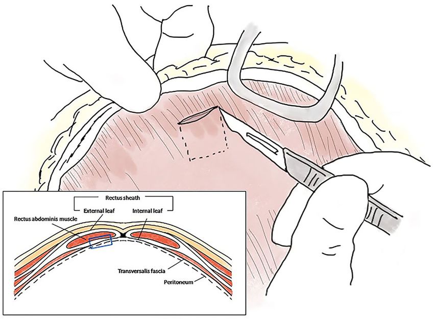
Han et al.                                                                                                       CDSL Correction Using ARS Graft

CASE PRESENTATION                                                         was sampled for bacterial culture, the effusion was removed by
                                                                          constant suction. After careful approach to the gall bladder fossa
A 2.7 kg, 13-year-old, castrated male Yorkshire Terrier dog               and CBD, complete transection of the cystic duct was identified at
was referred for further evaluation and treatment of suspected            the junction of the cystic duct and the CBD (Figures 1A,B). The
bile peritonitis following open cholecystectomy. According to             end of the cystic duct was completely open without any ligatures.
the referring veterinarian, the dog had been diagnosed with               A tied suture had slipped from the transected cystic duct and
gallbladder mucocele, which had not been ruptured, and                    was found separately. Other visible hepatic ducts and the CBD
underwent open cholecystectomy with single circumferential                were intact without bile leakage. CBD patency was confirmed
ligation of the cystic duct using 3–0 polyglyconate (Maxon,               by antegrade cannulation, from the transected end of the cystic
Covidien, Mansfield, Massachusetts) 2 days before referral.               duct through the common bile duct into the duodenal papilla,
The CBD lavage was performed by antegrade flushing without                and saline flushing with a 5 Fr feeding tube [F.D.T. (D), HMS
duodenotomy. The veterinarian also reported that abdominal                Inc]. There was no remnant of the cystic duct stump (CDS) to
fluid aspirated 2 days after the open cholecystectomy, exhibited          be ligated, because the almost entirely cystic duct was transected,
a total bilirubin (TBIL) level of 27.9 mg/dL, which was at least 20       and the severed end was very close to the bilateral hepatic ducts
times the level in the serum (1.1 mg/dL; reference range, 0–0.4           and the CBD. The length of CDS remnant seemed to be  210 µmol/L; reference range,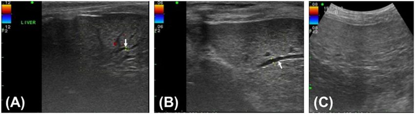
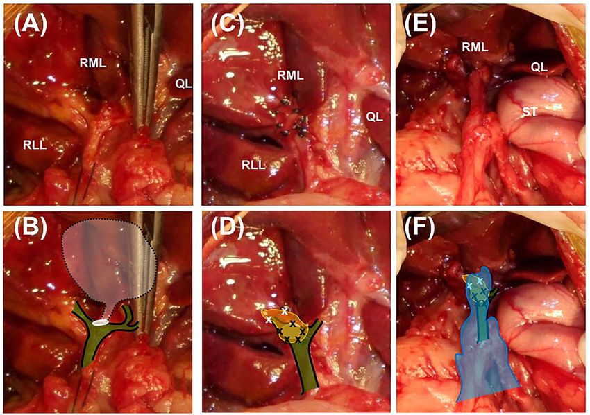
Han et al.                                                                                                                              CDSL Correction Using ARS Graft

 FIGURE 1 | Closure of the cystic duct stump using an autologous rectus sheath graft in a Yorkshire Terrier. Images (A,C,E) are intaoperative photographs; (B,D,F)
 incorporate schematic overlays to highlight the relevant anatomy and location of suture placement. (A,B) The transected end of the cystic duct is completely open
 (white circle) and is close to the base of the bilateral hepatic ducts and the common bile duct (green color plane with black solid line). The white semi-transparent
 plane with black dotted line indicates the location of the removed gall bladder and cystic duct. (C,D) The 1 × 1 cm rectus sheath graft (yellow plane) is sutured to the
 end of the cystic duct stump with simple interrupted sutures using 5–0 polyglyconate (black × marks). The abundant dorsal part of the sheath is sutured to the fibrous
 capsule of the surrounding right medial liver lobe (white × marks). (E,F) After confirmation of complete closure of the cystic duct, greater omentum is tacked over the
 patched cystic duct stump by suturing with the fibrous capsule of the right medial liver lobe (blue plane). RML, right medial lobe; RLL, left lateral lobe; QL, quadrate
 lobe; ST, stomach.

day 25 after surgery (Figures 3A,B). No dilatation was detected                         ranges on day 25 after surgery (WBC, 11.42 × 10³ /µL; CRP,
by ultrasound on day 55 after surgery (Figure 3C). Ultrasound                           38 µmol/L).
revealed minimal residual abdominal effusion immediately after                              For 6 months after surgery, the dog’s general condition
surgery, which subsequently disappeared. All serum liver enzyme                         and hepatobiliary enzyme levels were re-examined monthly
levels were increased immediately after surgery compared with                           or bimonthly at the Konkuk University Veterinary Medical
the levels prior to surgery, except for the TBIL level, which                           Teaching Hospital. No significant findings were noted during
decreased post-operatively but was still higher than the normal                         that period. Telephone follow-up was conducted for 3 years
range (ALT, 311 U/L; AST, 316 U/L; ALKP, 781 U/L; TBIL, 2.99                            following the surgery. The owner reported that the dog remained
mg/dL). All levels had returned to near normal levels on day 25                         healthy during that period without evidence of clinical signs
after surgery (ALT, 117 U/L; AST, 24 U/L; ALKP, 309 U/L; TBIL,                          associated with hepatobiliary disease.
0.12 mg/dL). The GGT level exhibited a transient post-operative
increase (15.1 mg/dL) and subsequently fell to near the normal                          DISCUSSION
range by 3 days after surgery (8.6 mg/dL). The GGT level was
again increased to 73.5 mg/dL on day 25 after surgery but had                           Because untreated CDSL can induce fatal complications
returned to normal level on day 55 after surgery (5.2 mg/dL).                           associated with bile leaks in humans and animals, such as
Inflammatory markers, including WBCs and CRP, consistently                              bile peritonitis and hepatic inflammation (2, 4, 8), immediate
improved after surgery and had returned to within reference                             detection and repair of CDSL is a key factor in positive outcomes

Frontiers in Veterinary Science | www.frontiersin.org                               3                                          February 2021 | Volume 8 | Article 584975
Han et al.                                                                                                                          CDSL Correction Using ARS Graft

 FIGURE 2 | Schematic diagram of the autologous rectus sheath graft. The internal leaf of the rectus sheath was approached via the coeliotomy site, and a 1 × 1 cm
 sheath with peritoneum was peeled off at the level of the midline, ∼1 cm from the incision (blue empty square in the box).

 FIGURE 3 | Post-operative ultrasound images of the hepatic parenchyma of a Yorkshire Terrier with unusual cystic duct stump leakage treated using an autologous
 rectus sheath graft. Color Doppler imaging shows intrahepatic bile duct dilatation in images (A,B) (white arrow). The intrahepatic bile duct diameter is 0.7 mm on
 post-operative day 5 (A), 1.4 mm on post-operative day 25 (B), and not measurable on post-operative day 55 (C).

(8). Some differences in the management of CDSL between                               early detection and minimally invasive treatment of CDSL
humans and dogs have been reported. In humans, despite                                can result in a good prognosis (9). Diversion of the biliary
significant morbidity and mortality caused by bile peritonitis,                       flow by endoscopic methods, including sphincterotomy with or

Frontiers in Veterinary Science | www.frontiersin.org                             4                                         February 2021 | Volume 8 | Article 584975
Han et al.                                                                                                            CDSL Correction Using ARS Graft

without stenting and nasobiliary drain placement for reduction                or non-absorbable suture was used to patch the graft to the
of bile flow resistance, are the preferred treatments for CDSL                CBD (16, 17, 19, 22). The use of sealant to prevent leakage
in humans (4, 10). Those treatments facilitate spontaneous                    from the grafting site was not considered, because a previous
healing of the cystic duct, resulting in a 94–100% cure rate                  study reported that a fibrin sealant could act as an infectious
(3, 4, 9). Re-exploratory surgery has an 83–100% success rate                 nidus, causing post-grafting acute cholangitis (17). ARS grafting
in human patients, but it is not commonly selected because                    to the circumference of the open end of the CDS, patching of
of its invasiveness (4). In dogs, the cure rate and mortality                 the remnant of the graft to the surrounding fibrous capsule, and
of patients with CDSL have not been investigated, but the                     traditional omental grafting were enough to seal the short CDS
mortality rate is 28–39% among dogs that are treated surgically               completely without any bile leakage form the grafting site.
for bile peritonitis (5, 11). For the treatment of CDSL in dogs,                  No implant to prevent post-grafting CBD stricture was placed,
biliary diversion surgery has not been well-developed because                 because there was a low possibility of post-grafting stricture,
of technical challenges and the unclear relationship between                  considering the anatomical location of the graft. According to
increased CBD pressure and CDSL (12). Therefore, open surgical                previous studies, a graft that bridges a CBD defect is likely to
treatment remains the preferred approach for CDSL in dogs                     develop post-grafting CBD stricture, so implants such as a biliary
(2, 6, 13).                                                                   stent or tube are usually recommended to prevent stricture (16–
    For open surgical treatment of CDSL, direct closure using                 22, 24, 26, 27). On the other hand, because the graft in this case
suture, surgical clips, or a vessel sealing device has been                   report was placed in the CDS, which was at the proximal end
introduced into the veterinary field to seal the cystic duct (2, 6, 7).       of the CBD and not the middle of the CBD, post-grafting CBD
If there is a spatial constraint in the CDS, however, treatment               stricture was not a major concern, because a graft at the proximal
by direct closure can have a high risk of slippage in case of                 end of the CBD is unlikely to impede the bile flow through the
suture ligation. Furthermore, if the cystic duct is ligated, clipped,         CBD even if stricture occurs.
or sealed too distally, the proximal CBD can be compromised,                      ARS graft for the reconstruction of the bile duct in humans
resulting in obstruction of the hepatic duct tributaries (4, 14). In          was first reported in 1917 (23). Several experimental applications
this case report, the CDS was too short to be ligated or sealed,              of ARS graft in dogs were reported between 1998 and 2005
and the hepatic ducts were located very close to the transected               (16, 24, 26). ARS graft was highly successful with a 83–91%
end of the cystic duct and therefore highly likely to be damaged              success rate in experimental dogs in several previous studies
by direct closure of the CDS. A previous study reported that it is            (16, 24, 26). Those clinical and experimental trials resulted in
feasible to sacrifice one or more hepatic ducts without removing              reconstructions of the biliary tract with no leakage, grossly similar
the affected liver lobe because collateral drainage will develop (2).         appearance between the grafts and the bile duct, and biliary
In this case report, the two most proximal bilateral hepatic ducts            columnar epithelium covering the inner surface of the grafts
were involved, however, accounting for more than 50% of the                   (16, 23, 24, 26). The same morphological change of ARS graft
total hepatic ducts in the animal. Because dogs usually have a total          tissue was observed in a study that applied ARS graft for arterial
of 3 or 4 major hepatic tributaries (15), impingement or stricture            replacement in a dog model (28). In that study, the fibroblast
of the hepatic ducts might have made the overall hepatic flow                 cells of the ARS graft changed into myofibroblast cells similar to
insufficient, resulting in clinically significant cholestasis. Because        the smooth muscle cells of intact arteries. Thus, several previous
of those risks, direct closure was not performed. As a result, there          studies demonstrated morphological and functional remodeling
was no possibility of ligation slippage, and cholestasis caused by            of ARS grafts, which became similar to the host vessels via
hepatic duct stricture was avoided, except for transient cholestasis          arterialization (28). Fascial grafts have other benefits as well,
due to post-operative biliary edema.                                          including ease of harvesting, low rates of adverse reaction, and
    Bile flow diversion, as used in humans, was not considered                lower cost compared with synthetic grafts, which are more
because of the small size of the dog and the luminal patency of               thrombogenic, more susceptible to infection, and more likely to
the CDS. No biliary stent or tube for diversion small enough to be            be rejected than fascial grafts (20). In this case study, the ARS
applicable to the dog was available, and even if bile flow diversion          graft was readily available and relatively easily harvested in a
was possible, it was not considered the best option, because                  situation that arose quickly and unexpectedly. On the basis of
diversion would not completely prevent bile leakage from the                  the results from previous studies, the peritoneum attaching to
open cystic duct.                                                             the internal leaf of the ARS was expected to promote biliary
    As an alternative option for CDSL closure, an autologous                  epithelization of the graft, helping to settle the graft to the CDS.
graft was used to cover the transected end of the cystic duct.                Unfortunately, it was not possible to histologically confirm the
That grafting for CDSL treatment is the most inventive part                   ARS remodeling because of the invasive procedure that would be
of this case report, because there are no other reports of such               required in order to do so.
grafting attempted in a dog with CDSL, although there have                        There were no major post-operative complications, except for
been reports of the use of biomaterials or grafts to repair CBD               post-operative cholestasis due to the increased levels of ALT,
injury by proximal and distal end-to-end anastomosis (16–24)                  AST, ALKP, and GGT and the dilatation of the intrahepatic bile
or primary closure of choledochal wall defects (25, 26). In order             ducts after surgery. The TBIL level rapidly decreased after the
to attach the autologous graft to the CDS, the graft was sutured              surgery but was still higher than the normal range. Post-grafting
with simple interrupted 5–0 absorbable monofilament, similarly                cholestasis can be caused by post-operative biliary edema, post-
to previous reports in which interrupted 5–0 to 6–0 absorbable                operative CBD stricture, or implant-associated problems such

Frontiers in Veterinary Science | www.frontiersin.org                     5                                    February 2021 | Volume 8 | Article 584975
Han et al. CDSL Correction Using ARS Graft
as biliary stent migration and T-tube obstruction due to bile for bacterial cholangitis, and resistance to fluoroquinolone in
sludge and stones (17, 19). Because there was not any implant Escherichia coli is less common than resistance to penicillin and
inside the patient’s biliary tract, and the TBIL level and size of cephalosporins, although there is a risk that fluoroquinolone
the intrahepatic bile ducts returned to normal after the surgery, resistance will develop over time (29). Because of that risk,
implant-associated problems and post-operative stricture could fluoroquinolone was used only for a short period, until
be ruled out. Therefore, post-operative biliary edema was the post-operative culture results showed that there was
considered to be the most likely cause of the temporary no infection.
cholestasis after surgery. A previous study suggested that post-
operative biliary edema could be caused by perioperative biliary CONCLUDING REMARKS
manipulation and suture placement, finally leading to stenosis
and elevation of the hepatobiliary enzyme levels. The stenosis This is the first report of the successful treatment of CDSL using
and elevated enzyme levels were usually temporary, however, an ARS graft in a dog. The grafting technique using an ARS
with enzymes returning to normal levels within 3 weeks (17). created a seal over the CDS, which could not be directly sealed
In this case report, considerable manipulation of the CBD was because of a short residual stump.
unavoidable during surgery, so there was a strong possibility
of post-operative biliary edema leading to transient stenosis DATA AVAILABILITY STATEMENT
of the biliary tract. Because it was a transient event, dilated
intrahepatic bile ducts were no longer observed on the ultrasound The original contributions presented in the study are included
performed on day 55 after surgery. Furthermore, most of the in the article/supplementary material, further inquiries can be
hepatobiliary enzyme levels returned to normal by 25 days after directed to the corresponding author/s.
surgery. The transient increase in GGT observed 25 days after
surgery might have been associated with biliary tract hyperplasia ETHICS STATEMENT
or waxing and waning of cholestasis. It was not considered a
major complication, however, because there were no associated Ethical review and approval was not required for the animal
clinical signs or abnormal findings consistent with increased study because this is a case report. Written informed consent was
GGT. Therefore, post-operative cholestasis should be considered obtained from the owners for the participation of their animals
an inevitable, short-term, non-life-threatening complication of in this study.
the surgery.
Antibiotics were used preoperatively and post-operatively AUTHOR CONTRIBUTIONS
because of the possibility of infectious peritonitis from bile
leakage. There was concern that the patient might have resistance H-JH contributed to preparing the conception and design of
to penicillin and cephalosporin antibiotics, because the patient the work, analyzing the data, and writing the manuscript.
had a history of long-term administration of clavamox and K-CK assisted with surgery, was involved with aftercare and
first-generation cephalosporins to manage dermatitis and otitis acquisition of data for the work. H-YY performed surgery and
externa. Therefore, fluoroquinolone was selected as an alternative supervised aftercare and critically revised the manuscript. All
drug to cover Gram-negative bacilli and Gram-positive cocci. those designated as authors contributed substantially to the study
Fluoroquinolone is one of the most commonly used drugs design, preparation and final approval of the manuscript.
REFERENCES 7. Marvel S, Monnet E. Use of a vessel sealant device for cystic duct ligation in
the dog. Vet Surg. (2014) 43:983–7. doi: 10.1111/j.1532-950X.2014.12274.x
1. Mehler SJ, Mayhew PD, Drobatz KJ, Holt DE. Variables associated with 8. Lau WY, Lai ECH. Classification of iatrogenic bile duct injury. Hepatobiliary
outcome in dogs undergoing extrahepatic biliary surgery: 60 cases (1988– Pancreat Dis Int. (2007) 6: 459–63. Available online at: http://www.hbpdint.
2002). Vet Surg. (2004) 33:644–9. doi: 10.1111/j.1532-950X.2004.04087.x com/EN/Y2007/V6/I5/459
2. Mehler SJ. Complications of the extrahepatic biliary surgery in companion 9. Wise Unger S, Glick GL, Landeros M. Cystic duct leak after laparoscopic
animals. Vet Clin North Am Small Anim Pract. (2011) 41:949–67. cholecystectomy: a multi-institutional study. Surg Endosc. (1996) 10:1189–93.
doi: 10.1016/j.cvsm.2011.05.009 doi: 10.1007/s004649900276
3. Woods MS, Shellito JL, Santoscoy GS, Hagan RC. Cystic duct leaks 10. Agarwal N, Sharma BC, Garg S, Kumar R, Sarin SK. Endoscopic management
in laparoscopic cholecystectomy. Am J Surg. (1994) 168:560–5. of postoperative bile leaks. Hepatobiliary Pancreat Dis Int. (2006) 5:273–7.
doi: 10.1016/S0002-9610(05)80122-9 Available online at: http://www.hbpdint.com/EN/Y2006/V5/I2/273
4. Eisenstein S, Greenstein AJ, Kim U, Divino CM. Cystic duct stump 11. Worley DR, Hottinger HA, Lawrence HJ. Surgical management of gallbladder
leaks: after the learning curve. Arch Surg. (2008) 143:1178–83. mucoceles in dogs: 22 cases (1999–2003). J Am Vet Med Assoc. (2004)
doi: 10.1001/archsurg.143.12.1178 225:1418–22. doi: 10.2460/javma.2004.225.1418
5. Pike FS, Berg J, King NW, Penninck DG, Webster CR. Gallbladder mucocele 12. Berent A, Weisse C, Schattner M, Gerdes H, Chapman P, Kochman M. Initial
in dogs: 30 cases (2000–2002). J Am VetMed Assoc. (2004) 224:1615–22. experience with endoscopic retrograde cholangiography and endoscopic
doi: 10.2460/javma.2004.224.1615 retrograde biliary stenting for treatment of extrahepatic bile duct obstruction
6. Scott J, Singh A, Mayhew PD, Case JB, Runge JJ, Gatineau M, et al. in dogs. J Am Vet Med Assoc. (2015) 246:436–46. doi: 10.2460/javma.246.4.436
Perioperative complications and outcome of laparoscopic cholecystectomy in 13. Simon A, Monnet E. Laparoscopic cholecystectomy with single port access
20 dogs. Vet Surg. (2016) 45 (S1):O49–59. doi: 10.1111/vsu.12534 system in 15 dogs. Vet Surg. (2020) 49 (S1):O156–62. doi: 10.1111/vsu.13289
Frontiers in Veterinary Science | www.frontiersin.org 6 February 2021 | Volume 8 | Article 584975Han et al. CDSL Correction Using ARS Graft
14. Matthiesen DT. Complications associated with surgery of the extrahepatic 24. Güler O, Aydin M, Dilek H, Bakir B, Irmak H, Aras A. Biliary tract
biliary system. Probl Vet Med. (1989) 1:295–315. reconstruction: autologous rectus sheat graft in the repair of common bile
15. Imagawa T, Ueno T, Tsuka T, Okamoto Y, Minami S. Anatomical variations of duct defects: an experimental study. Eastern J of Med. (2000) 1:58–60.
the extrahepatic ducts in dogs: knowledge for surgical procedures. J Vet Med Available online at: https://jag.journalagent.com/ejm/pdfs/EJM_5_2_58_60.
Sci. (2010) 72:339–41. doi: 10.1292/jvms.09-0196 pdf
16. Aydin M, Bakir B, Kösem M, Kisli E, Gençcelep M. Biliary tract 25. Mutter D, Aprahamian M, Evrard S, Damgé C, Marescaux J. Biomaterials for
reconstruction with autologous rectus sheath graft - an experimental study. primary closure of a choledochotomy in dogs. Eur Surg Res. (1996) 28:32–8.
Hepatogastroenterology. (2005) 52:1019–22. doi: 10.1159/000129437
17. Nau P, Liu J, Ellison EC, Hazey JW, Henn M, Muscarella P, et al. Novel 26. Akkus MA, Çifter C, Ilhan YS, Erdogan M, Cetinkaya Z, Bulbuller N.
reconstruction of the extrahepatic biliary tree with a biosynthetic absorbable Fascioperitoneal graft with T-tube drainage for patching bile duct defects. Res
graft. HPB. (2011) 13:573–8. doi: 10.1111/j.1477-2574.2011.00337.x Exp Med. (1998) 197:263–8. doi: 10.1007/s004330050075
18. Helmy AA, Hamad MA, Aly AM, Sherif T, mostafa H, El-sers DA, et al. 27. Chang EG. Repair of common bile duct injury with the round and falciform
Novel technique for biliary reconstruction using an isolated gastric tube ligament after clip necrosis: case report. JSLS. (2000) 4:163–5.
with a vascularized pedicle: a live animal experimental study and the 28. Csébi P, Jakab C, Patonai A, Arany-Tóth A, Kóbori L, Németh T.
first clinical case. Ann Surg Innov Res. (2011) 5:8. doi: 10.1186/1750- Morphological evaluation of experimental autologous rectus fascia sheath
1164-5-8 vascular grafts used for arterial replacement in a dog model. Acta Vet Hung.
19. Rosen M, Ponsky J, Petras R, Fanning A, Brody F, Duperier F. Small intestinal (2014) 62:429–38. doi: 10.1556/avet.2014.025
submucosa as a bioscaffold for biliary tract regeneration. Surgery. (2002) 29. Tamborini A, Jahns H, McAllister H, Kent A, Harris B, Procoli F, et al. Bacterial
132:480–6. doi: 10.1067/msy.2002.126505 cholangitis, cholecystitis, or both in dogs. J Vet Intern Med. (2016) 30:1046–55.
20. Lorenzana-Bautista I, Flores-Placencia A, Barrios-Pineda FJ, Alderete- doi: 10.1111/jvim.13974
Vázquez G, Sánchez-Valdivieso EA. Repair of bile duct injuries using
autologous peritoneal grafts. Cir Cir. (2013) 81:350–8. Conflict of Interest: The authors declare that the research was conducted in the
21. Xie B, Luo H, Yang X, Zhao Y, He C, Wan X, et al. Repair of bile duct injury absence of any commercial or financial relationships that could be construed as a
with autologous vein graft and stent. Exp Clin Transplant. (2018) 16:696–700. potential conflict of interest.
doi: 10.6002/ect.2016.0309
22. Capitanich P, Herrera J, Iovaldi ML, Balbuena R, Casas G, Malizia P, Copyright © 2021 Han, Kim and Yoon. This is an open-access article distributed
et al. Bile duct replacement using an autologous femoral vein graft: an under the terms of the Creative Commons Attribution License (CC BY). The use,
experimental study. Preliminary results. J Gastrointest Surg. (2005) 9:369–73. distribution or reproduction in other forums is permitted, provided the original
doi: 10.1016/j.gassur.2004.09.024 author(s) and the copyright owner(s) are credited and that the original publication
23. Ginsburg N, Speese J. Autogenous Fascial Reconstruction of the Bile-Duct. in this journal is cited, in accordance with accepted academic practice. No use,
Ann Surg. (1917) 65:79–88. doi: 10.1097/00000658-191701000-00007 distribution or reproduction is permitted which does not comply with these terms.
Frontiers in Veterinary Science | www.frontiersin.org 7 February 2021 | Volume 8 | Article 584975You can also read