A morphometric study of the human fetal heart on post-mortem 3-tesla magnetic resonance imaging - UZ Leuven
←
→
Page content transcription
If your browser does not render page correctly, please read the page content below
DOI: 10.1002/pd.4070

ORIGINAL ARTICLE

A morphometric study of the human fetal heart on post-mortem
3-tesla magnetic resonance imaging
Inga Sandaite1,5, Luc De Catte2, Philippe Moerman3, Marc Gewillig4, Luigi Fedele5, Jan Deprest2 and Filip Claus1*

1
 Department of Radiology, University Hospitals Leuven, Leuven, Belgium
2
 Department of Obstetrics and Gynaecology, University Hospitals Leuven, Leuven, Belgium
3
 Department of Pathology, University Hospitals Leuven, Leuven, Belgium
4
 Department of Pediatric and Congenital Cardiology, University Hospitals Leuven, Leuven, Belgium
5
 Department of Obstetrics and Gynaecology, Fondazione Ca’ Granda, Ospedale Maggiore Policlinico, Milan, Italy
*Correspondence to: Filip Claus. E-mail: filip.claus@uzleuven.be

ABSTRACT
Objective To report on the feasibility of assessing cardiac structures on post-mortem 3-tesla MRI (pmMRI) and to
provide morphometric data in fetuses without cardiac abnormalities.

Methods Retrospective single center study on 3T pmMRI of 39 consecutive fetuses without cardiac abnormalities
(13–38 weeks of gestation). Fetal cardiac anatomy was assessed and measurements of cardiac structures were
performed on T2-weighted 3D multiplanar reconstructed images. Linear regression analysis was performed to
examine changes of cardiac dimensions during gestation.

Results The four-chamber view of the fetal heart could be obtained and the measurements of cardiac chambers and
ventricular walls could be performed in all 39 cases. The aorta and the pulmonary artery were visualized and their
diameters were measured in 38 (97.4%) fetuses, ductus arteriosus in 32 (82%). All measurements showed strong linear
correlation with gestational age. The relationship of the diameters of the pulmonary artery, aorta, and ductus arteriosus
remained constant over pregnancy. All these observations are consistent with what is known from prenatal ultrasound.

Conclusions The present study proves the feasibility of visualizing normal cardiac structures on 3-tesla pmMRI in
fetuses beyond 14 weeks. We provide morphometric data that may enable diagnostic evaluation of cardiac
abnormalities on pmMRI. © 2013 John Wiley & Sons, Ltd.

Funding sources: JAD beneficiates from a fundamental clinical research grant of the Fonds Wetenschappelijk Onderzoek Vlaanderen (1.8.012.07.N.02). IS is
beneficent of a research grant from the Klinisch Onderzoeksfonds (KOF) of the UZ Leuven.
Conflicts of interest: None declared

INTRODUCTION                                                                  whereas identifying cardiovascular anomalies seemed to be
Conventional autopsy represents at present the gold standard                  problematic.8,9,12–16 However, in the recent years, technical
for post-mortem investigation of the fetus. The contribution                  advances, such as three-dimensional and high-field strength
of fetal and neonatal post-mortem examination to clinical                     (9.4T) MRI, improved cardiac evaluation on pmMRI. With
practice is widely recognized and well-documented.1–4 Despite                 regard to structural abnormalities in small fetuses, 9.4T MRI
the proven value of autopsy, the rate of post-mortem                          was shown to have a good diagnostic accuracy.17 One group
examinations of newborns and children has declined over the                   reported on the diagnostic usefulness of such technique
last three decades.5–7 This is probably due to combination of                 compared with low-field pmMRI in human fetuses with
several reasons; one of the major objections making many                      congenital heart disease.18 However, only fetuses between 11
families declining to consent is the invasive nature of the                   and 20 weeks were examined, because the bore diameter of
procedure.8                                                                   the 9.4T magnets does not accommodate larger fetuses. To
   Therefore, post-mortem magnetic resonance imaging                          our knowledge, at present time, there are no studies reporting
(pmMRI) was proposed more than a decade ago as an                             on the assessment of the normal fetal heart on pmMRI.
alternative to conventional autopsy.9,10 pmMRI theoretically                  Therefore, we aimed to document the ability of 3-tesla pmMRI
provides detailed anatomic insight in a non-invasive way.                     to visualize relevant cardiac structures in a group of fetuses
One recent study showed that pmMRI is accepted by nearly                      with proven normal cardiac anatomy and to provide
all mothers.11 Several studies showed that pmMRI is a valuable                morphometric data that could be helpful for diagnostic
adjunct for the examination of central nervous system,                        evaluation of cardiac abnormalities on pmMRI.

Prenatal Diagnosis 2013, 33, 1–10                                                                                         © 2013 John Wiley & Sons, Ltd.
I. Sandaite et al.
MATERIALS AND METHODS Magnetic resonance imaging analysis
The fetal heart analysis was performed offline on MPR images
Study population using Picture Archiving and Communication System (PACS,
The study protocol was approved by the local ethics committee. Agfa-Gevaert N.V., Mortsel, Belgium). To investigate different
We retrospectively reviewed the fetal pmMRI that were cardiac structures, we generated multiple views corresponding
performed in our center during a two and a half year period to those prenatally obtained by ultrasound: upper abdomen
(December 2008–June 2011) after termination of pregnancy view, four-chamber view, five-chamber view, as well as main
(TOP) for chromosomal or severe structural abnormalities pulmonary artery (PA) with bifurcation into left and right PAs,
diagnosed by detailed prenatal imaging or following intrauterine and three-vessel and trachea view. In addition, by using
fetal death. multiplanar imaging, we had a freedom to recreate numerous
For the present study, only fetuses with normal cardiac images not only from the standard axial, sagittal, and coronal
anatomy were selected. Inclusion criteria were: (1) singleton planes, but also from different oblique planes.
pregnancies with known exact gestational age (GA) based on first Each heart was assessed by a sequential segmental approach
trimester ultrasound (US); (2) absence of fetal congenital heart as suggested by Carvalho et al, 2005.19 First, visceroatrial situs,
anomalies either on prenatal ultrasound or on pathological cardiac position, size, and axis were established. Second,
examination; (3) normal fetal growth reported on prenatal number, arrangement, and relative size of the cardiac chambers
ultrasound and (4) image quality sufficient to define most of were analyzed. Moreover, position of the atrioventricular valves,
the fetal cardiac structures. We purposely excluded fetuses as well as continuity of the interventricular septum, was
with abnormalities that are likely to alter cardiac dimensions assessed. The inflow veins (superior and inferior vena cava
or confound biometric measurements, such as congenital [SVC/IVC], pulmonary veins) and the outflow vessels (aorta
diaphragmatic hernia, chest anomalies in skeletal dysplasia, [Ao], PA, ductus arteriosus [DA]) were evaluated with emphasis
pulmonary hypoplasia in fetuses with renal anomalies, and fetal on their relative size, origin, and course. In addition, the presence
hydrops. Moreover, fetuses with chromosomal anomalies that of the aortic and pulmonary valves was determined.
are known to be associated to cardiac abnormalities were also
excluded from this study. Only three fetuses were excluded Measurements
because of poor image quality, two of them were 14 weeks of All the measurements of fetal cardiac structures were performed
gestation and one 13 weeks and 5 days. Moreover, one of these on MPR images by a single operator. First, on a transverse plane
fetuses was imaged on MRI 35 h after delivery. of the fetal chest at the four-chamber view, cardiac and thoracic
For TOP prostaglandin induction of labor alone was used for circumferences were measured (Figure 1). They were expressed
early or midterm pregnancies. For terminations at 24 weeks as a cardiothoracic (C/T) circumference ratio, by dividing cardiac
or beyond, intracardiac injection of potassium chloride or circumference by thoracic circumference. Further, on the same
injection of lidocaine hydrochloride (Linisol) and fentanyl four-chamber view, transverse internal diameters of ventricular
(Fentanyl–Janssen) was used before induction of labor with and atrial cavities, as well as the thickness of ventricular walls
prostaglandins. Fetuses were kept in the delivery ward at 4 C and interventricular septum, were assessed (Figure 2). The
until MRI was performed. transverse internal diameter of each atrium was measured at
After the MRI and in case informed consent was obtained the level where the diameter was largest, from the lateral wall
from the parents, fetuses were referred for conventional to the septum atrium primum. The transverse internal diameter
autopsy carried out by an experienced fetal pathologist
according to a predesigned protocol.
Post-mortem magnetic resonance imaging
Magnetic resonance autopsy imaging (unfixed tissue) was
performed on 3.0-tesla units (either Magnetom Trio, Siemens
Medical Systems, Erlangen, Germany; or Achieva TX, Philips
Medical Systems, Best, the Netherlands). The fetus was
positioned supine and imaged with the wrist, knee, or head coil
according to the size of the fetus. Small fetuses were placed in a
60 mL syringe filled with iso-osmolar solution (to increase
sample size on MRI). For assessment of fetal anatomy, we used
T2-weighted images, obtained using a 3D turbo spin echo
(HASTE) sequence with the following parameters: TE 131 ms,
TR 1000 ms, slice thickness 0.3 to 0.8 mm (according to the size
of the fetus), no intersection gap, field-of-view > 200 200 mm.
These parameters allowed acquiring images with isotropic
voxel resolution, suitable for high-resolution multiplanar Figure 1 MPR PMMR image (transverse plane) in a 13 weeks and
2 days fetus with encephalocele showing measurements of cardiac
reconstruction (MPR). All fetuses were imaged according to
and thoracic circumferences at four-chamber view. The fetus is
a predesigned full body MRI protocol, with a maximum
scanned in a 60 mL syringe filled with iso-osmolar solution
acquisition time of 40 min, independently from GA.
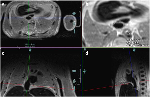
Prenatal Diagnosis 2013, 33, 1–10 © 2013 John Wiley & Sons, Ltd.Fetal heart on post-mortem MRI Figure 2 MPR PMMR image (transverse plane) demonstrating four-chamber view in a 33 week fetus with meningocele. The arrows indicate the points for measurements of (a) cardiac cavities (atria and ventricles) as well as (b) the thickness of ventricular walls and interventricular septum of each ventricle (endocardial-to-endocardial) was determined Statistical analysis just below the atrioventricular valve. The thickness of the Statistical analysis was performed using Excel 2010 (Microsoft ventricular wall (endocardial-to-epicardial surface) and the Corp., Remond Wash., USA). Linear regression analysis was ventricular septum were measured just below the atrioven- used to determine the relationship between the measurements tricular valves. Internal diameters of Ao, PA, DA, and SVC were of different cardiac structures and the GA. assessed on multiple views. The diameter of each vessel was measured in different planes (multiplanar view with three RESULTS orthogonal planes), and the mean of two measurements was Out of 160 pmMRI examinations, 39 matched the inclusion and taken for further analysis (Figure 3). The diameters of ascending exclusion criteria. The median GA was 24 weeks (range 13 w Ao, as well as PA, were assessed above the aortic and pulmonary 2 d–38 w 6 d). Of the 39 fetuses, 17 were female fetuses, and 22 valves at the level of the sinotubular junction. Moreover, the ratio were male fetuses. Thirty-seven fetuses underwent TOP, and of the diameter of the PA to the diameter of the ascending Ao two were intrauterine fetal death. The medical indications for (PA/Ao ratio) was calculated. The diameter of the descending the TOP are displayed in Table 1. Feticide was performed in Ao was taken just below the entrance of the DA. Further, the TOP in 26 cases. A prostaglandin induction of labor was diameter of the DA was obtained at its middle, half the distance performed in all cases. Among the 39 fetuses, four had no between its union with the Ao and the PA (Figure 3). The conventional autopsy (10.3%): three cases were terminated measurement of the SVC was carried out at its entry into the right because of a genetic disorder, and in one, a cytomegalovirus atrium. In most cases, on the transverse view, the IVC appears as fetopathy was present. pmMRI was performed within the an oval-shaped structure slightly compressed antero-posteriorly. interval of 1 h 10 min to 20 h 30 min (median 6 h 30 min) after Assuming that it was more circular in vivo, the diameter of delivery of the fetus. The median isotropic voxel size was the IVC was calculated from the circumference (for the purpose 0.5 mm (range 0.3–0.8 mm). of graphic presentation). The latter was taken on the axial A four-chamber view could be obtained, and the heart and view where the IVC enters into the right atrium (Figure 4b). the thoracic circumferences were measured in all 39 cases In all cases, measurements were performed only when the (Appendix 1). The cardiothoracic ratio was fairly constant image quality was good enough to allow clear definition of throughout gestation with a median of 51% (range 46–57%) the structures. (Figure 5). The widths of the cardiac chambers and the Figure 3 MPR PMMR image on sagittal/oblique (a, b) and transverse (c) planes of 18 weeks fetus with segmental spinal dysgenesis. (a) long-axis view of the right ventricle (RV); (b) long-axis view of the aortic arch; (c) three-vessel view. Arrows indicate the measurements of the diameters of aorta (Ao) and pulmonary artery (PA). Ductus arteriosus (asterix) connects the PA to the Ao. RPA right pulmonary artery; SVC superior vena cava; S spine Prenatal Diagnosis 2013, 33, 1–10 © 2013 John Wiley & Sons, Ltd.
I. Sandaite et al.

Figure 4 MPR PMMR images demonstrates inferior vena cava (IVC) on (a, b) transverse, (c) coronal, and (d) sagittal planes in a 24-week fetus
with genetic disorder. (b) IVC appears as an oval shaped structure slightly compressed antero-posteriorly. (b) measurement of the perimeter of
IVC at her entry into the right atrium

ventricular wall thickness, as well as of the interventricular            A significant correlation between GA and the dimensions of
septum, were successfully assessed in all cases. The diameter of        cardiac structures was found, with linear growth over time
the left atrium ranged between 2.0 and 12.8 mm (median                  (Appendix 3[a,b]).
7.6 mm), being slightly smaller than the right atrium (range
2.0–19.7 mm, median 10.0 mm). Both ventricles were almost               DISCUSSION
equal in size ranging between 1.0 and 12.3 mm, with the median          Herein, we demonstrate the feasibility of visualizing fetal
value of 4.8 mm for the left ventricle and 4.7 mm for the right         cardiac structures on 3-tesla pmMRI. With sufficient
ventricle. The median value of the ventricular wall thickness           resolution, normal cardiac structures can be discriminated
was 3.8 mm for the left ventricle (range 1.2–6.4 mm), 3.5 mm for        and measured as early as 14 weeks of gestation.
the right ventricle (range 1.2–6.2 mm), and 3.1 mm for the                 We report on the assessment of the normal heart on 3-tesla
interventricular septum (range 1.1–5.5 mm). The great arteries          pmMRI in fetuses between 13 and 38 weeks of gestation. In
(Ao and PA) were visualized and measured in 38 cases (97%).             our study, we used 3D data sets with the slice thickness that
The only one where it was not possible was a fetus of 13 weeks          ranged between 0.3 and 0.8 mm. As a result, we obtained
and 2 days of gestation. The DA was successfully assessed in            isometric voxel sizes for MPR in nearly all cases, clearly
32 cases (82%) (Appendix 2). The PA (median 3.9 mm, range               improving the MPR image quality and resulting in better
1.3–6.5 mm) had the largest diameter throughout gestation,              visualization of the cardiac structures. As previous post-
whereas the ascending Ao (median 3.3 mm, range 1.1–5.6 mm)              mortem studies have primarily used low field strengths and
followed the same growth curve but was slightly smaller than            2D data sets,20–22 this could explain the difficulty to assess the
PA, whereas the DA (median 1.3 mm, range 0.6–3.0 mm) had                fetal heart. The use of higher field strength and isotropic 3D
the smallest diameter (Figure 6). The PA/Ao ratio was calculated        sequences optimizes the image quality for multiplanar
in 38 cases and plotted against GA (range 0.85–1.58, median 1.16)       evaluation of the cardiac structures.
(Figure 6). SVC and IVC have been visualized and measured in all           One recent study compared the diagnostic utility of 1.5T, 3T,
39 fetuses. The pulmonary veins were not seen in one fetus of           and 9.4T MRI in fetuses at or under 20 weeks of gestation.18
13 weeks of gestation. The presence of the aortic and pulmonary         The authors concluded that only by using a 9.4T MRI fetal
valves was noted respectively in 32 (82%) and in 33 (84.6%) cases.      cardiac structures could be visualized irrespective of GA. The
On four-chamber view, the insertion of the tricuspid valve and          former study mainly focused on fetuses with congenital heart
the mitral valve was visualized in 26 (66.7%) and 30 (76.9%) cases,     disease. However, to our knowledge, to date, no dedicated
respectively. Five of these fetuses, in which both atrioventricular     studies are available on cardiac evaluation on pmMRI in
valves could not have been assessed on pmMRI, were fetuses              fetuses without cardiac pathologies. Moreover, our study
below 18 weeks of gestation. In the remaining fetuses at different      provides biometric data of cardiac structures on pmMRI over
GA, fluid levels due to post-mortem blood sedimentation                  a wide GA range. Our results show that all cardiac dimensions
were observed.                                                          have a strong linear correlation with GA as previously

Prenatal Diagnosis 2013, 33, 1–10                                                                                 © 2013 John Wiley & Sons, Ltd.
Fetal heart on post-mortem MRI

Table 1 Medical indications for termination for pregnancy in 37                   echocardiography.23,26,27 Prenatal sonographic measurements
fetuses                                                                           are taken at a definite point in the cardiac cycle (end-diastole),
                                                                                  when the chamber size and vessel diameter are at their
 Type of anomaly                                                Number of cases
                                                                                  maximal size, and the ventricular wall dimensions are the
 Intrauterine infection:                                                          thinnest. Moreover, on prenatal ultrasound, some authors
    Cytomegalovirus infection                                          11         measure the diameters of the great vessels at the level of the
    Toxoplasmosis                                                       1         valve,23,26 what also could result in slightly higher values.
 Neural tube defects                                                              Because in some cases on PMMR images, the valve is not
                                                                                  clearly visible, we chose to measure the diameters of Ao and
    Meningocele                                                         2
                                                                                  PA just above the assumed valvular level. Even reference charts
    Encephalocele                                                       1
                                                                                  derived from morphological studies are often unreliable for
    Lumbosacral spina bifida                                             2
                                                                                  in vivo measurements.24,28
 Other nervous system anomalies:                                                     Although individual values for the measurement of different
    Callosal agenesis (one case associated with pes equinus)            2         cardiac structures seem to differ from those reported in the
    Intracranial bleeding                                               1         literature, the C/T ratio was concordant with prenatal
                                                                                  findings,29 about 50%. Wong et al. (2007)30 assessed PA/Ao
    Hydrocephaly (post-hemorrhagic; by third ventricular                2
    obstruction)                                                                  ratio at the level of three-vessel view in a group of 966 fetuses
                                                                                  between 16–24 weeks of gestation, and it was comparable to
    Rhombencephalosynapsis                                              1
                                                                                  the one in our study (1.14 and 1.16, respectively), highlighting
    Megalencephaly polymicrogyria, and hydrocephalus                    1
                                                                                  that although post-mortem changes might slightly alter the
    syndrome
                                                                                  dimensions of cardiac structures, the relationship between
    Spinal dysgenesis                                                   1
                                                                                  the dimensions is maintained.
 Fetal hypokinesia (migration disorder; suspect of valproic             2            Two major strengths of our study are (1) the short time delay
     acid embryopathy)
                                                                                  between delivery and pmMRI and (2) the use of 3D sequences
 Distal arthrogryposis                                                  2
                                                                                  with isovolumetric voxel size on a 3T MRI. This allowed us to
 Cloacal dysgenesis                                                     1         acquire good quality images for MPR and provided us with
 Harlequin ichthyosis                                                   1         an unlimited choice of image orientation to view the cardiac
 Amniotic band syndrome                                                 1         structures in any desirable plane. Moreover, as 3T MR units
                                                                                  have no limitations regarding the size of the fetus and are
 Microphthalmia and ventriculomegaly                                    1
                                                                                  clinically more readily available, we could include in our study
 Genetic disorders:
                                                                                  fetuses from 13 weeks of gestation up to term. As shown by
    Mucoviscidosis (=cystic fibrosis)                                    1         other groups, the use of 9.4-tesla MRI provides an even better
    Unbalanced translocation 5 and 6 (palatoschisis/cleft lip           1         tissue contrast and spatial resolution for evaluating the fetus
    on US);
                                                                                  up to 20 weeks of gestation.17,18 Although the assessment of
    PGRN mutation, known to be associated with                          1         actual diagnostic value of this technique was beyond the aim
     frontotemporal dimension
                                                                                  of this study, we showed that the main cardiac structures can
 PROM at 18 weeks of gestation                                          1         be identified on 3T pmMRI in fetuses beyond 14 weeks.
                                                                                  Moreover, this technique allows examination of the fetal heart
                                                                                  in situ, where connections with other structures are
                                                                                  maintained and can easily be assessed. Furthermore, very
                                                                                  small structures can be assessed.
                                                                                     Our study has several limitations. First, although we
                                                                                  focused on pregnancies terminated for various non-cardiac
                                                                                  abnormalities, they may not be fully representative for a
                                                                                  normal healthy fetal population. Therefore, only fetuses with
                                                                                  anomalies not interfering with the cardiac anatomical
                                                                                  development and function and with documented normal
                                                                                  growth were included. The fetuses in this study group were
                                                                                  not homogeneously distributed throughout gestation. We are
                                                                                  aware that accurate morphometry may prove more difficult
                                                                                  in non-preselected cases or fetuses with cardiac pathology,
Figure 5 Cardiothoracic (C/T) ratio throughout gestation in 39
                                                                                  but the aim of this study was to investigate the ability of
fetuses without cardiac abnormalities
                                                                                  pmMRI to visualize and measure the structures of the normal
                                                                                  fetal heart. Further studies in fetuses with cardiac or chest
described for measures by prenatal ultrasound23 and post-                         abnormalities are necessary to validate the usefulness of the
natally on conventional autopsy.24,25 Interestingly, the                          provided normal reference data. Second, as one of the
absolute values do not match perfectly; they seem to be slightly                  objectives of the study was to provide reference values for
lower than those of reference ranges derived from 2D fetal                        cardiac measurements, we limited our population to cases

Prenatal Diagnosis 2013, 33, 1–10                                                                                        © 2013 John Wiley & Sons, Ltd.
I. Sandaite et al.
Figure 6 Scatter-plots of pulmonary artery (left top image), aorta (right top image), and ductus arteriosus (left bottom image) demonstrating
their growth (diameters measured on MPR 3T PMMR images) throughout gestation in fetuses without cardiac abnormalities. Pulmonary artery/
aorta (PA/Ao) ratio throughout gestation (right bottom image)
with good quality MR images (based on a subjective visual To conclude, cardiac pmMRI evaluation using 3T equipment
assessment). Among all cases that met the other inclusion allows the demonstration of normal cardiac anatomy and
criteria, only three cases had an unacceptable image quality. biometry in fetuses beyond 14 weeks. The presented data could
All three of them had a GA less than 15 weeks. At last, post- serve as references for the detection of cardiac pathology on
mortem changes related to cessation of circulation and post- post-mortem MR images.
mortem muscle contractions might limit the interpretation
and influence the correct biometry on PMMR images. As in
WHAT’S ALREADY KNOWN ABOUT THIS TOPIC?
our case, we experienced difficulties to visualize the cardiac
valves and the continuity of the DA due to blood • Post-mortem fetal magnetic resonance imaging (pmMRI) is a
valuable addition or alternative for conventional autopsy
sedimentation in cardiac chambers. Post-mortem contraction
examination. Studies investigating the normal fetal anatomy on
of the fetal heart muscle might alter cardiac dimensions, pmMRI are sparse.
resulting in smaller ventricle cavities and thicker ventricular
walls. In the total post-mortem population of 160 fetuses,
cardiac contraction was observed on pmMRI in only two cases. WHAT DOES THIS STUDY ADD?
Both of them had a GA of 24 weeks and were not included in
• This study (1) comprehensively describes the methodology of the
the study population, on the basis of the inclusion criteria. assessment of the normal fetal heart on pmMRI and (2) provides
The measurements of the ventricular walls and the ventricular morphometric data helpful for the diagnostic evaluation of cardiac
cavities for these two cases were clear outliers on the presented abnormalities on pmMRI.
cardiac reference graphs.
REFERENCES
1. Burton JL, Underwood J. Clinical, educational, and epidemiological 7. Brodlie M, Laing IA, Keeling JW, et al. Ten years of neonatal autopsies in
value of autopsy. Lancet 2007;369(9571):1471–80. tertiary referral centre: retrospective study. BMJ 2002;324:761–3.
2. Faye-Petersen OM, Guinn DA, Wenstrom KD. Value of perinatal 8. Griffiths PD, Paley MN, Whitby EH. Post-mortem MRI as an adjunct to
autopsy. Obstet Gynecol 1999;94(6):915–20. fetal or neonatal autopsy. Lancet 2005;365(9466):1271–3.
3. Cartlidge PH, Dawson AT, Stewart JH, Vujanic GM. Value and quality of 9. Brookes JA, Hall-Craggs MA, Sams VR, Lees WR. Non-invasive
perinatal and infant postmortem examinations: cohort analysis of 400 perinatal necropsy by magnetic resonance imaging. Lancet 1996;348
consecutive deaths. BMJ 1995;310(6973):155–8. (9035):1139–41.
4. Porter HJ and Keeling JW. Value of perinatal necropsy examination. J 10. Ros PR, Li KC, Vo P, Baer H, Staab EV. Pre-autopsy magnetic resonance
Clin Pathol 1987;40(2):180–4. imaging: initial experience. Magn Reson Imaging 1990;8(3):303–8.
5. Adappa R, Paranjothy S, Roberts Z, Cartlidge PHT. Perinatal and infant 11. Cannie M, Votino C, Moerman P, et al. Acceptance, reliability and
autopsy. Arch Dis Child Fetal Neonatal 2007;Ed 92:F49–50. confidence of diagnosis of fetal and neonatal virtuopsy compared with
6. Khong TY, Tanner AR. Foetal and neonatal autopsy rates and conventional autopsy: a prospective study. Ultrasound Obstet Gynecol
use of tissue for research: the influence of ‘organ retention’ 2012;39(6):659–65. doi: 10.1002/uog.10079. Epub 2012 May 22.
controversy and new consent process. J Paediatr Child Health 12. Breeze AC, Jessop FA, Set PA, et al. Minimally-invasive fetal autopsy
2006;42(6):366–9. using magnetic resonance imaging and percutaneous organ biopsies:
Prenatal Diagnosis 2013, 33, 1–10 © 2013 John Wiley & Sons, Ltd.Fetal heart on post-mortem MRI
clinical value and comparison to conventional autopsy. Ultrasound 22. Breeze AC, Cross JJ, Hackett GA, et al. Use of a confidence scale in
Obstet Gynecol 2011;37(3):317–23. doi: 10.1002/uog.8844. reporting postmortem fetal magnetic resonance imaging. Ultrasound
13. Thayyil S, Chandrasekaran M, Chitty LS, et al. Diagnostic accuracy of post- Obstet Gynecol 2006;28:918–24.
mortem magnetic resonance imaging in fetuses, children and adults: a 23. Shapiro I, Degani S, Leibovitz Z, et al. Fetal cardiac measurements
systematic review. Eur J Radiol 2010;75(1):e142–8. Epub 2009 Nov 11. Review. derived by transvaginal and transabdominal cross-sectional
14. Huisman TA. Magnetic resonance imaging: an alternative to autopsy in echocardiography from 14 weeks of gestation to term. Ultrasound
neonatal death? Semin Neonatol 2004;9(4):347–53. Obstet Gynecol 1998;12:404–18.
15. Griffiths PD, Variend D, Evans M, et al. Postmortem MR imaging of the 24. Ursell PC, Byrne JM, Fears TR, et al. Growth of the great vessels in the
fetal and stillborn central nervous system. AJNR Am J Neuroradiol normal human fetus and in the fetus with cardiac defects. Circulation
2003;24(1):22–7. 1991;84:2028–33.
16. Woodward PJ, Sohaey R, Harris DP, et al. Postmortem fetal MR imaging: 25. Castillo EH, Arteaga-Martínez M, García-Peláez I, et al. Morphometric
comparison with findings at autopsy. AJR Am J Roentgenol 1997;168(1):41–6. study of the human fetal heart. I. Arterial segment. Clin Anat 2005;
17. Thayyil S, Cleary JO, Sebire NJ, et al. Post-mortem examination of human 18(4):260–8.
fetuses: a comparison of whole-body high-field MRI at 9.4 T with 26. Lee W, Riggs T, Amula V, et al. Fetal echocardiography: z-score
conventional MRI and invasive autopsy. Lancet 2009;374(9688):467–75. reference ranges for a large patient population. Ultrasound Obstet
18. Votino C, Jani J, Verhoye M, et al. Postmortem examination of human Gynecol 2010;35(1):28–34.
fetal hearts at or below 20 weeks’ gestation: a comparison of high-field 27. Schneider C, McCrindle BW, Carvalho JS, et al. Development of Z-scores
MRI at 9.4 T with lower-field MRI magnets and stereomicroscopic for fetal cardiac dimensions from echocardiography. Ultrasound Obstet
autopsy. Ultrasound Obstet Gynecol 2012;40:437–44. Gynecol 2005;26(6):599–605.
19. Carvalho JS, Ho SY, Shinebourne EA. Sequential segmental analysis in 28. Alvarez L, Aránega A, Saucedo R, et al. Morphometric data on the
complex fetal cardiac abnormalities: a logical approach to diagnosis. arterial duct in the human fetal heart. Int J Cardiol 1991;31(3):337–44.
Ultrasound Obstet Gynecol 2005;26(2):105–11. 29. Paladini D, Chita SK, Allan LD. Prenatal measurement of
20. Huisman TA, Wisser J, Stallmach T, et al. MR autopsy in fetuses. Fetal cardiothoracic ratio in evaluation of heart disease. Arch Dis Child
Diagn Ther 2002;17(1):58–64. 1990;65:20–3.
21. Alderliesten ME, Peringa J, van der Hulst VP, et al. Perinatal mortality: 30. Wong SF, Ward C, Lee-Tannock A, et al. Pulmonary artery/aorta ratio in
clinical value of postmortem magnetic resonance imaging compared simple screening for fetal outflow tract abnormalities during the second
with autopsy in routine obstetric practice. BJOG 2003;110(4):378–82. trimester. Ultrasound Obstet Gynecol 2007;30(3):275–80.
APPENDIX1
Prenatal Diagnosis 2013, 33, 1–10 © 2013 John Wiley & Sons, Ltd.I. Sandaite et al.
APPENDIX 2
Prenatal Diagnosis 2013, 33, 1–10 © 2013 John Wiley & Sons, Ltd.Fetal heart on post-mortem MRI
APPENDIX 3a
20
20 18
Right atrium width, mm
18
Left atrium width, mm
16
16 14
14 12
12
10
10
8
8
6 6
4 R2 = 0.7612
4 2
R = 0,6219
2 2
0 0
12 14 16 18 20 22 24 26 28 30 32 34 36 38 40 12 14 16 18 20 22 24 26 28 30 32 34 36 38 40
Gestational age, weeks Gestational age, weeks
Right ventricle width, mm
14
14
Left ventricle width, mm
12
12
10 10
8 8
6 6
4 4
2
R = 0,6723 R2 = 0.784
2 2
0 0
12 14 16 18 20 22 24 26 28 30 32 34 36 38 40 12 14 16 18 20 22 24 26 28 30 32 34 36 38 40
Gestational age, weeks Gestational age, weeks
7 7
Right ventricle thickness, mm
Left ventricle thickness, mm
6 6
5 5
4 4
3 3
2
R = 0,7715
2 2 2
R = 0,8184
1 1
0 0
10 12 14 16 18 20 22 24 26 28 30 32 34 36 38 40 12 14 16 18 20 22 24 26 28 30 32 34 36 38 40
Gestational age, weeks Gestational age, weeks
Prenatal Diagnosis 2013, 33, 1–10 © 2013 John Wiley & Sons, Ltd.I. Sandaite et al.
APPENDIX 3b
7 7
Interventricular septum
6 6
Aorta descendens
thickness, mm
diameter, mm
5 5
4 4
3 3
2 2
R2 = 0,7657 R2 = 0,7891
1 1
0 0
12 14 16 18 20 22 24 26 28 30 32 34 36 38 40 10 12 14 16 18 20 22 24 26 28 30 32 34 36 38 40
Gestational age, weeks Gestational age, weeks
12 12
10
Vena cava superior
10
Vena cav ainferior
diameter, mm
diameter, mm
8 8
6 6
4 4
R2 = 0,6897
2 2 2
R = 0,466
0 0
12 14 16 18 20 22 24 26 28 30 32 34 36 38 40 12 14 16 18 20 22 24 26 28 30 32 34 36 38 40
Gestational age, weeks Gestational age, weeks
Prenatal Diagnosis 2013, 33, 1–10 © 2013 John Wiley & Sons, Ltd.You can also read