Implementing Compression Models of OA in Mice and Rats
←
→
Page content transcription
If your browser does not render page correctly, please read the page content below
Implementing Compression Models of OA in Mice and Rats
Organizers
Blaine A. Christiansen, Ph.D., Associate Professor, Orthopaedic Surgery, UC Davis Health
Deva D. Chan, Ph.D., Assistant Professor, Biomedical Engineering, Rensselaer Polytechnic Institute
Abstract
Rodent models are critical tools for osteoarthritis (OA) and post-traumatic osteoarthritis (PTOA) research.
All rodent models of OA are able to initiate joint degradation, but many current models do not reproduce
clinically-relevant injury conditions, due to invasive and non-physiologic injury methods. In addition, many
of these injury methods cause an injury response due to invasive surgical procedures, complicating
studies of the inflammatory response associated with the injury itself. Compression models of OA/PTOA
are able to non-invasively induce knee degeneration using externally-applied mechanical loads, closely
mimicking injury conditions relevant to humans. The goal of this workshop is to present various
compression models of OA/PTOA in mice and rats, and discuss practical/technical considerations of
each of these models.
The three speakers in this workshop have considerable expertise developing and using compression
models of OA. Dr. Blaine Christiansen will present his work using the anterior cruciate ligament rupture
(ACLR) compression model in mice. Dr. Tristan Maerz will present his work translating the ACLR model
to rats. Dr. Marjolein van der Meulen will present her work using the cyclic compression loading model in
mice. Group discussion will focus on implementation of these models, strengths and limitations of each
method, potential sources of variability, and common hurdles encountered with these models.
Significance and Purpose
There is emerging interest in compression models of OA that can non-invasively induce joint
degeneration in mice and rats using externally-applied mechanical loads. These models can be
implemented using a variety of methods, each with its own advantages, limitations, and technical
considerations. Given the recent development and characterization of these new models, the literature
published with these models to date demonstrates expected variability in terms of disease phenotype,
severity, and rate of onset. At present, it remains unclear how much of this variability is due to intrinsic
biological effects (i.e. strain-, age-, or sex-specific effects) versus implementation of the mechanical
loading regimen. The current lack of standardization in the implementation of compression OA models
highlights the need for improved standardization and reproducibility. To this end, the primary purpose of
this workshop is to discuss the development and implementation of compression models of OA in mice
and rats, and present common hurdles encountered with methods. The goal of this workshop is to
provide insight to investigators who are using compression models of OA or are considering these
models for their research.
Educational Need
Compression models are powerful research tools that can provide novel insights into the joint injury
response, even at the earliest time points post-injury. However, these loading models often require
specialized equipment and custom-made fixtures that are not universally available, and not always
consistent between research groups. Therefore, there is a need to educate researchers about key
differences between these models, technical challenges associated with their implementation, and the
careful selection of an appropriate compression model for a particular research question. There is also a
need to define standardization of each model to facilitate greater reproducibility, and multiple aspects of
the mechanical loading regimen warrant discussion (e.g. preloading and preconditioning, loading rate,
targeting a load vs targeting a displacement, using break detection, etc.).
Learning Outcomes
After this workshop, attendees will be familiar with multiple compression models that have been used for
studies of OA in mice and rats, including previous findings using these models and practical
considerations for successful implementation. They will understand the strengths, limitations, and
technical challenges associated with each model, and will have the opportunity to discuss these models
with established experts who have pioneered the use of compression models in rodents. This workshop
will stimulate important discussion regarding model-dependent aspects of the loading regimen that are
anticipated to be contributors to model variability and therefore potential targets for standardization.
Compression-Induced ACL Injury in Mice
                                                                Blaine A. Christiansen, Ph.D.
                                                   Associate Professor, Department of Orthopaedic Surgery
                                                             University of California Davis Health
Introduction:
This injury method uses a single, externally-applied tibial compression overload to non-invasively induce
anterior cruciate ligament (ACL) injury in a mouse knee (Christiansen 2012). In this way, this injury model
closely recapitulates injury conditions relevant to humans, and allows for analysis of inflammation and
other biological processes immediately after injury, since the adaptive response is not confounded by an
injection or invasive surgical procedure.
Description of the Loading Setup:

                               0                                                             0
         Displacement (mm)

                                                                                             -2
                             -0.4
                                                                                             -4
                                                                                                   Force (N)

                             -0.8                                                            -6
                             -1.2                                                            -8
                                                                                             -10
                             -1.6                  Displacement                              -12
                              -2    20   21   22
                                                   Force
                                                    23    24   25   26   27   28   29   30
                                                                                             -14
                                                         Time (s)
The lower leg of the mouse is positioned between two loading platens that are aligned vertically in an
electromagnetic materials testing machine (ElectroForce 3200, TA Instruments, New Castle, DE). A
preload of 1-2 N is applied to hold the lower leg in place. A single compressive load is applied at 1 mm/s
to a target force (~12-15 N) or until ACL injury (noted by a release of force and an audible sound), after
which the load can be manually stopped.
Alternatively, the ACL can be injured by applied a considerably faster load (~200 mm/s) applied to a target
displacement (1.7 mm). This type of loading is more likely to induce midsubstance tear of the ACL, rather
than avulsion from the distal femur (Lockwood 2014).
For this type of injury, mice are anesthetized with 2-3% isoflurane, and are given a single injection of
buprenorphine (0.05 mg/kg SC) at the time of injury. Anesthesia time is typically less than 5 minutes.
Timeline of OA Progression and Common Observations:
• Following non-invasive ACL injury, joint degeneration progresses to severe OA by 6-8 weeks; the
   medial side of the joint is more affected than the lateral side
• ACL deficiency causes the articulation of the femur to move to a more posterior position on the
   proximal tibia; this can lead to dislocation of the posterior horn of the medial meniscus
• Joint inflammation, synovitis, and protease activity are observed primarily during the first 2-3 weeks,
   peaking around 1-3 days post-injury

ORS Preclinical Section Workshop                                                                               2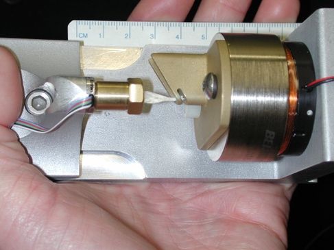
•   Considerable fibrosis and chondrophyte formation occurs within the first 1-2 weeks; mineralized
    osteophytes are by 4 weeks post-injury, these will continue to grow and mineralize at later time points
•   Osteophytes are commonly observed on the medial side of the distal femur, the posterior-medial
    proximal tibia, and the medial meniscus
•   Epiphyseal trabecular bone loss (~20-40% decrease in BV/TV) occurs during the first 2 weeks post-
    injury, primarily due to trabecular thinning; this bone loss is partially recovered at later time points
•   Thinning of the subchondral bone plate also occurs during the first 2 weeks post-injury; at later time
    points, this is reversed and subchondral bone plate thickness is increased in injured joints
•   No significant differences in overall activity level of mice following injury, though gait analysis
    indicates a shorter brake phase and longer swing phase in the injured limb (see below)

Advantages of this Injury Model:
• More clinically relevant to human injury than surgical or injection models
• Recapitulates many key features of PTOA in humans
• Once successfully set up, this method is easy to learn with not much expertise required
• Injury is highly reproducible (though faster loading rates are somewhat less reproducible), with a
   predictable pattern of joint degeneration
• Each injury takes very little time (only takes a few minutes), so can generate high throughput
• Allows for study of inflammation, joint biomechanics, or other processes at early time points, even
   only a few minutes after injury
Limitations of this Injury Model:
• Creates joint instability; OA progression may be largely driven by mechanical factors
• Severe OA develops within 6-8 weeks, model can’t be adjusted to produce a milder injury or slower
   PTOA progression
• Therapies may be “overpowered” by rapid OA progression, especially at later time points, making it
   difficult to evaluate long-term therapeutic benefit
• Expensive (and immobile) materials testing system and custom-made attachments usually required
Key Findings:
• No differences in PTOA progression between males and females (Satkunananthan 2014)
• Tibial compression that causes ACL rupture leads to PTOA, while the same load regimen without
   ACL rupture does not initiate PTOA (Onur 2014)
• Treatment with alendronate following injury inhibits subchondral bone resorption and slows PTOA
   progression (Khorasani 2015)
• Genetic difference in synovial response at early time points after injury in healer (LGXSM-6) vs. non-
   healer (LGXSM-33) mouse strains (Duan 2017)
• Differential gene expression following non-invasive ACL injury (Chang 2017)

ORS Preclinical Section Workshop                                                                          3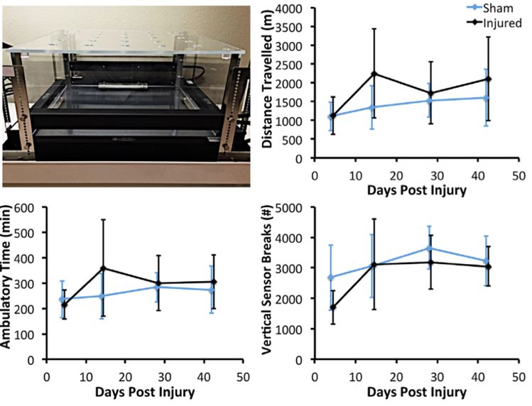
•   Joint range of motion and anterior-posterior joint laxity are increased immediately following injury, but
    are reduced at later time points, correlating with chondro/osteophyte formation (Hsia 2017)
•   Sclerostin (Sost) slows PTOA progression and reduces MMP2/3 protein levels in the joint following
    injury (Chang 2018)
•   MRL/MpJ mice are protected from articular cartilage degeneration following ACL injury, and had
    reduced expression of inflammatory cytokines and catabolic enzymes (Sebastian 2018)
•   Comprehensive analysis of joint inflammation after ACL injury, describing distinct phases of PTOA
    development, involved tissues/cells, and early molecular changes (Gilbert 2018)
•   ACL injury induces metabolomic changes, but inhibition of primary response gene activation with a
    Cdk9 inhibitor can largely prevent these metabolic changes (Haudenschild 2019)
•   Hindlimb unloading following joint injury reduces early protease activity and inhibits osteophyte
    formation and PTOA progression during subsequent reloading (Hsia, Poster #0600)
References
Anderson MJ, Diko S, Baehr LM, Baar K, Bodine SC, Christiansen BA. Contribution of Mechanical Unloading to
   Trabecular Bone Loss Following Non-Invasive Knee Injury in Mice. J Orthop Res. 2016;34(10):1680-7. PMID:
   26826014.
Chang JC, Christiansen BA, Murugesh DK, Sebastian A, Hum NR, Collette NM, Hatsell S, Economides AN,
   Blanchette CD, Loots GG. Sost/Sclerostin Improves Posttraumatic Osteoarthritis and Inhibits Mmp2/3
   Expression after Injury. J Bone Miner Res. 2018;33(6):1105-13. PMID: 29377313.
Chang JC, Sebastian A, Murugesh DK, Hatsell S, Economides AN, Christiansen BA, Loots GG. Global Molecular
   Changes in a Tibial Compression Induced Acl Rupture Model of Post-Traumatic Osteoarthritis. J Orthop Res.
   2017;35(3):474-85. PMID: 27088242.
Christiansen BA, Anderson MJ, Lee CA, Williams JC, Yik JH, Haudenschild DR. Musculoskeletal Changes
   Following Non-Invasive Knee Injury Using a Novel Mouse Model of Post-Traumatic Osteoarthritis. Osteoarthritis
   Cartilage. 2012;20(7):773-82. PMID: 22531459.
Duan X, Rai MF, Holguin N, Silva MJ, Patra D, Liao W, Sandell LJ. Early Changes in the Knee of Healer and Non-
   Healer Mice Following Non-Invasive Mechanical Injury. J Orthop Res. 2017;35(3):524-36. PMID: 27591401.
Gilbert SJ, Bonnet CS, Stadnik P, Duance VC, Mason DJ, Blain EJ. Inflammatory and Degenerative Phases
   Resulting from Anterior Cruciate Rupture in a Non-Invasive Murine Model of Post-Traumatic Osteoarthritis. J
   Orthop Res. 2018. PMID: 29453795.
Haudenschild DR, Carlson AK, Zignego DL, Yik JHN, Hilmer JK, June RK. Inhibition of Early Response Genes
   Prevents Changes in Global Joint Metabolomic Profiles in Mouse Post-Traumatic Osteoarthritis. Osteoarthritis
   Cartilage. 2019;27(3):504-12. PMID: 30572121.
Hsia AW, Anderson MJ, Heffner MA, Lagmay EP, Zavodovskaya R, Christiansen BA. Osteophyte Formation after
   Acl Rupture in Mice Is Associated with Joint Restabilization and Loss of Range of Motion. J Orthop Res.
   2017;35(3):466-73. PMID: 27031945.
Hsia AW, Tarke FD, Shelton TJ, Tjandra PM, Christiansen BA. Comparison of Knee Injury Threshold During Tibial
   Compression Based on Limb Orientation in Mice. J Biomech. 2018;74:220-4. PMID: 29678417.
Khorasani MS, Diko S, Hsia AW, Anderson MJ, Genetos DC, Haudenschild DR, Christiansen BA. Effect of
   Alendronate on Post-Traumatic Osteoarthritis Induced by Anterior Cruciate Ligament Rupture in Mice. Arthritis
   Res Ther. 2015;17:30. PMID: 25888819.
Lockwood KA, Chu BT, Anderson MJ, Haudenschild DR, Christiansen BA. Comparison of Loading Rate-Dependent
   Injury Modes in a Murine Model of Post-Traumatic Osteoarthritis. J Orthop Res. 2014;32(1):79-88. PMID:
   24019199.
Onur TS, Wu R, Chu S, Chang W, Kim HT, Dang AB. Joint Instability and Cartilage Compression in a Mouse Model
   of Posttraumatic Osteoarthritis. J Orthop Res. 2014;32(2):318-23. PMID: 24167068.
Satkunananthan PB, Anderson MJ, De Jesus NM, Haudenschild DR, Ripplinger CM, Christiansen BA. In Vivo
   Fluorescence Reflectance Imaging of Protease Activity in a Mouse Model of Post-Traumatic Osteoarthritis.
   Osteoarthritis Cartilage. 2014;22(10):1461-9. PMID: 25278057.
Sebastian A, Chang JC, Mendez ME, Murugesh DK, Hatsell S, Economides AN, Christiansen BA, Loots GG.
   Comparative Transcriptomics Identifies Novel Genes and Pathways Involved in Post-Traumatic Osteoarthritis
   Development and Progression. International journal of molecular sciences. 2018;19(9). PMID: 30205482.
Wu P, Holguin N, Silva MJ, Fu M, Liao W, Sandell LJ. Early Response of Mouse Joint Tissue to Noninvasive Knee
   Injury Suggests Treatment Targets. Arthritis Rheumatol. 2014;66(5):1256-65. PMID: 24470303.

ORS Preclinical Section Workshop                                                                              4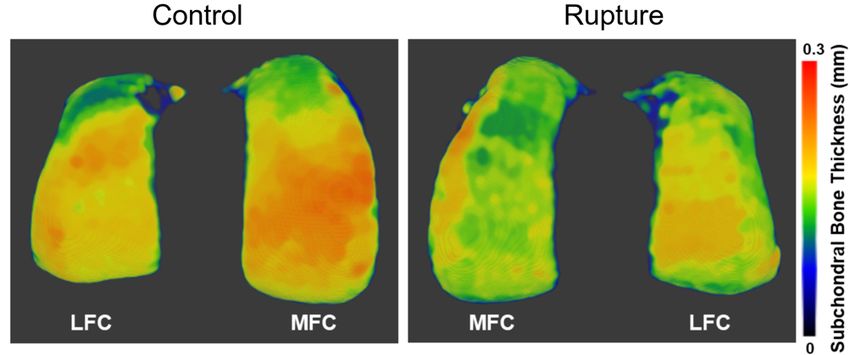
Compression-Induced ACL Injury in Rats
Tristan Maerz, PhD
Assistant Professor, Department of Orthopaedic Surgery
University of Michigan
Introduction:
Noninvasive induction of a complete ACL injury in rats enables studies relevant to post-traumatic
osteoarthritis (PTOA), acute intra-articular inflammation, and traumatic tissue responses in ligaments,
articular cartilage, and subchondral bone. The method in rats is adapted from a similar procedure in
mice, described by Christiansen et al (2012). Due to its larger skeletal size, the rat has some unique
advantages in both the types of studies (e.g. joint injections requiring volumes larger than 10-15 μL) and
types of assessments (e.g. articular cartilage imaging), as compared to similar studies in mice.
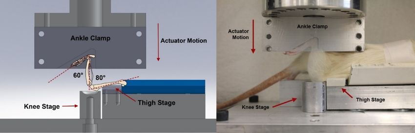
Fixtures, Positioning, Anesthesia & Analgesia:
Considerations
• Appropriately-sized knee stage that restricts medial-lateral motion
o Both “cup” and “trough/channel” shapes known to work
• Elevation of contralateral limb to height of ipsilateral is important. Pelvic rotation induces ipsilateral
femoral internal/external rotation.
• Completely-constrained ankle fixture facilitates injurious motion at the knee
• Quadriceps tendon injury can be avoided/mitigated with padding and/or rounding of edges
• Improper positioning and/or low loading speed may induce dislocation of distal femoral physis
Anesthesia & Analgesia
• Rats can be anesthetized with ketamine/xylazine or, preferably, with 2-2.5% inhaled isoflurane.
• Post-injury analgesia: 5 mg/kg SC Carprofen and/or 0.05 mg/kg SC buprenorphine.
• Total anesthesia time is 3-5 mins with an additional ~5 mins of recovery monitoring.
Loading Sequence and Parameters:
Loading Sequence (as described by Maerz et al Ann Biomed Eng (2015))
• 3N Preload for 10s
• 10x Preconditioning cycles 1N – 5N (0.5 Hz)
ORS Preclinical Section Workshop 5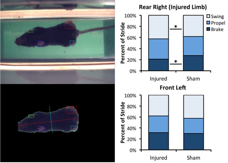
• 1 - 15N Preload Ramp (0.1 mm/s)
• Failure ramp: 3 mm downward displacement (8 mm/s)
Failure Statistics (200 – 200g Lewis rat):
• Failure Load: 60 – 70 N
• Failure Displacement: 2.2 – 2.5 mm
• Stiffness: 40 – 50 N/mm
*** Variations of rat ACL rupture models have been described by Ramme et al. (2016, 2018), Brown et
al. (2019), and Lepley et al. (2020) ***
Timeline of PTOA Progression and Observed Manifestations:
• General time course of PTOA progression (Maerz 2016, Maerz 2018, Maerz 2016):
o Early articular cartilage damage observed by 1-2 weeks (surface fibrillation, acute necrosis)
o Mid-grade OA severity by 4-6 weeks
o Severe OA by 10 weeks
• Sex differences remain largely uncharacterized
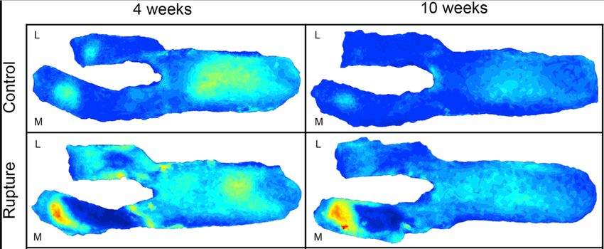
• Medial compartment damage more severe than lateral compartment (below, left) (Maerz 2016)
o Up to 2x OARSI grade on MFC compared to LFC at 4 weeks
• Synovitis observed as early as 3 days and persisting up to 16 weeks post-injury (Brown 2019, Maerz
2017)
• Acute epiphyseal trabecular bone loss (~20-30% decrease in BV/TV) and subchondral bone thinning
(~15-20%) (below, right) is observed within the first 2 weeks. Bone volume deficit of 5-10%
observable up to 10 weeks post-injury (Maerz 2016).
• Bony remodeling pattern in epiphyseal trabecular and subchondral bone is complex, compartment-
dependent (medial vs lateral), and temporally not unidirectional
o Importance of compartment-dependent analysis of microCT data
Advantages of this Injury Model:
• Completely eliminates potential confounding factors due to surgical injury induction
• Larger skeletal size of rats enables larger joint injection volumes (100-150 μL) and more sensitive
analysis of articular cartilage morphology using imaging
• Biomechanical analysis of relative tibiofemoral joint motion demonstrates clinically-relevant injury
mechanism similar to human non-contact injury (Maerz 2015)
• No wound or suture monitoring necessary
• Highly reproducible procedure with minimal training. No known operator-dependent effects.
• Short procedure time and use of isoflurane for anesthesia enables rapid recovery and minimal
anesthesia-dependent effects (i.e. prolonged joint offloading)
Limitations and Disadvantages:
• Materials testing system is required for accurate injury induction.
• Chronic destabilization may exacerbate PTOA onset and severity. While ACL reconstruction
procedures have been described in the rat, these are unlikely to mitigate and may even exacerbate
joint degeneration.
• Higher cost of rats relative to mice
• Uncharacterized but likely significant biomechanical compensation in quadrupeds
ORS Preclinical Section Workshop 6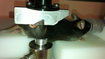
o   Use of contralateral limb as internal control requires careful consideration and comparison to
       healthy, age- and sex-matched limbs
Key Findings
• Injury loading induces anterior tibial translation and internal tibial rotation up to the point of ACL
   failure, at which point rapid external tibial rotation and anterior subluxation occurs (Maerz 2015).
• Rats exhibit less overall osteophyte and chondrophyte formation at similar time points compared to
   mice.
• Low loading rates (1 mm/s) induce avulsion-type injuries (at femoral side) (Maerz 2015).
• Articular cartilage on medial femoral condyle exhibits zones of severe necrosis and thinning (anterior)
   and zones of swelling and hypertrophy (posterior) (Maerz 2016).
• Metabolomic characterization of serum demonstrates injury-induced involvement of multiple
   inflammation and immune-related processes (Maerz 2018).
• Elevated CTX-II and TNF-α levels observed in synovial fluid longitudinally after injury (Brown 2019).
• Reduced bone formation rates and increased bone resorption rates observed by molecular imaging
   and dynamic histomorphometry acutely after injury (~1-2 weeks) (Maerz 2018, Ramme 2016).
• Progressively-increasing MMP13 expression by articular chondrocytes following injury (Ramme
   2016)
References
Brown SB, Hornyak JA, Jungels RR, Shah YY, Yarmola EG, Allen KD, Sharma B. Characterization of Post-
   Traumatic Osteoarthritis in Rats Following Anterior Cruciate Ligament Rupture by Non-Invasive Knee Injury
   (Niki). J Orthop Res. 2019. PMID: 31520482.
Christiansen BA, Anderson MJ, Lee CA, Williams JC, Yik JH, Haudenschild DR. Musculoskeletal Changes
   Following Non-Invasive Knee Injury Using a Novel Mouse Model of Post-Traumatic Osteoarthritis. Osteoarthritis
   Cartilage. 2012;20(7):773-82. PMID: 22531459.
Lepley LK, Davi SM, Butterfield TA, Shahbazmohamadi S. Visualization of Knee Joint Degeneration after Non-
   Invasive Acl Injury in Rats. Cambridge, MA: JoVE; 2020.
Maerz T, Fleischer M, Newton MD, Davidson A, Salisbury M, Altman P, Kurdziel MD, Anderson K, Bedi A, Baker
   KC. Acute Mobilization and Migration of Bone Marrow-Derived Stem Cells Following Anterior Cruciate Ligament
   Rupture. Osteoarthritis Cartilage. 2017;25(8):1335-44. PMID: 28284998.
Maerz T, Kurdziel M, Newton MD, Altman P, Anderson K, Matthew HW, Baker KC. Subchondral and Epiphyseal
   Bone Remodeling Following Surgical Transection and Noninvasive Rupture of the Anterior Cruciate Ligament as
   Models of Post-Traumatic Osteoarthritis. Osteoarthritis Cartilage. 2016;24(4):698-708. PMID: 26620090.
Maerz T, Kurdziel MD, Davidson AA, Baker KC, Anderson K, Matthew HW. Biomechanical Characterization of a
   Model of Noninvasive, Traumatic Anterior Cruciate Ligament Injury in the Rat. Ann Biomed Eng.
   2015;43(10):2467-76. PMID: 25777293.
Maerz T, Newton M, Fleischer M, Hartner S, Gawronski K, Baker K, editors. Anterior Cruciate Ligament Rupture Is
   Associated with Acutely Decreased Bone Anabolism and Increased Bone Catabolism. Orthopaedic Research
   Society; 2018; New Orleans, LA.
Maerz T, Newton MD, Kurdziel MD, Altman P, Anderson K, Matthew HW, Baker KC. Articular Cartilage
   Degeneration Following Anterior Cruciate Ligament Injury: A Comparison of Surgical Transection and
   Noninvasive Rupture as Preclinical Models of Post-Traumatic Osteoarthritis. Osteoarthritis Cartilage.
   2016;24(11):1918-27. PMID: 27349462.
Maerz T, Sherman E, Newton M, Yilmaz A, Kumar P, Graham SF, Baker KC. Metabolomic Serum Profiling after Acl
   Injury in Rats: A Pilot Study Implicating Inflammation and Immune Dysregulation in Post-Traumatic
   Osteoarthritis. J Orthop Res. 2018;36(7):1969-79. PMID: 29315787.
Ramme AJ, Lendhey M, Raya JG, Kirsch T, Kennedy OD. A Novel Rat Model for Subchondral Microdamage in
   Acute Knee Injury: A Potential Mechanism in Post-Traumatic Osteoarthritis. Osteoarthritis Cartilage.
   2016;24(10):1776-85. PMID: 27235904.
Ramme AJ, Lendhey MS, Strauss EJ, Kennedy OD. A Biomechanical Study of Two Distinct Methods of Anterior
   Cruciate Ligament Rupture, and a Novel Surgical Reconstruction Technique, in a Small Animal Model of
   Posttraumatic Osteoarthritis. J Knee Surg. 2018;31(1):43-9. PMID: 28355681.

ORS Preclinical Section Workshop                                                                              7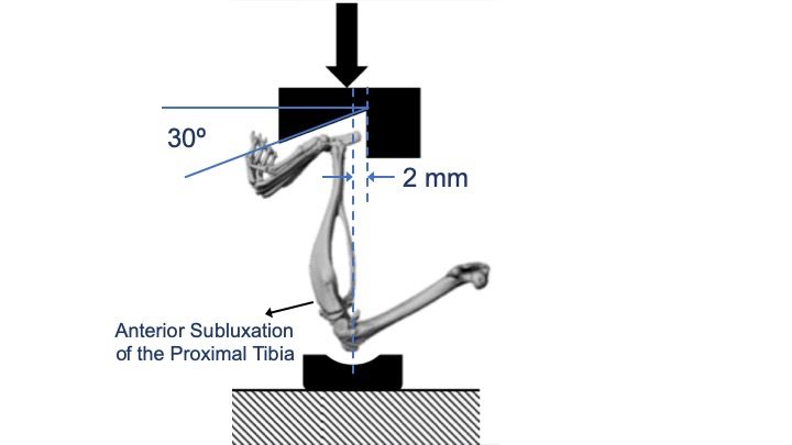
Load-Induced Joint Injury in Mice
                                  Marjolein C. H. van der Meulen, Ph.D.
                  Director, Nancy E & Peter C Meinig School of Biomedical Engineering
                    Professor, Sibley School of Mechanical & Aerospace Engineering
                                            Cornell University
Introduction:
Applying non-invasive compression across the mouse lower limb induces joint damage at the knee with
daily and single loading bouts. No surgery is required in this model. All joint structures remain intact, but
damage is present. The joint changes differ between the two loading approaches. Cartilage damage is
present in both loading regimens; daily loading also induces bone adaptation. This loading approach can
also be combined with other methods that induce OA-like changes in the knee.
Description of the Loading Setup:

The lower leg of the mouse is positioned between two loading platens. We load using a custom-made
device with an electromagnetic actuator and load cell with feedback control using LabView. The loading
can also be performed using an electromagnetic materials testing machine.
Cyclic compression is applied across the lower limb using a set                                        Time (s)
daily protocol. Loading is performed while the mouse is                                     0   0.25     0.5      0.75   1.0
anesthetized. A small preload of 0.5-1 N is applied to hold the                    –0.5 N

lower leg in place. Our waveform and timing are based on
                                                                     Applied Force (N)

osteogenic loading of the metaphysis. We apply loads at 4 Hz,
corresponding to the walking frequency of the mouse at ~4%
strain per second. To achieve this loading rate and frequency,
we insert a dwell between each load-unload cycle. Loads are
determined based on the inducing 1000-1200 µe on the mouse                         –9.0 N

cortex. The load magnitude varies with animal age and strain.
Generally loads are in the 7-10N range.
Timeline of OA Progression and Common Observations:
• With daily in vivo loading cartilage damage is present in both male and female 10-wk and 26-wk-old
   C57BL/6J mice. The changes are mouse-strain specific. Cartilage damage progresses over time. We
   have examined 1, 2 and 6 weeks of loading. The joint tissue effects described below are based
   primarily on male 26-wk-old C57Bl6 mice, unless stated otherwise.
• Bone mass changes are present in the subchondral, epiphyseal and metaphyseal bone. Over time
   daily loading enhances bone mass at the metaphysis. Changes closer to the joint surface are more
   variable and may initially decrease at early time points, followed by increased bone mass with time.
• Osteophytes/enthesophytes form on the medial aspect of the joint at the MCL insertion. These
   structures are cartilaginous after 1 and 2 weeks of loading, and are mineralized at 6 weeks of loading
• Joint fibrosis is present, not classical inflammation. The degree of fibrosis differs substantially across
   different mouse strains

ORS Preclinical Section Workshop                                                                                           8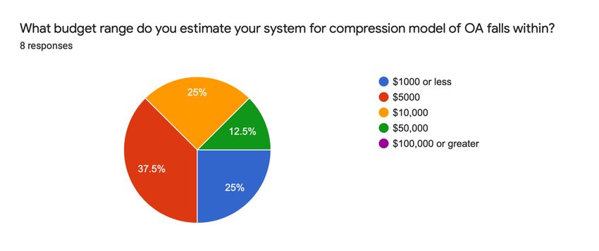
•   Loading induces compression and shear at the joint through AP translation of the tibia and rotation of
    the femur
•   Gait analysis shows no major differences in loaded animals.
•   A single bout of loading produces similar effects to daily loading after 1 and 2 weeks.
Advantages of this Injury Model:
• More clinically relevant to human injury than surgical or injection models
• Recapitulates many key features of OA and PTOA in humans
• The loading is non-invasive, and all joint ligaments remain intact
• Once successfully set up, this method is easy to learn with not much expertise required
• Altering the load magnitude modulates the amount of joint damage generated
• OA development is induced with loading that creates consistent and repeatable joint kinematics
• Implementation can be with small handheld device and laptop computer; however, displacement
   cannot be measured
Limitations of this Injury Model:
• OA progression is more rapid than surgical models such as destabilization of the medial meniscus or
   ACL transection
• Degree of knee flexion may influence joint damage
• Fibrosis is mouse strain-specific, and when severe may produce joint stiffness and likely alters joint
   loading
Key Findings:
• Cartilage and joint damage was similar in 10-week and 26-week old C57BL/6 male mice
• Treatment with alendronate during loading exacerbates or protects from joint injury in a mouse-strain
   specific fashion (Adebayo, et al., 2017)
• Load-induced damage is not additive in mice with spontaneous proteoglycan loss resulting from a
   mutation in collagen XI (Holyoak, et al., 2018)
• Severe obesity increased load-induced cartilage damage (Guss, et al., 2019)
• The gut microbiome may influence cartilage pathology (Guss, et al., 2019)
• Low magnitude loading can be beneficial and inhibit damage developed in response to surgically-
   induced damage following destabilization of the medial meniscus (Holyoak, et al., 2019)
• Increased subchondral bone mass and stiffness modulate cartilage damage
References:
Model Development and Information:
Adebayo OO, Ko FC, Goldring SR, Goldring MB, Wright TM, van der Meulen MC. Kinematics of Meniscal- and Acl-
   Transected Mouse Knees During Controlled Tibial Compressive Loading Captured Using Roentgen
   Stereophotogrammetry. J Orthop Res. 2017;35(2):353-60. PMID: 27153222.
Holyoak DT, Chlebek C, Kim MJ, Wright TM, Otero M, van der Meulen MCH. Low-Level Cyclic Tibial Compression
   Attenuates Early Osteoarthritis Progression after Joint Injury in Mice. Osteoarthritis Cartilage. 2019;27(10):1526-
   36. PMID: 31265883.
Ko FC, Dragomir C, Plumb DA, Goldring SR, Wright TM, Goldring MB, van der Meulen MC. In Vivo Cyclic
   Compression Causes Cartilage Degeneration and Subchondral Bone Changes in Mouse Tibiae. Arthritis
   Rheum. 2013;65(6):1569-78. PMID: 23436303.
Ko FC, Dragomir CL, Plumb DA, Hsia AW, Adebayo OO, Goldring SR, Wright TM, Goldring MB, van der Meulen
   MC. Progressive Cell-Mediated Changes in Articular Cartilage and Bone in Mice Are Initiated by a Single
   Session of Controlled Cyclic Compressive Loading. J Orthop Res. 2016;34(11):1941-9. PMID: 26896841.
Melville KM, Robling AG, van der Meulen MC. In Vivo Axial Loading of the Mouse Tibia. Methods Mol Biol.
   2015;1226:99-115. PMID: 25331046.
Poulet B, Hamilton RW, Shefelbine S, Pitsillides AA. Characterizing a Novel and Adjustable Noninvasive Murine
   Joint Loading Model. Arthritis Rheum. 2011;63(1):137-47. PMID: 20882669.

ORS Preclinical Section Workshop                                                                                    9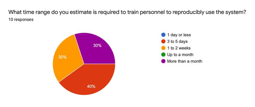
Application of Model:
Adebayo OO, Ko FC, Wan PT, Goldring SR, Goldring MB, Wright TM, van der Meulen MCH. Role of Subchondral
   Bone Properties and Changes in Development of Load-Induced Osteoarthritis in Mice. Osteoarthritis Cartilage.
   2017;25(12):2108-18. PMID: 28919430.
Guss JD, Ziemian SN, Luna M, Sandoval TN, Holyoak DT, Guisado GG, Roubert S, Callahan RL, Brito IL, van der
   Meulen MCH, Goldring SR, Hernandez CJ. The Effects of Metabolic Syndrome, Obesity, and the Gut
   Microbiome on Load-Induced Osteoarthritis. Osteoarthritis and Cartilage. 2019;27(1):129-39. PMID: 30240938.
Holyoak DT, Otero M, Armar NS, Ziemian SN, Otto A, Cullinane D, Wright TM, Goldring SR, Goldring MB, van der
   Meulen MCH. Collagen Xi Mutation Lowers Susceptibility to Load-Induced Cartilage Damage in Mice. J Orthop
   Res. 2018;36(2):711-20. PMID: 28898438.
Holyoak DT, Wheeler TA, van der Meulen MCH, Singh A. Injectable Mechanical Pillows for Attenuation of Load-
   Induced Post-Traumatic Osteoarthritis. Regenerative Biomaterials. 2019;6(4):211-9. PMID: 31402982.

Reviews:
Adebayo OO, Holyoak DT, van der Meulen MCH. Mechanobiological Mechanisms of Load-Induced Osteoarthritis in
  the Mouse Knee. J Biomech Eng. 2019;141(7). PMID: 31209459.
Christiansen BA, Guilak F, Lockwood KA, Olson SA, Pitsillides AA, Sandell LJ, Silva MJ, van der Meulen MC,
  Haudenschild DR. Non-Invasive Mouse Models of Post-Traumatic Osteoarthritis. Osteoarthritis Cartilage.
  2015;23(10):1627-38. PMID: 26003950.

Appendix: Selected Results from the Pre-Workshop Survey by Current Compression Model Users

This handout, the full pre-workshop survey results, and other documents from
this workshop are available for workshop participants to access at
bit.ly/ORS2020_PTOA or by scanning the QR code.

ORS Preclinical Section Workshop                                                                             10
You can also read