Lateral collagen meniscus implant (CMI): techniques and outcomes-a narrative review - Annals of Joint
←
→
Page content transcription
If your browser does not render page correctly, please read the page content below
Review Article
                                                                                                                                          Page 1 of 9

Lateral collagen meniscus implant (CMI): techniques and
outcomes—a narrative review
Alberto Grassi, Gian Andrea Lucidi, Piero Agostinone, Stefano di Paolo, Giacomo dal Fabbro,
Stefano Zaffagnini

II Clinica Ortopedica e Traumatologica, IRCCS Istituto Ortopedico Rizzoli, Bologna, Italy
Contributions: (I) Conception and design: GA Lucidi, A Grassi, S Zaffagnini; (II) Administrative support: S Zaffagnini; (III) Provision of study
materials or patients: GA Lucidi, A Grassi, P Agostinone; (IV) Collection and assembly of data: GA Lucidi, G dal Fabbro; (V) Data analysis and
interpretation: GA Lucidi, A Grassi; (VI) Manuscript writing: All authors; (VII) Final approval of manuscript: All authors.
Correspondence to: Gian Andrea Lucidi. Department of Orthopaedic Surgery II Clinic, Rizzoli Institute, via C. Pupilli 1, 40100, Bologna, Italy.
Email: gianandrea.lucidi@studio.unibo.it.

                 Abstract: The menisci increase the stability of the tibio-femoral joint, distribute axial load, absorb shock,
                 and provide nutrition and lubrification to the knee articular cartilage. Therefore, is it clear the importance of
                 the meniscus on the overall knee function and the need to preserve it during arthroscopic surgery. However,
                 according to many registry databases, meniscectomy is still the most performed meniscus surgery. In a
                 percentage of patients, knee pain and swelling, as well as bone edema on the tibial plateau, could follow
                 meniscus resection; this constellation of symptoms is known as “post-meniscectomy syndrome”. If this
                 syndrome is not promptly managed, a rapid worsening of the symptoms and develop of knee osteoarthritis
                 could be expected. While dealing with such condition, the clinician must perform first an accurate clinical
                 examination and a full radiological evaluation. If the patient is candidate for surgery, the replacement of
                 the resected tissue should be performed: a meniscus allograft should be implanted in case of previous total
                 meniscectomy or a meniscus scaffold if the patients has an history of a previous partial resection. The present
                 article represents a comprehensive review of the literature and aims to discuss basic science, preoperative
                 planning and evaluation, indication, surgical technique, and outcomes of the lateral collagen meniscus
                 implant (CMI), a biologic scaffold aimed at replacing partial meniscal defects.

                 Keywords: Meniscal scaffold; lateral meniscus; collagen meniscus implant (CMI); surgical technique

                 Received: 27 January 2021; Accepted: 23 March 2021.
                 doi: 10.21037/aoj-21-2
                 View this article at: http://dx.doi.org/10.21037/aoj-21-2

Introduction                                                                   role of the menisci on load transfer. A total meniscectomy
                                                                               reduces the contact area by 33 to 50 percent in a fully
The comprehension of the anatomy and biomechanics
                                                                               extended knee (1).
of the joint is mandatory to understand that the clinical                          Moreover, Walker et al. showed that the lateral
decision to perform a meniscectomy or a meniscal suture                        compartment is much more dependent on the meniscus
could have catastrophic consequences at a long-term follow-                    function than the medial one. The lateral meniscus carries
up. Moreover, the medial and the lateral compartment                           a higher percentage of load transfer than the medial
of the knee have different kinematic properties and the                        meniscus because in the medial compartment, a higher load
clinician must take into account the different degree of                       is transferred directly by the exposed cartilage surface (2).
mobility, bony structure and load distribution between these                   This could be explained by the different bony morphology
two compartments.                                                              of the tibiofemoral compartments: in the sagittal plane, in
    Biomechanical studies have demonstrated the essential                      the medial side the convexity of the femoral condyle and the

© Annals of Joint. All rights reserved.                                                          Ann Joint 2021 | http://dx.doi.org/10.21037/aoj-21-2
Page 2 of 9                                                                                                        Annals of Joint, 2021

                              A                                     B

Figure 1 Arthroscopic visualization of the medial (A) and lateral (B) compartment of a 14-year-old patient that underwent 2 years
before partial lateral meniscectomy for a discoid meniscus. Note the evidence of chondrolysis and cartilage degeneration in the lateral
compartment.

concavity of the medial tibial plateau give some degree of              The current treatment options include a meniscus allograft
congruity, even after a meniscectomy. On the contrary, on               transplantation or meniscal scaffold implant based on the
the lateral side, both the convexity of the femoral condyle             degree of the previous meniscectomy (13). There are three
and the lateral tibial plateau make this compartment much               different scaffold types described in the literature: the
more prone to an increase in peak contact pressures after               collagen meniscus implant (CMI) derived from a bovine
meniscus resection (3).                                                 collagen, the Actifit, a polyurethane scaffold, and the 3D
   From a clinical point of view, this aspect has been                  printed scaffolds (14,15). While the latter solution has been
confirmed by worse results reported after lateral                       recently proposed as an experimental treatment and only
meniscectomy rather than the medial meniscectomy at a                   a few case reports are available, CMI and Actifit have been
long-term follow-up (4,5). These findings are guaranteed                widely studied.
if the meniscectomy is performed during adolescence:                       This article aims to provide an overview regarding the
in a prospective 30 years of follow-up study, after medial              current indications, surgical technique, and outcomes of the
meniscectomy about 80% maintained good or excellent                     lateral CMI, the first meniscal scaffold developed.
clinical results, while if lateral meniscectomy was performed              We present the following article in accordance with the
these results dropped to less than 50% (6) (Figure 1).                  Narrative Review reporting checklist (available at http://
   Despite these clear evidences, the number of meniscus                dx.doi.org/10.21037/aoj-21-2).
surgeries performed in Europe and the United States
is increasing every year due to a more active and older
population (7-9). Even if the percentages of meniscus repair            Methods
procedures are increasing, meniscectomy is still the most               A research on PubMed
performed meniscal treatment.
   In fact, most of the meniscal lesions are in the white-              CMI—basic science
white zone or, especially in older patients, could involve              Since the accepted indication for meniscus allograft
degenerated tissue and could have a complex pattern.                    transplantation is a prior total or subtotal meniscectomy,
Moreover, complex dislocated bucket handle tear could be                an allograft is not a solution for the treatment of partial
difficult to reduce and suture (10).                                    meniscus defect. Moreover, the limited availability of
   A subgroup of patients experience pain and worsening of              meniscus allograft, the need for a precise sizing before the
symptoms due to the increased contact stress within the joint           surgery, and the potential disease transmission motivated
over the course of months or years after, a finding referred            some researchers to develop meniscus scaffold for the
to as “post-meniscectomy syndrome” (11) (Figure 2).                     regeneration of partial meniscus defect.
If these patients do not have advanced osteoarthritis (OA),                The CMI (Ivy Sports Medicine, Germany) is a
meniscal replacement surgery should be considered (12).                 porous biologic scaffold. It is composed of about 97% of

© Annals of Joint. All rights reserved.                                                Ann Joint 2021 | http://dx.doi.org/10.21037/aoj-21-2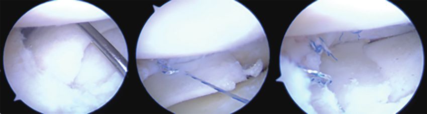
Annals of Joint, 2021                                                                                                       Page 3 of 9

                              A                                   B

                              C                                   D

Figure 2 A 20-year-old professional basketball player with progressive worsening of pain and swelling 3 years after partial lateral
meniscectomy. Preoperative MRI in coronal (A) and sagittal plane (B) and arthroscopic images before (C) and after (D) the lateral CMI
implantation. Note that the lateral meniscus remnant was partially extruded from the joint and the degenerative changes in lateral
compartment (red circles). CMI, collagen meniscus implant.

collagen type I purified from the bovine Achilles tendon.                 Histological analysis performed on both animals and
The remaining portion of the CMI is composed of                       humans also showed the healing of the implant with
glycosaminoglycan (GAG).                                              progressive reabsorption of the collagen fibrils within 6
   The size of the scaffold’s micropores have been                    to 18 months and an increased host tissue invasion and
specifically studied to increase the fibrocartilage maturation        vascularization with the final evidence of meniscus-like
while avoiding pseudo-capsule formation and foreign body              tissue (19,20).
reaction (16). Moreover, the scaffold has been demonstrated
to be safe in terms of cytotoxicity and carcinogenicity.              Indication for surgery
   The first studies on animals were performed by Stone
et al.: they demonstrated that a copolymeric collagen-based           Patients evaluation
scaffold can be constructed that is compatible with meniscal          Obtaining an accurate history of knee trauma and surgical
fibrochondrocyte growth in vitro and in vivo, that does not           procedures is mandatory during the initial evaluation of the
inhibit meniscal regeneration in an immature pig, and that            patients. A history of previous meniscectomy with recurrent
may induce regeneration of the meniscus in the mature                 knee pain, swelling and mechanical symptoms that worsen
dog (17). Moreover, animal models demonstrated no                     during light or moderate physical activity is typically
evidence of cartilage wear or damage and no immunological             consistent with a post-meniscectomy syndrome. The
reaction (18).                                                        evidence of bone marrow edema on MRI, further confirm

© Annals of Joint. All rights reserved.                                              Ann Joint 2021 | http://dx.doi.org/10.21037/aoj-21-2
Page 4 of 9                                                                                                   Annals of Joint, 2021

this diagnosis.                                                           the lateral meniscus;
   Height, weight and body mass index (BMI) should                 (III)  Intact rim (1 mm or greater) over the entire
be recorded because the morbidly obese could have less                    circumference of the involved meniscus (a
symptom relief from meniscal replacement surgery. With                    deficient popliteal hiatus can be allowed because,
the patient standing, the lower limb alignment must be                    the native meniscal rim has low vascularization
evaluated; the presence of varus or valgus thrust should                  and consequently low healing power at this
also be reported. Then, the physical examination must turn                location);
to ligament stability: varo-valgus laxity, as well as anterior     (IV) Diagnosis of Outerbridge grade I to III OA. In
and posterior drawer exam and the pivot shift test, must                  the presence of a focal cartilage defects, a cartilage
be performed. The presence of anterior cruciate ligament                  procedure such as microfracture, mosaicplasty
(ACL) laxity is not a strict contraindication for this type of            or a cartilage scaffold implantation could be
surgery. However, the clinician must consider an associated               performed as an associated surgery;
ACL reconstruction concomitant to the CMI implantation.            (V)    ACL deficiency could be present; however, an
   A full radiological evaluation must be performed before                ACL reconstruction should be performed as a
turning to surgery. It is mandatory to obtain weight-bearing              concomitant procedure;
X-rays of the whole limb to asses joint space narrowing,           (VI) Valgus alignment of the knee of less than 5°.
sign of osteonecrosis, advanced OA and measure the                        If the valgus exceeds this threshold, a distal
mechanical axis. While the evidence of osteonecrosis and                  femur osteotomy (DFO) must be performed as a
OA grade 4 represents a contraindication for surgery, if the              concomitant procedure.
patients present a valgus deformity of 5 degrees or more a         The contraindications for surgery are the following:
corrective osteotomy should be planned concomitantly or a          (I)    Concomitant posterior cruciate ligament
staged procedure performed.                                               insufficiency of the involved knee;
   An MRI is also essential to confirm the indications for         (II)   Diagnosis of Outerbridge grade IV in the affected
surgery. First of all, the percentage of meniscus previously              joint;
resected must be evaluated because the CMI should not              (III) Uncorrected malformations or axial malalignment
be implanted in cases of total or near-total meniscectomy.                greater than 5°;
Attention must then be turned to the anterior and                  (IV) Documented allergy to collagen or chondroitin
posterior horns of the meniscus, which must be intact.                    sulfate of animal origin;
Ligamentous injuries such as ACL tear must be evaluated            (V)    Systemic or local infection;
as well because they could modify the surgical planning.           (VI) History of an anaphylactic reaction;
Finally, the attention should be turned to the cartilage           (VII) Systemic administration of corticosteroid or
status and the presence of subchondral bone marrow. If                    immunosuppressive agents within 30 days of
the latter condition is present, there is also a radiological             surgery;
confirmation of the “post-meniscectomy syndrome”. While            (VIII) Evidence of osteonecrosis in the involved knee;
the cartilage status of the compartment could be useful            (IX) History of rheumatoid arthritis, inflammatory
to establish patients expectations and evidence of bone-                  arthritis, or autoimmune diseases;
bone contact could be an exclusion criteria for the surgery,       (X)    Neurological abnormalities or conditions that
the presence of a focal lesion is not a contraindication for              would preclude the patient’s requirements for the
surgery. Instead, a cartilage restoration procedure (such as              rehabilitation program;
microfracture or mosaicplasty) could be indicated in these         (XI) Pregnancy;
patients.                                                          (XII) A B M I o v e r 3 0 i s c o n s i d e r e d a r e l a t i v e
   The indications for surgery for lateral CMI implantation               contraindication (21).
consist of:
   (I)     Irreparable acute lateral meniscal tears requiring
                                                                 Surgical technique
           partial meniscectomy (acute pattern) or prior
           traumatic or degenerative loss of lateral meniscal    The technique for arthroscopic lateral CMI implantation is
           tissue (chronic pattern) greater than 25%;            clearly described in the literature.
   (II)    Intact anterior and posterior horn attachments of        The patient is positioned supine with a tourniquet

© Annals of Joint. All rights reserved.                                          Ann Joint 2021 | http://dx.doi.org/10.21037/aoj-21-2
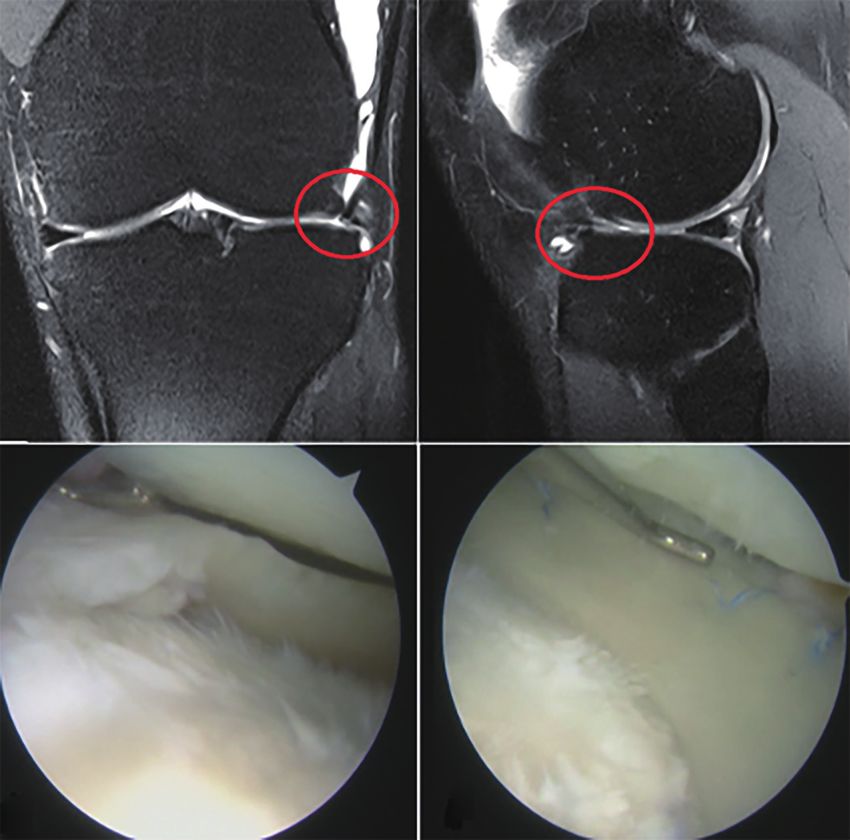
Annals of Joint, 2021                                                                                                           Page 5 of 9

A                                                B                                             C

Figure 3 Surgical pictures showing the surgical preparation of the meniscal defect prior to the CMI implantation. (A) The meniscal lesion
is identified under arthroscopy; the presence of an intact anterior and posterior horn is confirmed as well. (B) The degenerated or unstable
meniscal tissue is removed and the defect area is prepared for the insertion of the CMI. (C) The defect is measured using the specific
instrumentation; the CMI will be sized accordingly. CMI, collagen meniscus implant.

and the knee flexed at 90°. A leg holder is placed 5 cm                  enhanced by making puncture holes in the peripheral rim
proximal to the superior pole of the patella in order to allow           with a Steadman awl.
complete visualization of the medial compartment. The                        Once the defect area is prepared, the defect size should
opening of the lateral compartment could be achieved by                  be measured with the appropriate instrumentation through
flexing the leg in the “figure of four” position. If the lateral         the ipsilateral portal (Figure 3). Note that the lateral CMI
compartment is particularly tight, the second surgeon could              should be oversized by 10% to obtain primary press-fit
provide additional stress by internally rotating the leg and             stability that could help during the following surgical steps.
pushing against the medial part of the proximal tibia.                   If the popliteal hiatus is included in meniscectomized area,
   After the positioning of the patient, standard                        an additional oversizing by 20% could be indicated in order
anteromedial and anterolateral are made. Then a standard                 to prevent an excessive movement of the scaffold into the
diagnostic arthroscopy is performed. The ACL should be                   hiatus during the procedure. Once the correct length of
functionally intact or it should be reconstructed during                 the CMI has been estimated, the scaffold could be trimmed
the same procedure; the degree of cartilage degeneration                 using a scalpel. The CMI is then introduced inside the joint
in each compartment must be assessed as well. In the                     with a delivery clamp through an enlarged lateral portal and
presence of focal cartilage lesions, these could be treated              a blunt probe could be used in order to reach the correct
with standard cartilage procedures such as microfracture,                position.
mosaicplasty or scaffold implants.                                           Once a good press-fit stability has been achieved, the
   Then attention should be turned to the lateral                        scaffold could be sutured to the host meniscus remnant and
compartment. All the unstable and degenerated meniscal                   to the capsule with “all-inside” stitches (Figure 4).
tissue should be debrided to a stable rim, unless an acute                   Please note that suturing should begin at the posterior
irreparable tear or the sequelae of a previous meniscectomy              aspect of the CMI to achieve better visualization of
are noted. This procedure could be done with a basket or a               the position of the scaffold and prevent intraoperative
shaver based on the surgeon preference. It is important to               dislocation. It is also mandatory that the anterior and
ensure that the anterior and the posterior horn must both                posterior ends of the CMI are sutured with horizontal
be present and functionally stable.                                      sutures, while vertical mattress sutures are used throughout
   The area of the CMI implantation must be trimmed to                   the remained CMI every 5–7 mm (21). During the whole
a clean border to accept the scaffold easily; the prepared               procedure, the suture should not be overtightened because
defect size should maintain uniform width of the meniscus                this could damage the CMI structure and lead to vertical
rim and extend into the red/white or red/red zone.                       CMI tear or instability of the implant. Finally, the scaffold
Additionally, the blood supply from the capsule could be                 stability must be tested with a probe.

© Annals of Joint. All rights reserved.                                                  Ann Joint 2021 | http://dx.doi.org/10.21037/aoj-21-2
Page 6 of 9                                                                                                          Annals of Joint, 2021

Figure 4 Surgical pictures showing the last steps of the surgery. (A) The CMI is inserted into the joint with a clamp and released in the
correct position, a blunt probe is used to push the CMI into the defect area and obtain a primary “press-fit” stability. (B) The scaffold is
sutured with an all-inside technique to the capsule and the meniscus remnant. (C) Arthroscopic appearance of the lateral CMI at the end of
the surgical procedure. CMI, collagen meniscus implant.

Rehabilitation protocol                                                  differences were reported at 5 years of follow-up if the
                                                                         CMI was implanted in the acute group as a prophylactic
The following rehabilitation protocol must be applied
                                                                         procedure.
after the surgery to avoid early dislocation or failure of the
                                                                            As already highlighted, the outcomes of the lateral
sutures and the implant. A knee brace is applied, locked
                                                                         CMI have been investigated by a lower number of trials
in full extension and maintained for 6 weeks. Continuous
                                                                         with a shorter follow-up; a recent systematic review that
passive motion (CPM) exercises should be performed 4
                                                                         investigated 396 CMI found that only 10% of them were
times per day, up to 60° for the first 2 weeks and up to 90°
                                                                         implanted in the lateral compartment (23).
for the 2nd to 4th week; complete range of motion (ROM)
                                                                            Hirschmann et al. (24) investigated a series of 67 patients
is permitted at the 6th week. Progressive weight-bearing is              that underwent medial or lateral CMI implantation
allowed 3 weeks after surgery with about 30% of the body                 associated with ACL reconstruction (45%), high tibial
weight. A progressive increase of the weight bearing should              osteotomy (7.5%) or microfracture (4.5%). One year of
be encouraged from the 4th week to reach the total weight-               follow-up demonstrated a marked decrease of pain and an
bearing 6 weeks after the surgery. Muscle-strengthening                  improvement in the Tegner, IKDC and Lysholm score.
should start on the second postoperative day with isometric              Moreover, the results of the medial and the lateral group
exercises; cycling is allowed at the 4th postoperative week.             were comparable.
Full unrestricted activity as tolerated was permitted after                  In a recent multicenter study, Zaffagnini et al. (21)
6 months from surgery (22).                                              reported the outcomes of 43 patients clinically evaluated
                                                                         24±1.9 months after lateral CMI implant. In this study the
Outcomes                                                                 Lysholm score improved from 64.3±18.4 preoperatively to
                                                                         93.2±7.2 at final follow-up. Similarly, the pain experienced
The outcomes after the medial CMI have been extensively                  both during strenuous activity and at rest was significantly
studied since the medial scaffold was made commercially                  reduced. Interestingly, the clinical scores improved from
available about 10 years before the lateral one. For this                6 months after surgery to 12 months of follow-up, thus
reason, the long-term results of the CMI are available only              demonstrating a better knee function after the CMI
for the medial procedure (22). A milestone in the literature is          maturation. At 2 years of follow-up, about 60% of patients
the randomized controlled trial of medial CMI versus medial              reported activity levels similar to their preinjury values and
meniscectomy performed by Rodkey et al. in 2008 (20).                    the satisfaction rate was 95%. Interestingly, the presence of
The authors reported better clinical scores and a lower                  a higher BMI, the need for concomitant procedures, and a
reoperation rate in the group of patients treated because of             chronic injury pattern resulted in reduced outcomes.
a chronic meniscal deficiency. On the contrary, no clinical                 Therefore, this conclusion must be taken into account

© Annals of Joint. All rights reserved.                                                  Ann Joint 2021 | http://dx.doi.org/10.21037/aoj-21-2
Annals of Joint, 2021 Page 7 of 9
Lateral CMI survivorship
100
90
80
Survival probability (%)
70
60
50
40
30
20
10
0
0 2 4 6 8 10 12 14
Time (years)
Figure 5 Kaplan-Maier curve of survivorship of lateral CMI. Failures were considered partial or total CMI removal for any reasons,
meniscus allograft transplantation, unicompartmental knee arthroplasty or total knee arthroplasty. CMI, collagen meniscus implant.
during the patient’s selection and could help set patient higher compared with the pre-operative status, except for
expectations accordingly. Also, the safety of the device was the Tegner score.
confirmed: adverse events of the scaffold leading to CMI
explantation were reported only in 6% of the patients.
Conclusions
In another study, the same author investigated a cohort
of 24 patients that underwent lateral CMI for acute The lateral CMI represents an attractive surgical option
irreparable meniscal tears (7 patients) or for a chronic lateral for treating a “post-meniscectomy syndrome” following a
meniscectomy (17 patients). At a minimum 2 year of follow- partial arthroscopic meniscal resection. In-vitro and in-vivo
up, all the clinical scores investigated significantly improved studies demonstrated the progressive reabsorption of the
from preoperative evaluation to final follow-up. Moreover, CMI and the substitution with a meniscus-like tissue with
the MRI evaluation demonstrated no progression of a potential chondroprotective effect. Satisfactory clinical
cartilage damage; 87.5% of implants were reduced in size, results have been reported in the vast majority of patients at
and in 3 cases (12.5%), they were completely resorbed (25). a short-term follow-up, however, patient selection and the
Grassi et al. (unpublished data) investigated the long- treatment of concomitant knee pathology is mandatory in
term results of the lateral CMI in 19 patients at 12.4 years order to achieve a clinical improvement. Additional long-
of mean follow-up. Their results showed that the scaffold term studies are needed to evaluate possible cartilaginous
provided good long-term results, with a 10-year survival protection and could help to better identify the long-term
rate of 85% and a 14-year survival rate of 64% (Figure 5). benefits and failures of the procedure.
Additionally, 58% of the patients were rated as “good” or
“excellent” according to the Lysholm score. Finally, 78%
Acknowledgments
were satisfied by the procedure, even though a general
decrease of the clinical scores was reported from the Funding: None.
2-year timepoint with respect to the long-term follow-up.
Specifically, the Lysholm score decreased to 82±14 at the
Footnote
final follow-up and the visual analog scale (VAS) for pain
was reduced to 3.1±3.1 points at 12 years. Moreover, only Provenance and Peer Review: This article was commissioned
3/19 patients (16%) reported to be completely without pain. by the editorial office, Annals of Joint for the series “The
Notably, all the average clinical scores were significantly Lateral Meniscus”. The article has undergone external peer
© Annals of Joint. All rights reserved. Ann Joint 2021 | http://dx.doi.org/10.21037/aoj-21-2Page 8 of 9 Annals of Joint, 2021
review. of open total and partial meniscectomy related to the
quantity and site of the meniscus removed. Int Orthop
Reporting Checklist: The authors have completed the 1992;16:122-5.
Narrative Review reporting checklist. Available at http:// 6. McNicholas MJ, Rowley DI, McGurty D, et al. Total
dx.doi.org/10.21037/aoj-21-2 meniscectomy in adolescence. A thirty-year follow-up. J
Bone Joint Surg Br 2000;82:217-21.
Conflicts of Interest: All authors have completed the ICMJE 7. Herzog MM, Marshall SW, Lund JL, et al. Trends in
uniform disclosure form (available at http://dx.doi. Incidence of ACL Reconstruction and Concomitant
org/10.21037/aoj-21-2). The series “The Lateral Meniscus” Procedures Among Commercially Insured Individuals in the
was commissioned by the editorial office without any United States, 2002-2014. Sports Health 2018;10:523-31.
funding or sponsorship. SZ served as the unpaid Guest 8. Jacquet C, Pujol N, Pauly V, et al. Analysis of the trends in
Editor of the series and serves as an unpaid editorial board arthroscopic meniscectomy and meniscus repair procedures
member of Annals of Joint from Mar. 2021 to Feb. 2023. AG in France from 2005 to 2017. Orthop Traumatol Surg Res
served as the unpaid Guest Editor of the series. SZ receives 2019;105:677-82.
the consulting fees from Depuy-Synthes and Smith and 9. Kawata M, Sasabuchi Y, Taketomi S, et al. Annual trends
Nephew, SZ receives payments from Depuy-Synthes and in arthroscopic meniscus surgery: Analysis of a national
Smith and Nephew). The authors have no other conflicts of database in Japan. PloS One 2018;13:e0194854.
interest to declare. 10. Hulet C, Pereira H, Peretti G, et al. editors. Surgery
of the Meniscus. 1st edition. Springer-Verlag Berlin
Ethical Statement: The authors are accountable for all Heidelberg, 2016.
aspects of the work in ensuring that questions related 11. Drobnič M, Ercin E, Gamelas J, et al. Treatment options
to the accuracy or integrity of any part of the work are for the symptomatic post-meniscectomy knee. Knee Surg
appropriately investigated and resolved. Sports Traumatol Arthrosc 2019;27:1817-24.
12. Getgood A, LaPrade RF, Verdonk P, et al. International
Open Access Statement: This is an Open Access article Meniscus Reconstruction Experts Forum (IMREF) 2015
distributed in accordance with the Creative Commons Consensus Statement on the Practice of Meniscal Allograft
Attribution-NonCommercial-NoDerivs 4.0 International Transplantation. Am J Sports Med 2017;45:1195-205.
License (CC BY-NC-ND 4.0), which permits the non- 13. Filardo G, Andriolo L, Kon E, et al. Meniscal scaffolds:
commercial replication and distribution of the article with results and indications. A systematic literature review. Int
the strict proviso that no changes or edits are made and the Orthop 2015;39:35-46.
original work is properly cited (including links to both the 14. Filardo G, Petretta M, Cavallo C, et al. Patient-specific
formal publication through the relevant DOI and the license). meniscus prototype based on 3D bioprinting of human
See: https://creativecommons.org/licenses/by-nc-nd/4.0/. cell-laden scaffold. Bone Joint Res 2019;8:101-6.
15. Ghodbane SA, Brzezinski A, Patel JM, et al. Partial
Meniscus Replacement with a Collagen-Hyaluronan
References
Infused Three-Dimensional Printed Polymeric Scaffold.
1. Zhang AL, Miller SL, Coughlin DG, et al. Tibiofemoral Tissue Eng Part A 2019;25:379-89.
contact pressures in radial tears of the meniscus treated 16. Klompmaker J, Jansen HW, Veth RP, et al. Porous
with all-inside repair, inside-out repair and partial implants for knee joint meniscus reconstruction: a
meniscectomy. Knee 2015;22:400-4. preliminary study on the role of pore sizes in ingrowth and
2. Walker PS, Hajek JV. The load-bearing area in the knee differentiation of fibrocartilage. Clin Mater 1993;14:1-11.
joint. J Biomech 1972;5:581-9. 17. Stone KR, Rodkey WG, Webber R, et al. Meniscal
3. McDermott ID, Amis AA. The consequences of regeneration with copolymeric collagen scaffolds. In vitro
meniscectomy. J Bone Joint Surg Br 2006;88:1549-56. and in vivo studies evaluated clinically, histologically, and
4. Jørgensen U, Sonne-Holm S, Lauridsen F, et al. Long- biochemically. Am J Sports Med 1992;20:104-11.
term follow-up of meniscectomy in athletes. A prospective 18. Stone KR, Steadman JR, Rodkey WG, et al. Regeneration
longitudinal study. J Bone Joint Surg Br 1987;69:80-3. of meniscal cartilage with use of a collagen scaffold.
5. Hede A, Larsen E, Sandberg H. The long term outcome Analysis of preliminary data. J Bone Joint Surg Am
© Annals of Joint. All rights reserved. Ann Joint 2021 | http://dx.doi.org/10.21037/aoj-21-2Annals of Joint, 2021 Page 9 of 9
1997;79:1770-7. a minimum 10-year follow-up study. Am J Sports Med
19. Rodkey WG, Steadman JR, Li ST. A clinical study of 2011;39:977-85.
collagen meniscus implants to restore the injured meniscus. 23. Grassi A, Zaffagnini S, Marcheggiani Muccioli GM, et
Clin Orthop Relat Res 1999;(367 Suppl):S281-92. al. Clinical outcomes and complications of a collagen
20. Rodkey WG, DeHaven KE, Montgomery WH, et al. meniscus implant: a systematic review. Int Orthop
Comparison of the collagen meniscus implant with partial 2014;38:1945-53.
meniscectomy. A prospective randomized trial. J Bone 24. Hirschmann MT, Keller L, Hirschmann A, et al. One-
Joint Surg Am 2008;90:1413-26. year clinical and MR imaging outcome after partial
21. Zaffagnini S, Grassi A, Marcheggiani Muccioli GM, meniscal replacement in stabilized knees using a collagen
et al. Two-Year Clinical Results of Lateral Collagen meniscus implant. Knee Surg Sports Traumatol Arthrosc
Meniscus Implant: A Multicenter Study. Arthroscopy 2013;21:740-7.
2015;31:1269-78. 25. Zaffagnini S, Marcheggiani Muccioli GM, Bulgheroni
22. Zaffagnini S, Marcheggiani Muccioli GM, Lopomo N, et P, et al. Arthroscopic collagen meniscus implantation for
al. Prospective long-term outcomes of the medial collagen partial lateral meniscal defects: a 2-year minimum follow-
meniscus implant versus partial medial meniscectomy: up study. Am J Sports Med 2012;40:2281-8.
doi: 10.21037/aoj-21-2
Cite this article as: Grassi A, Lucidi GA, Agostinone P, di
Paolo S, dal Fabbro G, Zaffagnini S. Lateral collagen meniscus
implant (CMI): techniques and outcomes—a narrative review.
Ann Joint 2021.
© Annals of Joint. All rights reserved. Ann Joint 2021 | http://dx.doi.org/10.21037/aoj-21-2You can also read