Usefulness of the Hybrid RFR-FFR Approach: Results of a Prospective and Multicenter Analysis of Diagnostic Agreement between RFR and FFR-The ...
←
→
Page content transcription
If your browser does not render page correctly, please read the page content below
Hindawi
Journal of Interventional Cardiology
Volume 2021, Article ID 5522707, 8 pages
https://doi.org/10.1155/2021/5522707

Research Article
Usefulness of the Hybrid RFR-FFR Approach: Results of a
Prospective and Multicenter Analysis of Diagnostic
Agreement between RFR and FFR—The RECOPA (REsting
Full-Cycle Ratio Comparation versus Fractional Flow Reserve
(A Prospective Validation)) Study

          Juan Casanova-Sandoval ,1,2 Diego Fernández-Rodrı́guez ,1,2 Imanol Otaegui,3
          Teresa Gil Jiménez,4 Marcos Rodrı́guez-Esteban,5 Kristian Rivera,1,2
          Francisco Torres-Saura,6 Vı́ctor Jiménez Dı́az,7 Raymundo Ocaranza-Sánchez,8
          Vicente Peral Disdier,9 Guillermo Sánchez-Elvira,10 and Fernando Worner1,2
          1
            Hospital Universitari Arnau de Vilanova, Lleida, Spain
          2
            Institut de Recerca Biomédica de Lleida (IRBLleida), Lleida, Spain
          3
            Hospital Universitari Vall d´Hebron, Barcelona, Spain
          4
            Hospital Universitario Clı́nico San Cecilio de Granada, Granada, Spain
          5
            Hospital Universitario Nuestra Señora de Candelaria, Tenerife, Spain
          6
            Hospital Universitario de Vinalopó, Elche, Spain
          7
            Hospital Universitario Alvaro Cunqueiro, Vigo, Spain
          8
            Hospital Universitario Lucus Augusti, Lugo, Spain
          9
            Hospital Son Espases, Palma de Mallorca, Spain
          10
             Complejo Hospitalario de Navarra, Pamplona, Spain

          Correspondence should be addressed to Juan Casanova-Sandoval; jucasanova@yahoo.es

          Received 11 January 2021; Revised 10 March 2021; Accepted 20 March 2021; Published 31 March 2021

          Academic Editor: Andrea Rubboli

          Copyright © 2021 Juan Casanova-Sandoval et al. This is an open access article distributed under the Creative Commons Attribution
          License, which permits unrestricted use, distribution, and reproduction in any medium, provided the original work is properly cited.

          Background. The resting full-cycle ratio (RFR) is a novel resting index which in contrast to the gold standard (fractional flow
          reserve (FFR)) does not require maximum hyperemia induction. The objectives of this study were to evaluate the agreement
          between RFR and FFR with the currently recommended thresholds and to design a hybrid RFR-FFR ischemia detection strategy,
          allowing a reduction of coronary vasodilator use. Materials and Methods. Patients subjected to invasive physiological study in 9
          Spanish centers were prospectively recruited between April 2019 and March 2020. Sensitivity and specificity studies were made to
          assess diagnostic accuracy between the recommended levels of RFR ≤0.89 and FFR ≤0.80 (primary objective) and to determine the
          RFR “grey zone” in order to define a hybrid strategy with FFR affording 95% global agreement compared with FFR alone
          (secondary objective). Results. A total of 380 lesions were evaluated in 311 patients. Significant correlation was observed (R2 � 0.81;
          P < 0.001) between the two techniques, with 79% agreement between RFR ≤ 0.89 and FFR ≤ 0.80 (positive predictive value, 68%,
          and negative predictive value, 80%). The hybrid RFR-FFR strategy, administering only adenosine in the “grey zone” (RFR: 0.86 to
          0.92), exhibited an agreement of over 95% with FFR, with high predictive values (positive predictive value, 91%, and negative
          predictive value, 92%), reducing the need for vasodilators by 58%. Conclusions. Dichotomous agreement between RFR and FFR
          with the recommended thresholds is significant but limited. The adoption of a hybrid RFR-FFR strategy affords very high
          agreement, with minimization of vasodilator use.
2 Journal of Interventional Cardiology
1. Introduction (ii) With culprit lesions in non-ST segment elevation
acute coronary syndrome (NSTEACS)
Fractional flow reserve (FFR) is the coronary resistance (iii) With nonculprit lesions in NSTEACS
index with the greatest body of supporting evidence and is (iv) With nonculprit lesions in ST segment elevation
considered the gold standard in the invasive detection of acute coronary syndrome (STEACS) subjected to
ischemia [1–5]. Nonhyperemic pressure ratios (NHPRs) that second step evaluation
do not require maximum hyperemia induction have grad-
ually been introduced, with the instantaneous wave-free
ratio (iFR) being the most widely used index [6, 7]. 2.1.2. Exclusion Criteria
The resting full-cycle ratio (RFR) is a new NHPR that (1) Allergy to the contrast medium not amenable to
assesses the hemodynamic significance of coronary stenoses, premedication
identifying the lowest distal arterial pressure (Pd)/arterial
(2) Severe bronchial asthma or intolerance to adenosine
pressure (Pa) ratio over the entire cardiac cycle. In contrast
to other NHPRs, its measurements would be independent of (3) Atrioventricular block (≥ second grade)
the morphology of the pressure waves, the electrical signal, (4) Cardiogenic shock
and the phasic variations in microcirculatory resistance (5) Women of child-bearing potential
[8, 9]. Initial validation of the RFR was performed by
(6) Any other medical condition which in the opinion of
Svanerud et al. [8], showing RFR values ≤0.89 to be in good
the investigator could pose patient safety problems
agreement with iFR values ≤0.89, through indirect analysis
or alter the study results
of registries from other studies [10–13]. Recently, Kumar
et al. have again validated these RFR thresholds against iFR
[14]. However, the validation of diagnostic tests without 2.2. Diagnostic Procedure. Following diagnostic coronary
comparison against the gold standard, using data from angiography, an analysis of coronary lesions, in terms of
nonspecifically designed studies and choosing dichotomous percentage of stenosis and length of the lesion by visual
thresholds, may limit assessment of the usefulness of a di- estimation, was performed. Subsequently, a pressure-
agnostic test. guided functional assessment of the coronary lesions was
Therefore, a specifically designed, prospective multi- carried out, first measuring RFR and then FFR, in order to
center study [the RECOPA (REsting full-cycle ratio COm- avoid the interference of coronary vasodilatation on RFR
paration versus fractional flow reserve: a Prospective values. More than one lesion could be evaluated in the
vAlidation) Study] was carried out to directly assess global same patient. The measurements were obtained posi-
agreement of the recommended values of RFR (≤0.89) and ™
tioning the PressureWire X Guidewire 0.014 (Abbott
FFR (≤0.80). We likewise compared the usefulness of a Vascular Inc., Santa Clara, CA, USA) distal to the lesion.
hybrid RFR and FFR guided ischemia detection strategy Adenosine, the most widely used vasodilator, could be
versus a strategy guided by FFR alone in reducing the need administered via both the intravenous and intracoronary
for coronary vasodilators, maintaining high agreement. routes. The recommendations for conduction of invasive
studies are specified more in detail in Supplementary
Material 1.
2. Materials and Methods
2.1. Study Population. In the period between April 2019 and 2.3. Study Variables and Objectives. The RFR values con-
March 2020, in 9 Spanish centers, we prospectively recruited sidered to be positive for ischemia were ≤0.89. With regard
patients with ischemic heart disease referred to the hemo- to the gold standard, the FFR values considered to be
dynamics laboratory for diagnostic coronary angiography, in positive for ischemia were ≤0.80. Based on these values, the
which functional assessment of the coronary lesions was primary study objective was to dichotomously determine the
considered necessary. Patients with both intermediate coro- diagnostic accuracy of RFR ≤ 0.89 against FFR ≤ 0.80.
nary lesions (40%–69%) and severe coronary lesions (≥70%) Due to the inherent variability of the sensitivity and
were included, and invasive pressure-guided physiological specificity values of the selected cut-off points, the secondary
coronary studies were performed to assess RFR and FFR study objective was to determine an interval of RFR values
values. If ad hoc percutaneous coronary intervention was affording high agreement, making it possible to reduce the
proved necessary, the clinical decision was made based on the administration of vasodilators in the context of a hybrid
result corresponding to FFR. RFR-FFR ischemia detection strategy versus a strategy
The eligibility criteria are detailed below. guided by FFR alone.
2.1.1. Inclusion Criteria 2.4. Data Collection and Ethical Considerations. All data
(1) Age ≥ 18 years were compiled on a prospective basis and entered into a
specifically designed database. Each center entered demo-
(2) Coronary lesions amenable to invasive physiological
graphic, clinical, laboratory test, angiographic, and physi-
evaluation in patients
ological data in the database. The study was approved by the
(i) With stable ischemic heart disease Clinical Research Ethics Committee of each center andJournal of Interventional Cardiology 3
abided with the requirements and standards of the Decla- main coronary artery with 1.1%. Most of the lesions were
ration of Helsinki and its subsequent amendments regarding under 12 mm in length.
research studies in humans, as well as with the data pro-
tection regulations applicable in Spain.
3.3. Physiological Characteristics of the Coronary Lesions.
The physiological characteristics of the coronary lesions and
2.5. Statistical Analysis. The statistical analyses were per- final treatment are described in Table 3. Most procedures
formed using the R version 3.4.2 package (R Foundation for were performed with a 6 F guide catheter (96.8%), using
Statistical Computing, Vienna, Austria), with statistical adenosine via the intracoronary (i.c.) route (67.1%). The
significance being considered for p < 0.05. Categorical var- median RFR was 0.91 (range: 0.86–0.95), the median Pd/Pa
iables were reported as numbers and relative frequencies at baseline was 0.93 (0.90–0.96), and the median FFR after
(percentages) and continuous variables as the mean (stan- adenosine administration was 0.84 (0.77–0.89). For the
dard deviation [SD]) or median and range or interquartile recommended cut-off value of RFR (≤0.89), the total pro-
range (IQR), depending on their distribution, which was portion of positive values was 40.0%, versus 35.8% for FFR
assessed using the Kolmogorov–Smirnov test. The data were (cut-off value ≤ 0.80).
evaluated per patient for clinical variables and per lesion for
the angiographic and physiological characteristics. 3.4. Agreement between the Recommended Values of RFR
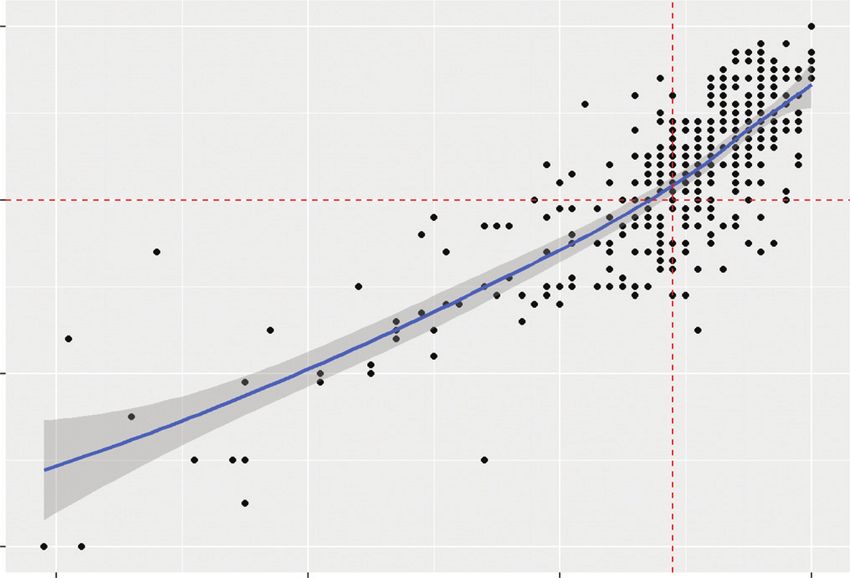
Sensitivity, specificity, and diagnostic accuracy analyses (≤0.89) and FFR (≤0.80). Figure 1 shows the histograms of the
were made in relation to the primary study objective, that is, distribution of the RFR and FFR values. Figure 2 in turn shows
evaluation of the agreement of the recommended values of the distribution of the RFR and FFR values after the admin-
RFR (≤0.89) and FFR (≤0.80). Likewise, we estimated the istration of adenosine for each lesion; a significant correlation
optimum cut-off point of RFR in our sample for an FFR was observed between the two measures (R2 � 0.81; P < 0.001).
value (≤0.80), based on analysis of the receiver operating For the recommended values (RFR ≤ 0.89 and FFR ≤ 0.80), the
characteristic (ROC) curves, with determination of the diagnostic accuracy was 79%. The sensitivity and specificity
Youden index. The coefficient of determination (R2) was also values were 76% and 80%, respectively, with a PPV of 0.68 and
estimated. an NPV of 0.86.
In relation to the secondary study objective, exploratory The overall analysis for sensitivity and specificity and the
analyses were made to define an interval of values capable of stratified analyses according to the route of adenosine ad-
optimizing the positive and negative predictive values (PPV ministration are shown in Supplementary Material 3.
and NPV) of RFR, evaluating the global agreement between
a hybrid RFR-FFR strategy and an exclusive FFR strategy in
determining the functionally significant lesions. A total 3.5. Optimal Cut-Off Point of RFR. The ROC curve (Figure 3)
agreement of at least 95% was considered ideal. presented an area under the curve (AUC) of 0.873
(0.836–0.911; P < 0.001). We found RFR ≤ 0.88 to have the
greatest discriminant power in determining FFR ≤0.80, with
3. Results a Youden index of 0.59. The mentioned value had a diag-
nostic accuracy of 81%, with a sensitivity of 71% and a
A total of 311 patients and 380 lesions were included in the specificity of 87%. The PPV was 0.75 and the NPV was 0.84.
RECOPA Study. A single lesion was examined in most
patients, with 5 being the maximum number of lesions
evaluated in one patient. 3.6. Comparison between the Hybrid RFR-FFR Strategy versus
the Exclusive FFR Strategy. Figure 4 shows the dispersion
plot related to hybrid RFR-FFR strategy compared to FFR-
3.1. Baseline Clinical Characteristics. The baseline clinical only strategy. Two adenosine-free zones (blue) were
characteristics are described in Table 1. The mean patient age established, one below 0.86 and the other above 0.92. The
was 65.4 ± 11.5 years; 19.6% of the patients were women and adenosine zone (grey) lies between RFR values of 0.86 and
35.7% were diabetic. On the other hand, 66.2% of the 0.92, with both included. A PPV of 0.91 was obtained for the
measurements were made in the clinical setting of stable lower limit (RFR < 0.86), with an NPV of 0.92 for the upper
ischemic heart disease and 33.8% in the setting of acute limit (RFR > 0.92). This yielded a global agreement of 95.3%
coronary syndrome. between the two strategies (with only 18 lesions being er-
roneously classified out of a total of 380 lesions: 7 false
positive and 11 false negative). Compared to a FFR-only
3.2. Angiographic Characteristics of the Coronary Lesions. strategy that would require vasodilator administration in all
The angiographic characteristics of the coronary lesions are lesions, the hybrid RFR-FFR strategy would require vaso-
described in Table 2 and Supplementary Material 2. The dilator administration in the “adenosine zone,” representing
mean angiographic stenosis was 58 ± 11%, with a reference only 42% of the measurements (158 lesions). 58% of the
vessel diameter of 3.02 ± 0.53 mm. The main artery evaluated lesions (222 lesions from a total of 380 lesions) in the
was the left anterior descending artery, with 59.2% of the adenosine-free zones would not require vasodilators use (77
measurements, followed by the right coronary artery with lesions [20% of the total] below RFR < 0.86 and 145 lesions
21.6%, the left circumflex artery with 18.2%, and the left [38% of the total] above RFR > 0.92).4 Journal of Interventional Cardiology
Table 1: Baseline clinical characteristics. Table 3: Physiological characteristics and final treatment.
Patients (n � 311) Lesions (n � 380)
Age (years), mean (SD) 65.4 (11.5) Adenosine administration, n (%)
Female gender, n (%) 61 (19.6%) Adenosine i.c. 255 (67.1%)
BMI (kg/m2), mean (SD) 28.2 (4.8) Adenosine e.v. 125 (32.9%)
Hypertension, n (%) 220 (70.7%) Guideline catheter size, n (%)
Dyslipidemia, n (%) 221 (71.1%) 5 French 11 (2.9%)
Diabetes mellitus, n (%) 6 French 368 (96.8%)
No 200 (64.3%) 7 French 1 (0.3%)
Non-insulin-dependent 88 (28.3%) RFR (n), median (IQR) 0.91 (0.86–0.95)
Insulin-dependent 23 (7.4%) RFR results, n (%)
Smoking, n (%) Positive RFR (≤0.89) 152 (40.0%)
Not smoker 126 (40.5%) Negative RFR (>0.89) 228 (60.0%)
Ex-smoker 118 (37.9%) Basal Pd/Pa (n), median (IQR) 0.93 (0.90–0.96)
Current smoker 67 (21.5%) FFR (n), median (IQR) 0.84 (0.77–0.89)
Prior AMI, n (%) 82 (26.4%) FFR results, n (%)
Prior stroke, n (%) 24 (7.7%) Positive FFR (≤0.80) 136 (35.8%)
Atrial fibrillation, n (%) 30 (9.6%) Negative FFR (>0.80) 244 (64.2%)
Peripherical vasculopathy, n (%) 31 (10.0%) Final treatment by lesions, n (%)
COPD, n (%) 21 (6.8%) Medical management 256 (67.3%)
Chronic kidney disease, n (%) 92 (30.6%) PCI-DES 95 (25.0%)
Creatinine (mg/dL), mean (SD) 1.03 (0.61) PCI-BMS 4 (1.1%)
Glomerular filtration rate PCI-DEB 3 (0.8%)
76.0 (31.1)
(mL/min/1.73 m2), mean (SD) CABG 22 (5.8%)
Clinical indication, n (%) RFR, resting full-cycle ratio; IQR, interquartile range; Pd, distal pressure;
Stable ischemic heart disease 206 (66.2%) Pa, aortic pressure; FFR, fractional flow reserve; PCI, percutaneous coro-
NSTEACS culprit lesion 48 (15.4%) nary intervention; DES, drug-eluting stent; BMS, bare metal stent; DEB,
NSTEACS nonculprit lesion 31 (10.0%) drug-eluting balloon; CABG, coronary artery bypass grafting.
STEACS nonculprit lesion 26 (8.4%)
SD, standard deviation; BMI, body mass index; AMI, acute myocardial
infarction; COPD, chronic obstructive pulmonary disease; NSTEACS, non- 4. Discussion
ST segment elevation acute coronary syndrome; STEACS, ST segment el-
evation acute coronary syndrome. The main findings of the present study were the following:
(a) agreement between the recommended dichotomous
values for RFR (≤0.89) and FFR (≤0.80) is limited; and (b) a
hybrid RFR-FFR strategy for ischemia detection would re-
Table 2: Angiographic characteristics.
sult in very high agreement compared with an FFR guided
Patients strategy alone.
(n � 311)
Lesions/patient (n), median
1 (1–5)
(minimum-maximum) 4.1. Design and External Validity of the RECOPA Study.
Lesions (n � 380) The studies mainly conducted by Pijls and De Bruyne on
Affected vessel by syntax, n (%) coronary indices served to establish the physiological bases
Left main artery 4 (1.1%) of FFR, determine the expected cut-off points for the FFR
LAD 225 (59.2%)
technique, and allow its subsequent prospective validation,
LCx 69 (18.2%)
RCA 82 (21.6%) integrating information from different ischemia detection
Percentage of angiographic tests through a prospective multitesting Bayesian approach
58 (11) [1–3]. Thus, the authors were able to establish FFR as the
stenosis (%), mean (SD)
Grouped percentage of gold standard for the detection of myocardial ischemia
angiographic stenosis, n (%) related to coronary stenosis [3].
40–49% 33 (8.7%) Research on NHPRs has grown markedly in recent years,
50–59% 121 (31.8%) particularly in relation to iFR, with evidence of the non-
60–69% 152 (40.0%) inferiority of a revascularization strategy guided by iFR
≥70% 74 (19.5%) versus FFR in two randomized clinical trials [6, 7]. However,
Estimated vessel diameter (mm), mean (SD) 3.02 (0.53)
the lack of a validation strategy similar to FFR in the different
Length of lesion, n (%)
25 mm 44 (11.6%) for the invasive detection of myocardial ischemia and thus
SD, standard deviation; LAD, left anterior descending artery; LCx, left
for the validation of new coronary indices.
circumflex artery; RCA, right coronary artery. Affected segments are shown Initial RFR validation was made retrospectively and
in Supplementary Material 2. indirectly with respect to iFR [8]. The RFR thresholdJournal of Interventional Cardiology 5
50 60
40
40
30
n n
20
20
10
0 0
0.4 0.6 0.8 1.0 0.4 0.6 0.8 1.0
(a) (b)
Figure 1: Histograms of the distribution of the RFR and FFR values. (a) RFR; (b) FFR.
1.0 1.0
0.8
0.8 0.6
Sensitivity
FFR
0.4
0.6 0.2
0.0
0.4 0.0 0.5 1.0
1 – Specificity
0.4 0.6 0.8 1.0
RFR Figure 3: ROC curve of RFR versus FFR ≤0.80. The ROC curve
showed an AUC of 0.873 (0.836–0.911; P < 0.001). The optimal cut-
Figure 2: Distribution of the lesions according the RFR and FFR
off point was RFR ≤0.88, showing a Youden index of 0.59 and the
for the recommended cut-off points (RFR ≤ 0.89 and FFR ≤ 0.80).
following values: diagnostic accuracy: 0.81, sensitivity: 0.71; spec-
The RFR and FFR values showed a significant correlation (R2 � 0.81;
ificity: 0.87; positive predictive value: 0.75; negative predictive
P < 0.001). For the recommended cut-off points of the RFR (≤0.89)
value: 0.84.
and FFR (≤0.80), the following values were obtained: diagnostic
accuracy: 0.79; sensitivity: 0.76; specificity: 0.80; positive predictive
value: 0.68; negative predictive value: 0.86.
restrictive, allowing the inclusion of patients with ischemic
heart disease encompassing the entire spectrum of situations
obtained (≤0.89) was subsequently warranted by Kumar in which invasive physiological studies are used in routine
et al. on a prospective basis likewise against iFR [14]. clinical practice. Furthermore, in contrast to most published
However, the validation of one NHPR against another in- studies [9, 19, 21] that almost exclusively evaluate inter-
stead of against the gold standard (FFR) may limit valida- mediate lesions in stable patients, our study included a
tion. Although Lee et al. [9] reported excellent agreement modest percentage (19.5%) of lesions ≥70%. The lesions
among a number of resting indices (RFR, iFR, and diastolic included in this range of stenoses corresponded mainly to
pressure ratio) and between these indices and FFR, the data patients referred to the hemodynamics laboratory without
were obtained indirectly from information from other prior ischemia evaluation and patients with multivessel
studies [15–18]. To date, only two small studies have eval- disease amenable to surgical revascularization. Also, the
uated the degree of agreement between RFR and FFR RECOPA Study included a nonnegligible proportion of
[19, 20]. For this reason, the RECOPA prospective, multi- lesions in the acute coronary syndrome scenario (NSTEACS
center validation study was specifically designed to assess culprit lesion: 15.4%/NSTEACS nonculprit lesion: 10.0%/
agreement between the recommended cut-off values for RFR STEACS nonculprit lesion: 8.4%). These two facts allow us to
and FFR in different clinical scenarios with a large sample of hypothesize that the range of validation of the technique
patients and thus could provide valuable information. could extend beyond intermediate lesions in stable patients,
We consider the external validity of our results to be which is consistent with the data provided by recent pub-
high. This is because the inclusion criteria used were scantly lications [22, 23].6 Journal of Interventional Cardiology
specificity (80%), PPV (68%), and NPV (86%). Thus, the
Adenosine-free
diagnostic accuracy or agreement obtained was only 79%,
Adenosine
though this is consistent with the findings of Muroya et al.
(42%)
(38%)
Adenosine-free (20%)
0.92
[19] (81%), who also conducted a dichotomous evaluation of
RFR and FFR with the recommended values.
1.0
0.9
4.3. Hybrid RFR-FFR Strategy: Expanding Agreement and
0.8 Simplifying Physiological Assessment of Coronary Lesions.
The above results show that although the correlation be-
FFR
0.7
tween RFR and FFR proved significant and RFR has very
0.6
high discriminating power, the “all-or-nothing” assessment
0.5 of RFR levels may result in deficiencies in the diagnostic
0.4 accuracy of RFR. For this reason, we decided to establish an
adenosine administration “grey zone” to increase the pre-
0.4 0.5 0.6 0.7 0.8 0.9 1.0
RFR
dictive values of the technique. This approach, already de-
scribed for iFR [24], appears to be the most appropriate
Figure 4: Comparison of the hybrid RFR-FFR strategy versus the strategy, for although dichotomous tests are simpler to
exclusive FFR strategy for an agreement of at least 95%. The hybrid interpret and are widely used in standard practice, greater
RFR-FFR strategy only misclassified 18 lesions (7 false positives and precision of the results would be afforded by establishing an
11 false negatives), diminishing the percentage of lesions requiring
intermediate zone in which to assess FFR. Petraco et al. [24]
the administration of vasodilators to 42% (158 lesions from a total
of 380 lesions) compared to the exclusive FFR strategy. Red dots established iFR cut-off points of 0.86 and 0.93, establishing
represent the disagreement and black dots represent the agreement an NPV of 91% for excluding hemodynamic significance of
between the two strategies. Grey dots should be reclassified by the lesions and a PPV of 92% for identifying functionally
administration of vasodilators and determination of FFR. Two significant lesions. In our study, with the established limits of
adenosine-free zones (blue) were established (RFR < 0.86 and 0.86 and 0.92, similar predictive values would be obtained,
RFR < 0.92). The adenosine zone (grey) falls between the RFR with an NPV of 92% and a PPV of 91%. With this hybrid
values of 0.86 and 0.92, with both included. approach, and in accordance with the results of our study,
the use of adenosine would be limited to 42% of the
Another strength of our study was the induction of physiological studies performed, maintaining agreement
coronary vasodilatation with adenosine—which is the drug superior to 95% with respect to use of FFR alone.
most commonly used for this purpose—in contrast to other The evidence shows the benefits of revascularization
RFR validation studies involving other vasodilators (nitro- guided by the invasive assessment of coronary lesions, and
glycerin, nicorandil, etc.) [9, 19, 21]. although its use has increased slightly in recent years, it is still
little employed in clinical practice—with differences from one
country to another [25, 26]. One of the main obstacles to its
4.2. Diagnostic Precision and Agreement between RFR and expanded use in routine practice is the need to administer
FFR. The different diagnostic precision measures are related coronary vasodilators, which is linked to patient discomfort
to different aspects of the diagnostic procedures. Correlation and possible complications [25, 26]. The hybrid RFR-FFR
between RFR and FFR proved significant (R2 � 0.81; strategy (Figure 5) we recommend shows very good agree-
p < 0.001), with values similar to those recently reported by ment with the reference diagnostic technique and moreover
Lee et al. [9] (R2 � 0.82; P < 0.001). The ROC analysis per- allows an important decrease in drug use. It therefore could
formed in our study showed RFR to have very good dis- facilitate generalization of the invasive assessment of coronary
criminating power in measuring ischemia defined as an FFR lesions by simplifying the procedures and making them more
threshold ≤0.80 (AUC: 0.873 [0.836–0.911]; P < 0.001), convenient for the patient and operators.
determining an RFR cut-off point ≤0.88 as being optimal.
The results of our ROC analysis are similar to those reported 4.4. Limitations. A first limitation of our study is the fact
by Svanerud et al. [8] in the pivotal cohort study of RFR that it is a single-country study, which could limit extrap-
versus FFR, showing an AUC of 0.862 (0.834–0.889; olation of the findings to other populations. However, its
P < 0.001), though the mentioned authors found the optimal multicenter nature attenuates this limitation by including
cut-off value to be RFR ≤ 0.89. populations from different geographical areas. Second, the
However, although the determination of strict cut-off study protocol allowed the induction of maximum hyper-
points facilitates clinical decision-making and is supported emia via both the intravenous and the intracoronary routes,
by the revascularization guides, it may result in simplifi- which could influence the study results. However, the use of
cation of the significance of the different indices. On both forms of hyperemia induction is widely supported in
establishing a comparative dichotomous evaluation of the the literature and is considered to be equivalent [27]. Third,
previously recommended thresholds corresponding to RFR the inclusion criteria allowed the enrolment of patients with
(≤0.89) and FFR (≤0.80), as the main objective of our study, stable ischemic heart disease or acute coronary syndrome,
moderate values were obtained for sensitivity (76%), despite the fact that the invasive assessment of coronaryJournal of Interventional Cardiology 7
Hybrid RFR-FFR strategy reserve; RFR, resting full-cycle ratio; Pd, distal pressure; Pa,
aortic pressure. Supplementary Material 2: segments affected
according to syntax classification. RCA, right coronary ar-
RFR
tery; LAD, left anterior descending artery; LCx, left cir-
RFR < 0.86 RFR RFR > 0.92 cumflex artery. Supplementary Material 3: sensitivity and
(0.86 up to 0.92) specificity analyses for overall cohort and stratified by the
route of adenosine administration (RFR (≤0.89) and FFR
(≤0.80)). A: overall cohort (380 lesions). B: intracoronary
adenosine (255 lesions). C: endovenous adenosine (125 le-
FFR sions). (Supplementary Materials)
FFR ≤ 0.80 FFR > 0.80
References
[1] N. H. Pijls, J. A. van Son, R. L. Kirkeeide, B. De Bruyne, and
Treat Defer K. L. Gould, “Experimental basis of determining maximum
coronary, myocardial, and collateral blood flow by pressure
Figure 5: Diagnostic-therapeutic algorithm proposal according to
measurements for assessing functional stenosis severity before
a hybrid RFR-FFR strategy. Initially, the RFR will be performed to
and after percutaneous transluminal coronary angioplasty,”
assess the hemodynamic significance of the coronary lesions to be
Circulation, vol. 87, no. 4, pp. 1354–1367, 1993.
evaluated. If the RFR value is inferior to 0.86, the lesion will be
[2] B. De Bruyne, J. Bartunek, S. U. Sys, and G. R. Heyndrickx,
treated. In case the RFR value is superior to 0.92, the treatment of
“Relation between myocardial fractional flow reserve calcu-
the lesion will be deferred. In the intermediate RFR values (from
lated from coronary pressure measurements and exercise-
0.86 to 0.92, with both included), the significance of the coronary
induced myocardial ischemia,” Circulation, vol. 92, no. 1,
lesion will be reclassified by determining the FFR. Lesions with FFR
pp. 39–46, 1995.
values less than or equal to 0.80 will require treatment. In those
[3] N. H. J. Pijls, B. De Bruyne, K. Peels et al., “Measurement of
lesions with FFR values greater than 0.80, the treatment should be
fractional flow reserve to assess the functional severity of
deferred.
coronary-artery stenoses,” New England Journal of Medicine,
vol. 334, no. 26, pp. 1703–1708, 1996.
lesions is recommended primarily in patients with stable [4] P. A. L. Tonino, B. De Bruyne, N. H. J. Pijls et al., “Fractional
ischemic heart disease. Nevertheless, these techniques are flow reserve versus angiography for guiding percutaneous
also used in routine practice in the context of acute coronary coronary intervention,” New England Journal of Medicine,
vol. 360, no. 3, pp. 213–224, 2009.
syndrome, as supported by the literature [22, 23]. Finally, the
[5] B. De Bruyne, N. H. J. Pijls, B. Kalesan et al., “Fractional flow
percentage of stenoses ≥70% was limited, which reduces reserve-guided PCI versus medical therapy in stable coronary
validity in reference to angiographically significant lesions. disease,” New England Journal of Medicine, vol. 367, no. 11,
However, the inclusion of lesions ≥70% was greater than that pp. 991–1001, 2012.
in previous studies [9, 19, 21]. [6] J. E. Davies, S. Sen, H.-M. Dehbi et al., “Use of the instan-
taneous wave-free ratio or fractional flow reserve in PCI,” New
5. Conclusions England Journal of Medicine, vol. 376, no. 19, pp. 1824–1834,
2017.
The agreement between the currently recommended di- [7] M. Götberg, E. H. Christiansen, I. J. Gudmundsdottir et al.,
chotomous values of FRR and FFR is limited. However, the “Instantaneous wave-free ratio versus fractional flow reserve
adoption of a hybrid RFR-FFR strategy, with an RFR “grey to guide PCI,” New England Journal of Medicine, vol. 376,
zone” in which to determine FFR, allows for improved no. 19, pp. 1813–1823, 2017.
agreement between the two strategies, thus reducing the [8] J. Svanerud, J.-M. Ahn, A. Jeremias et al., “Validation of a
need for coronary vasodilators. novel non-hyperaemic index of coronary artery stenosis se-
verity: the Resting Full-cycle Ratio (VALIDATE RFR) study,”
EuroIntervention, vol. 14, no. 7, pp. 806–814, 2018.
Data Availability [9] J. M. Lee, K. H. Choi, J. Park et al., “Physiological and clinical
assessment of resting physiological indexes,” Circulation,
The original database can be made available from the cor- vol. 139, no. 7, pp. 889–900, 2019.
responding author upon request. [10] C. Berry, M. van ’t Veer, N. Witt et al., “VERIFY (VERification
of instantaneous wave-free ratio and fractional flow reserve
Conflicts of Interest for the assessment of coronary artery stenosis severity in
EverydaY practice),” Journal of the American College of
The authors have no conflicts of interest regarding this Cardiology, vol. 61, no. 13, pp. 1421–1427, 2013.
paper. [11] N. P. Johnson, A. Jeremias, F. M. Zimmermann et al.,
“Continuum of vasodilator stress from rest to contrast me-
Supplementary Materials dium to adenosine hyperemia for fractional flow reserve
assessment,” JACC: Cardiovascular Interventions, vol. 9, no. 8,
Supplementary Material 1: general recommendations for pp. 757–767, 2016.
FFR and RFR measurements (modified from Achenbach [12] B. Hennigan, K. G. Oldroyd, C. Berry et al., “Discordance
et al. [28] and Svanerud et al. [8]). FFR, fractional flow between resting and hyperemic indices of coronary stenosis8 Journal of Interventional Cardiology
severity: the VERIFY 2 study (A comparative study of resting [25] M. Götberg, C. M. Cook, S. Sen, S. Nijjer, J. Escaned, and
coronary pressure gradient, instantaneous wave-free ratio and J. E. Davies, “The evolving future of instantaneous wave-free
fractional flow reserve in an unselected population referred ratio and fractional flow reserve,” Journal of the American
for invasive angiography),” Catheterization and Cardiovas- College of Cardiology, vol. 70, no. 11, pp. 1379–1402, 2017.
cular Interventions, vol. 9, Article ID e004016, 2016. [26] R. V. Parikh, G. Liu, M. E. Plomondon et al., “Utilization and
[13] J.-M. Ahn, D.-W. Park, E.-S. Shin et al., “Fractional flow outcomes of measuring fractional flow reserve in patients with
reserve and cardiac events in coronary artery disease,” Cir- stable ischemic heart disease,” Journal of the American College
culation, vol. 135, no. 23, pp. 2241–2251, 2017. of Cardiology, vol. 75, no. 4, pp. 409–419, 2020.
[14] G. Kumar, R. Desai, A. Gore et al., “Real world validation of [27] G. W. M. Wijntjens, E. L. van Uffelen, M. Echavarrı́a-Pinto
the nonhyperemic index of coronary artery stenosis severity- et al., “Individual lesion-level meta-analysis comparing var-
Resting full-cycle ratio-RE-VALIDATE,” Catheterization and ious doses of intracoronary bolus injection of adenosine with
Cardiovascular Interventions, vol. 96, no. 1, 2019. intravenous administration of adenosine for fractional flow
[15] J. M. Lee, B.-K. Koo, E.-S. Shin et al., “Clinical implications of reserve assessment,” Circulation: Cardiovascular Interven-
three-vessel fractional flow reserve measurement in patients tions, vol. 13, Article ID e007893, 2020.
with coronary artery disease,” European Heart Journal, [28] S. Achenbach, T. Rudolph, J. Rieber et al., “Performing and
vol. 39, no. 11, pp. 945–951, 2018. interpreting fractional flow reserve measurements in clinical
[16] J. M. Lee, C. H. Kim, B.-K. Koo et al., “Integrated myocardial practice: an expert consensus document,” Interventional
perfusion imaging diagnostics improve detection of func- Cardiology Review, vol. 12, no. 2, pp. 97–109, 2017.
tionally significant coronary artery stenosis by 13 N-ammonia
positron emission tomography,” Circulation: Cardiovascular
Imaging, vol. 9, no. 9, Article ID e004768, 2016.
[17] D. Hwang, K.-H. Jeon, J. M. Lee et al., “Diagnostic perfor-
mance of resting and hyperemic invasive physiological indices
to define myocardial ischemia,” JACC: Cardiovascular In-
terventions, vol. 10, no. 8, pp. 751–760, 2017.
[18] J. M. Lee, D. Hwang, J. Park et al., “Exploring coronary
circulatory response to stenosis and its association with in-
vasive physiologic indexes using absolute myocardial blood
flow and coronary pressure,” Circulation, vol. 136, no. 19,
pp. 1798–1808, 2017.
[19] T. Muroya, H. Kawano, S. Hata et al., “Relationship between
resting full-cycle ratio and fractional flow reserve in assess-
ments of coronary stenosis severity,” Catheterization and
Cardiovascular Interventions, vol. 96, no. 6, 2020.
[20] C. Cortés, F. Rivero, E. Gutiérrez-Ibañes, Á. Aparisi, J. A. San
Román, and I. J. Amat-Santos, “Validación prospectiva y
comparación de los nuevos ı́ndices de evaluación de las
estenosis coronarias: resting full-cycle y quantitative flow
ratio,” Revista Española de Cardiologı́a, vol. 74, no. 1, p. 94,
2021.
[21] Y. Kawase, H. Oromi, T. Tanigaki et al., “In vivo validation of
resting full-cycle and diastolic pressure ratio: simultaneous
measurement with instantaneous wave-free ratio,” Cardio-
vascular Intervention and Therapeutics, vol. 36, no. 1,
pp. 74–80, 2020.
[22] M. Tebaldi, S. Biscaglia, A. Erriquez et al., “Comparison of
quantitative flow ratio, Pd/Pa and diastolic hyperemia-free
ratio versus fractional flow reserve in non-culprit lesion of
patients with non ST-segment elevation myocardial infarc-
tion,” Catheterization and Cardiovascular Interventions, 2020.
[23] E. Van Belle, S. B. Baptista, L. Raposo et al., “Impact of routine
fractional flow reserve on management decision and 1-year
clinical outcome of patients with acute coronary syndromes:
PRIME-FFR (insights from the POST-IT [Portuguese study
on the evaluation of FFR-guided treatment of coronary dis-
ease] and R3F [French FFRRegistry] integrated multicenter
registries—implementation of FFR [fractional flow reserve] in
routine practice),” Circulation: Cardiovascular Interventions,
vol. 10, Article ID e004296, 2017.
[24] R. Petraco, J. J. Park, S. Sen et al., “Hybrid iFR-FFR decision-
making strategy: implications for enhancing universal
adoption of physiology-guided coronary revascularisation,”
Eurointervention, vol. 8, no. 10, pp. 1157–1165, 2013.You can also read