Hip Pain in Adults: Evaluation and Differential Diagnosis - BINASSS
←
→
Page content transcription
If your browser does not render page correctly, please read the page content below
Hip Pain in Adults:Evaluation
and Differential Diagnosis
Rachel Chamberlain, MD, CAQSM, University of New Mexico, Albuquerque, New Mexico
Adults commonly present to their family physicians with hip pain, and diagnosing the cause is important for prescrib-
ing effective therapy. Hip pain is usually located anteriorly, laterally, or posteriorly. Anterior hip pain includes referred
pain from intra-abdominal or intrapelvic causes;extra-articular etiologies, such as hip flexor injuries;and intra-articular
etiologies. Intra-articular pain is often caused by a labral tear or femoroacetabular impingement in younger adults or
osteoarthritis in older adults. Lateral hip pain is most commonly caused by greater trochanteric pain syndrome, which
includes gluteus medius tendinopathy or tear, bursitis, and iliotibial band friction. Posterior hip pain includes referred pain
such as lumbar spinal pathology, deep gluteal syndrome with sciatic nerve entrapment, ischiofemoral impingement, and
hamstring tendinopathy. In addition to the history and physical examination, radiography, ultrasonography, or magnetic
resonance imaging may be needed for a definitive diagnosis. Radiography of the hip and pelvis should be the initial imaging
test. Ultrasound-guided anesthetic injections can aid in the diagnosis of an intra-articular cause of pain. Because femoro-
acetabular impingement, labral tears, and gluteus medius tendon tears typically have good surgical outcomes, advanced
imaging and/or early referral may improve patient outcomes. (Am Fam Physician. 2021;103(2):81-89. Copyright © 2021
American Academy of Family Physicians.)
Hip pain is common in adults of all ages and (Table 1).2-20 Diagnosing the cause of hip pain is
activity levels. In nonelite adult soccer players, important for prescribing effective therapy.
hip and groin injuries represent 28% to 45% of all The history should include personal history
injuries in women and 49% to 55% in men.1 The of developmental hip dysplasia, slipped capital
prevalence of the cam deformity (deformity of the femoral epiphysis, sports activities, and injuries;
femoral head) is 41% in nonelite male soccer play- family history of hip problems;and the location
ers and 17% in male nonathletes.2 In adults older and quality of pain, aggravating and alleviating
than 45 years, 6.7% to 9.7% have osteoarthritis factors, and mechanical symptoms.4,8 Physical
of the hip, and one in four adults will develop examination should include gait analysis with
symptomatic hip osteoarthritis in their lifetime.3 particular attention to antalgic or Trendelenburg
In the United States in 2009, hip replacements gait, evaluation of the range of motion in the hip
accounted for $13.7 billion in health care costs.3 joint and associated pain, strength testing of the
muscles overlying the hip joint, palpation of the
Approach to Evaluation painful area, and special tests as indicated.
Hip pain is often localized to one of three loca- If imaging is performed in the evaluation of a
tions:anterior, lateral, or posterior (Figure 14). patient with undifferentiated chronic hip pain,
A focused history and physical examination standing anteroposterior hip and pelvic radiog-
can help differentiate the causes of hip pain raphy is typically the initial imaging study.4,21
Magnetic resonance imaging (MRI) or ultra-
sonography may be helpful in the diagnosis,
CME This clinical content conforms to AAFP
depending on history and physical examination
criteria for CME. See CME Quiz on page 79.
findings.21-23
Author disclosure: No relevant financial
affiliations.
Anterior Hip Pain
Patient information: A handout on this
topic is available at https://w ww.aafp.org/
Intra-articular hip pain predominately presents
afp/2021/0101/p81-s1.html. anteriorly.4,5 In young adults, anterior hip or groin
pain that is aggravated by hip flexion or rotation
Downloaded
January 15,from
2021the◆ American Family
Volume 103, Physician
Number 2 website at www.aafp.org/afp. 2021 American Academy of Family Physicians.
Copyright ©
www.aafp.org/afp American ForFamily Physician
the private, 81
noncommer-
cial use of one individual user of the website. All other rights reserved. Contact copyrights@aafp.org for copyright questions and/or permission requests.
Descargado para Anonymous User (n/a) en National Library of Health and Social Security de ClinicalKey.es por Elsevier en febrero 24, 2021.
Para uso personal exclusivamente. No se permiten otros usos sin autorización. Copyright ©2021. Elsevier Inc. Todos los derechos reservados.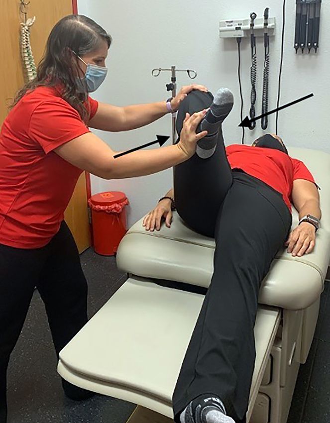
HIP PAIN IN ADULTS
SORT:KEY RECOMMENDATIONS FOR PRACTICE
Evidence
Clinical recommendation rating Comments
If imaging is performed in the evaluation of a patient with undiffer- C Expert opinion and
entiated chronic hip pain, standing anteroposterior hip and pelvic consensus guidelines
radiographs should be the first choice.4,21
For patients with anterior hip pain and history suggestive of a labral tear, C Expert opinion and
stress fracture of the femoral neck, or early avascular necrosis, magnetic reviews of prospective
resonance imaging should be performed for accurate diagnosis.5,11,12,21-23 and randomized trials
For intra-articular pain, ultrasound-guided anesthetic injection of the hip C Clinical review and
may be diagnostic, and corticosteroid injection may be therapeutic.30 expert opinion
For patients with greater trochanteric pain syndrome not responding to C Clinical reviews and
conservative therapy, ultrasonography or magnetic resonance imaging expert opinion
should be considered to evaluate for gluteus medius tendon tears.15,16,21
A = consistent, good-quality patient-oriented evidence;B = inconsistent or limited-quality patient-oriented evidence;
C = consensus, disease-oriented evidence, usual practice, expert opinion, or case series. For information about the SORT
evidence rating system, go to .
FIGURE 1
A B
Posterior
and buttock
Lateral
Lateral
Anterior
and groin
Localization of hip pain. (A) Anterior view. (B) Posterior view.
Illustration by Todd Buck
Reprinted with permission from Wilson JJ, Furukawa M. Evaluation of the patient with hip pain. Am Fam Physician.
2014;89(1):27-34.
warrants evaluation for intra-articular patholo- problems can present as anterior hip pain, the
gies. Hip flexor strains, tears, and avulsion frac- abdomen should be examined for gastrointesti-
tures can cause anterior hip pain, with the patient nal causes of pain, such as a mass;appendicitis;
history often including a sports-related or trau- hernia;or pain originating in the bladder (e.g.,
matic incident consistent with a flexor injury.5 from a mass) or the female reproductive system
Because referred pain from intra-abdominal (e.g., from ovarian cysts).4,6
82 American Family Physician www.aafp.org/afp Volume 103, Number 2 ◆ January 15, 2021
Descargado para Anonymous User (n/a) en National Library of Health and Social Security de ClinicalKey.es por Elsevier en febrero 24, 2021.
Para uso personal exclusivamente. No se permiten otros usos sin autorización. Copyright ©2021. Elsevier Inc. Todos los derechos reservados.
HIP PAIN IN ADULTS
TABLE 1
Differential Diagnosis of Hip Pain
Type of pain History Physical examination
Anterior
Referred
Intra-abdominal or Pain associated with urinary or bowel symptoms, Abdominal and/or pelvic examination
intrapelvic 4-6 cyclic pain associated with menses
Extra-articular
Flexor tendon5,6 Overuse activities, acute strain or injury with hip Pain over the hip bony prominence, anterior supe-
flexion activities rior iliac spine, anterior inferior iliac spine, or pubic
symphysis;pain with hip flexion strength testing
Intra-articular
Femoroacetabular Young, athletic patient;gradual onset;pain with Positive FADDIR and FABER test results
impingement 2,5,7,8 hip range of motion;history of slipped capital
femoral epiphysis or developmental dysplasia
Labral tear5,9 Young, athletic patient;acute injury (vs. gradual Positive FADDIR and FABER test results
onset);pain with hip range of motion;mechanical
symptoms
Femoral neck stress Overuse/overtraining, energy imbalance in Antalgic gait, pain with range of motion and
fracture5,10 athletes ambulating
Avascular necrosis11,12 Middle or older age, smoking, alcohol use, sys- Antalgic gait, pain with range of motion, limited
temic corticosteroid use, hemoglobinopathies, range of motion
chemotherapy, metabolic syndrome, and obesity
Osteoarthritis3,4,7,13 Older age, gradual onset, pain with sitting or Antalgic gait, pain with flexion and internal and
ambulating for long periods external rotation, limited range of motion
Hip fracture4,14 Older age, osteoporosis, fall/trauma Inability to walk on the affected limb;shortened,
externally rotated, abducted leg
Lateral
Greater trochanteric pain No injury, middle age, female sex, overweight, Tenderness to palpation over the lateral hip/
syndrome, including bursitis, pain with sleeping on affected hip, pain aggra- greater trochanter, Trendelenburg gait or positive
gluteus medius tendinopathy vated by physical activity or sitting for long Trendelenburg test, positive resisted external
or tear, external snapping, or periods derotation test
iliotibial band friction7,15,16
Posterior
Referred pain
Intra-abdominal or Pain associated with urinary or bowel symptoms, Abdominal and/or pelvic examination
intrapelvic4,17 cyclic pain associated with menses
Deep gluteal syndrome17,18 Deep buttock pain;no injury;worse with sitting, Seated piriformis stretch test
especially in a car;sciatica (burning pain shooting
down the leg)
Ischiofemoral Gradual onset of deep buttock pain that worsens Long-stride walking test
impingement19 with activities requiring a long stride (e.g., running)
Lumbar spine or muscle4,17 Pain in the low back (above L5) and hip/buttock, Tenderness over the lumbar spine or lumbar mus-
history of lumbar spinal problems culature above L5
Sacroiliac joint pain17 No history of lumbar spinal issues Tenderness over the sacroiliac joint, no tenderness
above L5
Proximal hamstring tendi- Overuse injury with hip extension activities (vs. Tenderness to palpation over the ischial tuberosity,
nopathy or tear20 acute injury with forceful hip extension) pain with hamstring strength testing;acute tears
cause ecchymosis of the posterior thigh
FABER = flexion abduction external rotation;FADDIR = flexion adduction internal rotation.
Information from references 2-20.
January 15, 2021 ◆ Volume 103, Number 2 www.aafp.org/afp American Family Physician 83
Descargado para Anonymous User (n/a) en National Library of Health and Social Security de ClinicalKey.es por Elsevier en febrero 24, 2021.
Para uso personal exclusivamente. No se permiten otros usos sin autorización. Copyright ©2021. Elsevier Inc. Todos los derechos reservados.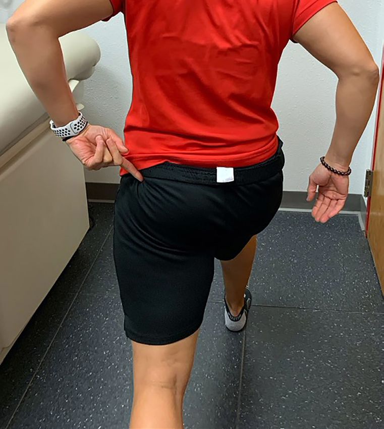
HIP PAIN IN ADULTS
FEMOROACETABULAR IMPINGEMENT adduction internal rotation and flexion abduc-
Femoroacetabular impingement is one of the most tion external rotation tests (Figures 3 and 4) are
common causes of hip pain in young adults.24 It indicative of intra-articular hip pathology.4,7
can be caused by a cam deformity, which is bony Radiography is used to help diagnose femoro-
overgrowth of the femoral head and neck, a pin- acetabular impingement, although the preferred
cer deformity of the acetabulum (too much cov- radiographic views may vary. Standing antero-
erage of the femoral head), or both (Figure 225). posterior radiographs of the pelvis may be most
Femoroacetabular impingement often has a grad- helpful for diagnosing of the pincer deformity,26
ual onset without a specific injury. It usually whereas Meyer lateral and 90-degree Dunn views
presents earlier in the disease process with less may be best to visualize the cam deformity.27
bony changes in athletes than in nonathletes and
is more bothersome to athletes whose activities LABRAL TEARS
require hyperflexion and wide range of motion Patients with labral tears may present with ante-
at the hip joint.2,5,9 Positive results on the flexion rior hip pain and a history of a sports-related or
traumatic injury. They may also be associated
with repetitive motions. Labral tears may cause
FIGURE 2
a popping, catching, or clicking sound associated
with activities such as dance, gymnastics, hockey,
basketball, and soccer.2,5,9 Physical examination
for labral tears should include flexion adduction
internal rotation and flexion abduction external
rotation tests.7
Pincer deformity
A standing radiograph should be the initial
imaging test.24 Previously, magnetic resonance
Cam deformity
arthrography (1.5 tesla) with gadolinium injection
of the hip was the diagnostic standard for labral
tears. However, with recent advances to 3-tesla
MRI and specialized hip protocols, noncontrast
3-tesla MRI is as sensitive and specific as magnetic
resonance arthrography and does not require a
procedure for contrast injection.22,23 Physicians
should consult with a local radiologist to deter-
mine the most appropriate test in their area.
Labral tears and femoroacetabular impinge-
ment are often comorbid conditions in young
active patients. Athletes are more likely to require
surgical intervention for these conditions, espe-
cially those with both conditions.9
FEMORAL NECK STRESS FRACTURES
Causes of femoroacetabular impingement. A pincer Stress fractures of the femoral neck are typically
deformity results from excessive overhang of the ace- associated with overuse and may also be associ-
tabulum, which causes the labrum to be impinged ated with energy imbalance in athletes. Femoral
between the acetabulum and femoral head when the
hip is flexed. A cam deformity occurs when exostosis
neck stress fractures are more common in women
along the femoral head and neck impinges the labrum than men but should not be excluded in men with
against the acetabulum. a history of overuse.10 Early in the disease process,
Illustration by Dave Klemm
stress fractures are typically not visible on radio-
Reprinted with permission from Kuhlman GS, Domb BG. Hip
graphs and therefore MRI is required for defini-
impingement:identifying and treating a common cause of hip pain. tive diagnosis. Early diagnosis of a femoral neck
Am Fam Physician. 2009;80(12):1431. stress fracture is important because conversion to
a complete fracture can be a devastating injury.5,28
84 American Family Physician www.aafp.org/afp Volume 103, Number 2 ◆ January 15, 2021
Descargado para Anonymous User (n/a) en National Library of Health and Social Security de ClinicalKey.es por Elsevier en febrero 24, 2021.
Para uso personal exclusivamente. No se permiten otros usos sin autorización. Copyright ©2021. Elsevier Inc. Todos los derechos reservados.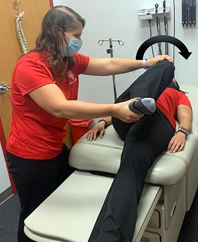
HIP PAIN IN ADULTS
               FIGURE 3

              A                                                                      B

               Flexion adduction internal rotation test. The examiner (A) passively flexes then (B) abducts and
               internally rotates the hip. The result is positive if pain is reproduced in the anterior hip/groin
               area. This test has been shown to have a sensitivity of 59% to 100% and specificity of 4% to 75%
               for intra-articular hip pathology.4,7

               FIGURE 4

              A                                                                      B

               Flexion abduction external rotation test. The examiner (A) passively flexes and then (B) abducts
               and externally rotates the hip. The result is positive if pain is reproduced in the anterior hip/
               groin area. This test has been shown to have 42% to 81% sensitivity and 18% to 25% specificity
               for intra-articular hip pathology.4,7

January 15, 2021   ◆   Volume 103, Number 2                        www.aafp.org/afp                                            American Family Physician 85

             Descargado para Anonymous User (n/a) en National Library of Health and Social Security de ClinicalKey.es por Elsevier en febrero 24, 2021.
             Para uso personal exclusivamente. No se permiten otros usos sin autorización. Copyright ©2021. Elsevier Inc. Todos los derechos reservados.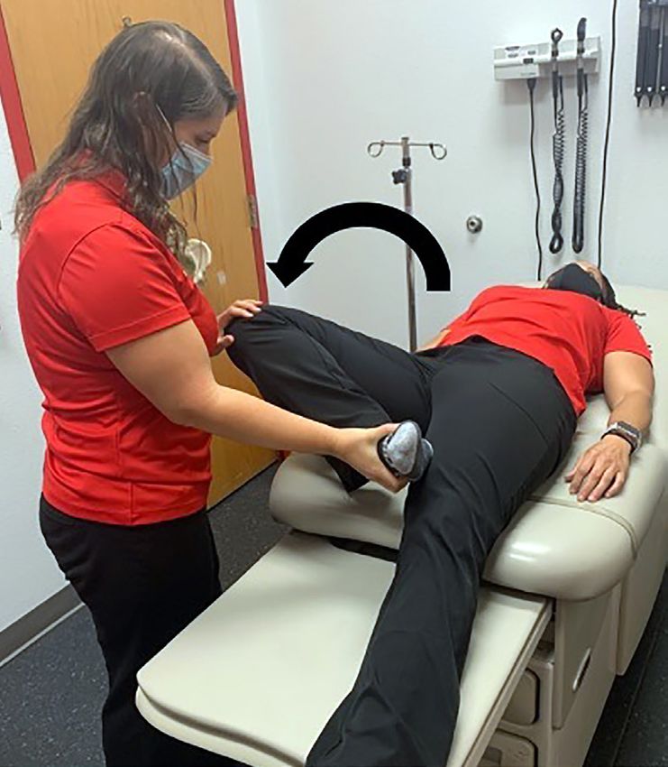
HIP PAIN IN ADULTS

          AVASCULAR NECROSIS                                                      this syndrome may be associated with bursitis,
          Avascular necrosis of the femoral head most com-                        gluteus medius tendinopathy or tears are now
          monly presents in middle-aged to older adults.                          thought to be more common.16,31 Iliotibial band
          Risk factors include alcohol use, smoking, sys-                         friction and external snapping hip can contribute
          temic corticosteroid use, hemoglobinopathies,                           to greater trochanteric pain syndrome.15
          chemotherapy, metabolic syndrome, and obesity.                             Greater trochanteric pain syndrome presents
          Early detection of avascular necrosis can be joint                      as lateral hip pain aggravated by ambulation or
          sparing, but no physical examination finding is                         other physical activities, sitting for long peri-
          specific for this diagnosis.11 Late-stage disease may                   ods, and sleeping on the affected hip. It most
          be visible on a radiograph, but earlier diagnosis                       commonly affects women 40 to 60 years of age.16
          often requires MRI or computed tomography.12,21                         There is typically no inciting injury. The patient
                                                                                  may walk with a Trendelenburg gait or have posi-
          OSTEOARTHRITIS                                                          tive findings on the Trendelenburg test or resisted
          In older adults, osteoarthritis of the femoroace-                       external derotation test.7,16 (Figure 5).
          tabular joint is the most common cause of ante-                            If a patient with greater trochanteric pain syn-
          rior hip pain.3 It can lead to significant morbidity                    drome does not improve with anti-inflammatory
          and decrease in physical activity. Osteoarthritis                       medications and physical therapy, a gluteus
          of the hip typically has a gradual onset, but some                      medius tendon tear should be considered. MRI
          patients recall a specific injury or fall. Patients                     and musculoskeletal ultrasonography performed
          with this condition may have pain with sitting                          by an experienced sonographer are sufficiently
          and ambulating for long periods and may have                            sensitive and specific for diagnosing a gluteus
          an antalgic gait. Physical examination maneuvers                        medius tendon tear.15,16,21 Referral to an orthope-
          such as flexion and internal and external rotation                      dic hip specialist is often indicated for large par-
          may reproduce pain, and range of motion may be                          tial or complete tears because surgery is typically
          decreased.4,6                                                           associated with good outcomes in patients with
             Standing anteroposterior radiography of the                          this condition.15,16
          pelvis is the radiologic test of choice and will
          show joint space narrowing and osteophyte for-                          Posterior Hip Pain
          mation. However, presence of these findings does                        The cause of posterior hip pain can be difficult
          not always correlate with symptom severity.7,13,29                      to diagnose. The differential diagnosis includes
          Ultrasound-guided anesthetic injection of the                           musculoskeletal causes and referred pain from
          hip joint may help differentiate an intra-articular                     intrapelvic and gynecologic issues. Patients with
          cause of pain from other causes (e.g., lumbar                           intrapelvic problems may have a history of cyclic
          spine or extra-articular pain), and corticosteroid                      pain associated with menses or urinary or bowel
          injection may be therapeutic for intra-articular                        symptoms.17
          pain.30
                                                                                  PIRIFORMIS AND DEEP GLUTEAL SYNDROME
          HIP FRACTURES                                                           Piriformis syndrome is thought to be a result of
          Hip fractures are more common in older adults                           the piriformis muscle entrapping the sciatic nerve,
          and often present after a fall or other trauma or                       causing hip and buttock pain and sciatica (burn-
          may be associated with osteoporosis. Physical                           ing pain shooting down the leg). Piriformis syn-
          examination usually reveals an inability to walk                        drome is a subset of deep gluteal syndrome, which
          on the affected limb and a shortened, externally                        includes entrapment of the sciatic nerve and/or
          rotated, abducted leg while in the supine posi-                         pudendal nerve by the piriformis muscle, gemelli-
          tion.14 Most hip fractures are visible on a radio-                      obturator internus, or proximal hamstrings.18
          graph and require surgical fixation.4                                   Patients with deep gluteal syndrome have deep
                                                                                  buttock pain that is aggravated by sitting and sci-
          Lateral Hip Pain                                                        atica symptoms. The seated piriformis stretch test
          The most common cause of lateral hip pain is                            (Figure 6) may reproduce this pain.18
          greater trochanteric pain syndrome (previously                             Although conservative treatments are
          called greater trochanteric bursitis).15 Although                       often helpful, MRI may be needed to identify

86 American Family Physician                                     www.aafp.org/afp                                  Volume 103, Number 2        ◆   January 15, 2021

           Descargado para Anonymous User (n/a) en National Library of Health and Social Security de ClinicalKey.es por Elsevier en febrero 24, 2021.
           Para uso personal exclusivamente. No se permiten otros usos sin autorización. Copyright ©2021. Elsevier Inc. Todos los derechos reservados.
HIP PAIN IN ADULTS
pathology in the deep gluteal muscles or sciatic long-stride walking test (Figure 7) is the most
and pudendal nerves. Additionally, electrodiag- sensitive and specific test for this condition.18,19
nostic nerve testing can help localize the area of
nerve entrapment.18 LUMBAR SPINE AND SACROILIAC JOINT
PATHOLOGY
ISCHIOFEMORAL IMPINGEMENT Lumbar spinal issues can present as posterior
Ischiofemoral impingement is impingement of hip pain. Patients typically have pain in the lum-
the quadratus femoris muscle and nerve between bar spine or musculature and in the posterior
the proximal femur at the level of the lesser tro- hip/buttock area and may report previous lum-
chanter and the ischial tuberosity.19 Patients with bar spinal problems. Radiography of the lum-
ischiofemoral impingement have gradual onset of bar spine may show degenerative disease, and
deep buttock pain that is worsened with activi- MRI can help identify disk herniation or nerve
ties requiring a long stride, such as running. The entrapment.17
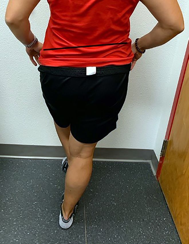
FIGURE 5
A B
Tests for gluteal tendinopathy. (A) Modified Trendelenburg test. Patients are instructed to stand on the affected leg/
hip and lift the other leg for 30 seconds. The result is positive if the iliac crest falls below the standing side, showing
weakness in hip abductors (gluteus muscles). A positive finding on the Trendelenburg test has been shown to have
23% to 97% sensitivity and 77% to 96% specificity for gluteal tendinopathy.4,7 (B) Resisted external derotation test.
While the patient lies on a table, the hip is passively flexed to 90 degrees, then internally rotated. The patient is asked
to return the leg to the same axis as the table (0 degrees rotation), pushing against the examiner’s (resisting) hand. The
result is positive if pain is reproduced over the lateral hip. This test has been shown to have 88% sensitivity and 97%
specificity for gluteal tendinopathy.7
January 15, 2021 ◆ Volume 103, Number 2 www.aafp.org/afp American Family Physician 87
Descargado para Anonymous User (n/a) en National Library of Health and Social Security de ClinicalKey.es por Elsevier en febrero 24, 2021.
Para uso personal exclusivamente. No se permiten otros usos sin autorización. Copyright ©2021. Elsevier Inc. Todos los derechos reservados.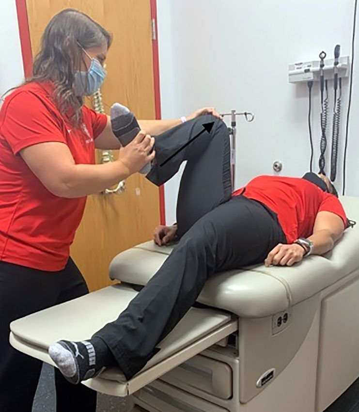
HIP PAIN IN ADULTS
FIGURE 6 FIGURE 7
Seated piriformis stretch test. The patient is seated Long-stride walking test. The patient is instructed to
with 90 degrees of hip flexion. The examiner extends take a long step on the unaffected leg with the hip
the knee and passively moves the hip into adduction pointed forward. This narrows the space between
and internal rotation while palpating just lateral to the the lesser trochanter and ischium on the posterior
ischium. The result is positive if pain is reproduced at (affected) hip, which can reproduce pain caused by
the piriformis muscle. This test has been shown to ischiofemoral impingement. This test has been shown
have a 91% sensitivity and 80% specificity for deep to have a sensitivity of 94% and specificity of 85% for
gluteal syndrome. 18 ischiofemoral impingement. 18,19
Sacroiliac joint dysfunction and/or arthritis A complete hamstring tear or avulsion often
may also present as posterior hip pain. The most causes ecchymosis of the posterior thigh.20 Ham-
common physical examination finding is tender- string tendinopathy or partial tears are typically
ness to palpation over the sacroiliac joint. Sacro- exacerbated by hamstring strength testing but do
iliac pain typically does not occur above the L5 not cause obvious bruising or deformity. Patients
level, which would indicate a lumbar spinal eti- with acute tears should be referred to an orthope-
ology of pain.17 Radiography may show sacroiliac dic surgeon.20 If it is unclear whether the patient
joint arthritis. Image-guided injections may be a has a hamstring injury, MRI may be helpful in
helpful diagnostic and therapeutic tool. If diag- determining the diagnosis.17
nosis is uncertain, MRI can show inflammation
or arthritis at the sacroiliac joint.17 This article updates previous articles on this topic by
Wilson and Furukawa,4 and O’Kane.6
HAMSTRING INJURIES Data Sources: A PubMed search was conducted
using the following key words, with filters set for the
Posterior hip/buttock pain around the ischium
previous five years and humans only:hip pain, ante-
may be indicative of a hamstring strain, tear, rior hip pain, femoroacetabular impingement, gluteus
or avulsion. Patients may have a history of a medius tendinitis, hip physical exam, 3-tesla hip MRI,
traumatic, sports-related, or overuse injury. ultrasound guided hip injection, greater trochanteric
88 American Family Physician www.aafp.org/afp Volume 103, Number 2 ◆ January 15, 2021
Descargado para Anonymous User (n/a) en National Library of Health and Social Security de ClinicalKey.es por Elsevier en febrero 24, 2021.
Para uso personal exclusivamente. No se permiten otros usos sin autorización. Copyright ©2021. Elsevier Inc. Todos los derechos reservados.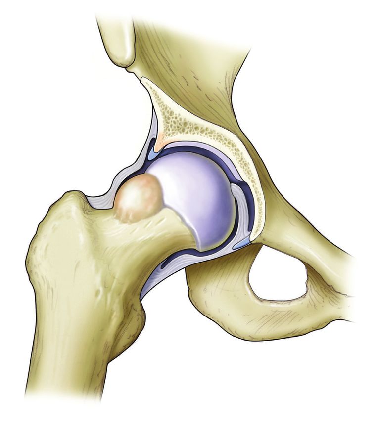
HIP PAIN IN ADULTS
pain syndrome, deep gluteal syndrome, ischiofemoral 14. LeBlanc KE, Muncie HL Jr., LeBlanc LL, et al. Hip frac-
syndrome, hamstring avulsion. Additional searches ture:diagnosis, treatment, and secondary prevention. Am
included the Cochrane Database of Systematic Fam Physician. 2014;89(12):945-951. Accessed August 19,
2020. https://w ww.aafp.org/afp/2014/0615/p945.html
Reviews and Essential Evidence Plus. Search dates:
November 8, 2019, to September 30, 2020. 15. Redmond JM, Chen AW, Domb BG. Greater trochan-
teric pain syndrome. J Am Acad Orthop Surg. 2016;24(4):
231-240.
The Author 16. LaPorte C, Vasaris M, Gossett L, et al. Gluteus medius tears
of the hip:a comprehensive approach. Phys Sportsmed.
RACHEL CHAMBERLAIN, MD, CAQSM, is the 2019;47(1):15-20.
associate program director of the Sports Medi- 17. Gómez-Hoyos J, Martin RL, Martin HD. Current concepts
cine Fellowship and an assistant professor in the review:evaluation and management of posterior hip pain.
Department of Family and Community Medicine at J Am Acad Orthop Surg. 2018;26(17):597-609.
the University of New Mexico, Albuquerque. 18. Park JW, Lee Y-K, Lee YJ, et al. Deep gluteal syndrome as
a cause of posterior hip pain and sciatica-like pain. Bone
Address correspondence to Rachel Chamberlain, Joint J. 2020;102-B(5):556-567.
MD, CAQSM, 1 University of New Mexico, Albuquer- 19. Hernando MF, Cerezal L, Pérez-Carro L, et al. Evaluation
que, NM 87131 (email:rchamberlain@salud.unm. and management of ischiofemoral impingement:a patho-
edu). Reprints are not available from the author. physiologic, radiologic, and therapeutic approach to
a complex diagnosis. Skeletal Radiol. 2016;45(6):7 71-787.
20. Degen RM. Proximal hamstring injuries:management of
References tendinopathy and avulsion injuries. Curr Rev Musculo-
1. Langhout R, Weir A, Litjes W, et al. Hip and groin injury is skelet Med. 2019;1 2(2):1 38-146.
the most common non-time-loss injury in female ama- 21. American College of Radiology ACR Appropriateness Cri-
teur football. Knee Surg Sports Traumatol Arthrosc. 2019; teria. Chronic hip pain. Accessed June 5, 2020. https://
27(10):3133-3141. acsearch.acr.org/docs/69425/Narrative/
2. Thorborg K, Rathleff MS, Petersen P, et al. Prevalence and 22. Carstensen SE, McCrum EC, Pierce JL, et al. Magnetic res-
severity of hip and groin pain in sub-elite male football: onance imaging (MRI) and hip arthroscopy correlations.
a cross-sectional cohort study of 695 players. Scand Sports Med Arthrosc Rev. 2017;25(4):199-209.
J Med Sci Sports. 2017;27(1):107-114. 23. Crespo-Rodríguez AM, De Lucas-Villarrubia JC, Pastrana-
3. Murphy L, Helmick CG. The impact of osteoarthritis in the Ledesma M, et al. The diagnostic performance of
United States:a population-health perspective. Am J Nurs. non-contrast 3-tesla magnetic resonance imaging (3-T
2012;1 12(3 suppl 1):S13-S19. MRI) versus 1.5-tesla magnetic resonance arthrogra-
4. Wilson JJ, Furukawa M. Evaluation of the patient with phy (1.5-T MRA) in femoro-acetabular impingement. Eur
hip pain. Am Fam Physician. 2014;89(1):27-34. Accessed J Radiol. 2017;88:109-116.
August 19, 2020. https://w ww.aafp.org/afp/2014/0101/ 24. Haldane CE, Ekhtiari S, de Sa D, et al. Preoperative physical
p27.html examination and imaging of femoroacetabular impinge-
5. Dick AG, Houghton JM, Bankes MJK. An approach to hip ment prior to hip arthroscopy—a systematic review. J Hip
pain in a young adult. BMJ. 2018;361:k 1086. Preserv Surg. 2017;4(3):201-213.
6. O’Kane JW. Anterior hip pain. Am Fam Physician. 1999; 25. Kuhlman GS, Domb BG. Hip impingement:identifying
60(6):1687-1696. Accessed August 19, 2020. https://w ww. and treating a common cause of hip pain. Am Fam Phy-
aafp.org/afp/1999/1015/p1687.html sician. 2009;80(12):1429-1434. Accessed August 19, 2020.
7. Reiman MP, Goode AP, Hegedus EJ, et al. Diagnostic https://w ww.aafp.org/afp/2009/1215/p1429.html
accuracy of clinical tests of the hip:a systematic review 26. Jackson TJ, Estess AA, Adamson GJ. Supine and standing
with meta-analysis. Br J Sports Med. 2013;47(14):893-902. AP pelvis radiographs in the evaluation of pincer femo-
8. Kamegaya M, Saisu T, Nakamura J, et al. Drehmann sign roacetabular impingement. Clin Orthop Relat Res. 2016;
and femoro-acetabular impingement in SCFE. J Pediatr 474(7):1692-1696.
Orthop. 2011;31(8):853-857. 27. Atkins PR, Shin Y, Agrawal P, et al. Which two-dimensional
9. Cianci A, Sugimoto D, Stracciolini A, et al. Nonoperative radiographic measurements of cam femoroacetabular
management of labral tears of the hip in adolescent ath- impingement best describe the three-dimensional shape
letes:description of sports participation, interventions, of the proximal femur? Clin Orthop Relat Res. 2019;477(1):
comorbidity, and outcomes. Clin J Sport Med. 2019;29(1): 242-253.
24-28. 28. Adkins SB III, Figler RA. Hip pain in athletes. Am Fam Phy-
10. Kupferer KR, Bush DM, Cornell JE, et al. Femoral neck sician. 2000;61(7):2109-2118. Accessed August 19, 2020.
stress fracture in Air Force basic trainees. Mil Med. 2014; https://w ww.aafp.org/afp/2000/0401/p2109.html
179(1):56-61. 29. Kim C, Nevitt MC, Niu J, et al. Association of hip pain with
11. Lamb JN, Holton C, O’Connor P, et al. Avascular necrosis radiographic evidence of hip osteoarthritis:diagnostic test
of the hip. BMJ. 2019;365:l2178. study. BMJ. 2015;351:h5983.
12. Guerado E, Caso E. The physiopathology of avascular 30. Lynch TS, Oshlag BL, Bottiglieri TS, et al. Ultra-
necrosis of the femoral head:an update. Injury. 2016; sound-guided hip injections. J Am Acad Orthop Surg.
47(suppl 6):S16-S26. 2019;27(10):e451-e461.
13. Beumer L, Wong J, Warden SJ, et al. Effects of exercise 31. Brennan KL, Allen BC, Maldonado YM. Dry needling versus
and manual therapy on pain associated with hip osteoar- cortisone injection in the treatment of greater trochan-
thritis:a systematic review and meta-analysis. Br J Sports teric pain syndrome:a noninferiority randomized clinical
Med. 2016;50(8):458-463. trial. J Orthop Sports Phys Ther. 2017;47(4):232-239.
January 15, 2021 ◆ Volume 103, Number 2 www.aafp.org/afp American Family Physician 89
Descargado para Anonymous User (n/a) en National Library of Health and Social Security de ClinicalKey.es por Elsevier en febrero 24, 2021.
Para uso personal exclusivamente. No se permiten otros usos sin autorización. Copyright ©2021. Elsevier Inc. Todos los derechos reservados.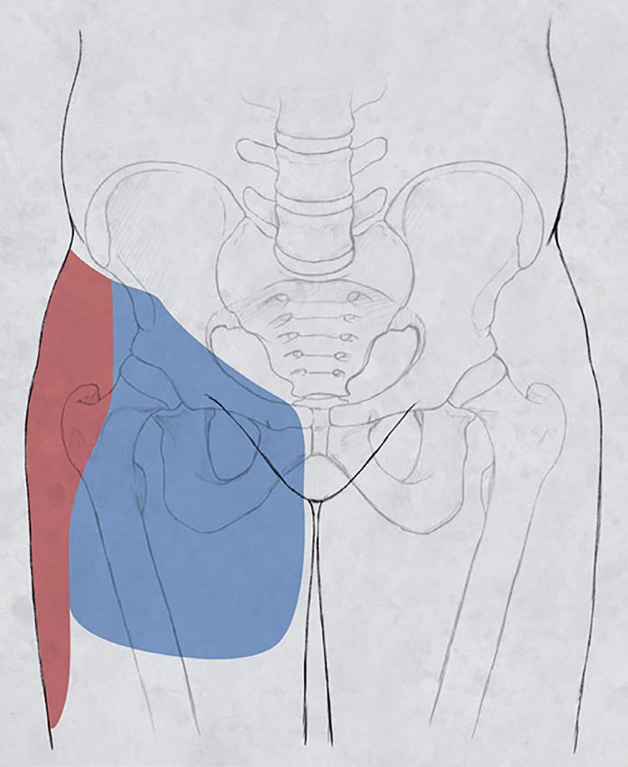
You can also read