Long-term management of canine disseminated granulomatous meningoencephalitis with imatinib mesylate: a case report
←
→
Page content transcription
If your browser does not render page correctly, please read the page content below
Case Report                                                       Veterinarni Medicina, 64, 2019 (02): 92–99

                                                                    https://doi.org/10.17221/70/2018-VETMED

Long-term management of canine disseminated
granulomatous meningoencephalitis with imatinib
mesylate: a case report
Joong Hyun Song1, Tae Sung Hwang1, Hee Chun Lee1, Do Hyeon Yu1, Byung
Joon Seung2, Jung Hyang Sur2, Dong In Jung1*
1
  Institute of Animal Medicine, College of Veterinary Medicine, Gyeongsang National University,
  Jinju, Republic of Korea
2
  College of Veterinary Medicine, Konkuk University, Seoul, Republic of Korea
*Corresponding author: jungdi@gnu.ac.kr

Citation: Song JH, Hwang TS, Lee HC, Yu DH, Seung BJ, Sur JH, Jung DI (2019): Long-term management of canine dis-
seminated granulomatous meningoencephalitis with imatinib mesylate: a case report. Veterinarni Medicina 64, 92–99.

Abstract: A seven-year-old Toy Poodle was presented for progressive ataxia and seizure episodes. Magnetic
resonance imaging revealed inflammatory lesions in the cerebrum and brainstem. Management with imatinib
mesylate, prednisolone and hydroxyurea were initiated and resulted in complete resolution of the clinical signs. In
regular magnetic resonance imaging scans, the overall appearance of the lesions deteriorated but improved again
after an increase in the imatinib mesylate dose. The patient had not shown any neurological signs until death and
survived for 1052 days after initial presentation. On histopathological examination, the patient was diagnosed
with disseminated granulomatous meningoencephalitis involving the cerebrum and brainstem. Immunohisto-
chemical staining was performed on the five types of tyrosine kinase (PDGFR-α, PDGFR-ß, VEGFR-2, c-Kit and
c-Abl proteins), which constitute therapeutic targets for conventional multitargeted tyrosine kinase inhibitors.
The immunohistochemical analysis revealed that all these tyrosine kinases were expressed in the brain samples.
The present report describes the first case of the use of imatinib mesylate therapy for granulomatous meningoen-
cephalitis in the dog. Therapy with imatinib mesylate plus glucocorticoids appears promising as a new therapeutic
intervention in meningoencephalitis of unknown aetiology.

Keywords: dog; immunohistochemical staining; magnetic resonance imaging; unknown aetiology; tyrosine kinase;
tyrosine kinase inhibitor

  Granulomatous meningoencephalitis (GME) is a             Systemic medical therapy with a combination of a
common non-infectious inflammatory disease of the          glucocorticoid and an immunosuppressant is the
central nervous system (CNS) in dogs and is thought        current mainstay of treatment despite the poorly
to be an autoimmune disorder (Fisher 2002; Matsuki         understood pathogenic mechanisms. Although the
et al. 2004; Granger et al. 2010). GME belongs to a        efficacy of these therapies varies depending on the
category of diseases termed meningoencephalitis of         patient and the clinicopathological subtype, the
unknown aetiology (MUE) and is characterised by            overall reported median survival time for dissemi-
focal or disseminated granulomatous lesions with           nated GME in dogs ranges from weeks to months
perivascular mononuclear cuffing within the CNS.           (Munana and Luttgen 1998; O’Neill et al. 2005).

Supported by the Basic Science Research Program through the National Research Foundation of Korea (NRF) funded by
the Ministry of Science, ICT & Future Planning, Republic of Korea (NRF-2017R1D1A1B03034904).

92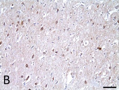
Veterinarni Medicina, 64, 2019 (02): 92–99                                                         Case Report

https://doi.org/10.17221/70/2018-VETMED

  Tyrosine kinases (TKs) are enzymes that phos-          needle). T1-weighted (T1W) images, T2-weighted
phorylate other proteins on tyrosine residues and        (T2W) images, fluid-attenuated inversion recov-
are key players in normal cell signal transduction,      ery (FLAIR) images and contrast-enhanced T1-
acting to tightly regulate cell growth and differen-     weighted (CET1W) images were obtained on the
tiation. It is well recognised that specific TKs are     MRI scan. The CET1W images were obtained after
abnormally activated in malignant neoplasms and          the intravenous injection of Omniscan (Gadolinium
inflammatory processes (Mirshafiey et al. 2014;          EDTA; GE-Healthcare, Little Chalfont, United
Bonkobara 2015). The efficacy of tyrosine kinase         Kingdom) in a dosage of 0.20 mmol/kg body weight.
inhibitors (TKIs) in a variety of cancers in both        On MRI images (Figure 1), it was possible to iden-
human and veterinary medicine has also been well         tify a focal, ill-defined lesion within the right thala-
demonstrated. Recently, inhibition of specific TKs       mus region. This lesion appeared hyperintense on
has shown clinical efficacy for the treatment of         T2W and FLAIR images and iso- to hypointense
autoimmune diseases in numerous animal models            on T1W images. There was moderate non-uniform
and human studies (Akashi et al. 2011; Azizi and         enhancement on CET1W images. On slightly right
Mirshafiey 2013).                                        parasagittal T2W and T1W images, a broad, ill-
  Up to now, no studies have investigated the role       defined brainstem lesion that extended from the
of TKs in dogs with immune-mediated diseases and         pons to the medulla oblongata and longitudinal
there was no clinical trial to assess the efficacy of    cervical syringomyelia which were suspected to be
TKIs in the canine immune-mediated CNS inflam-           secondary to the existing lesions were identified.
matory response. We here report the first case of        Examination of the CSF revealed an increased nu-
the use of imatinib mesylate therapy for dissemi-        cleated cell count of 18 cells/µl (reference range,
nated GME in a dog based on its medical record           < 5 cells/µl) and a protein concentration of 30 mg/
including clinical status, magnetic resonance im-        dl (reference range, < 25 mg/dl). Cytological exami-
aging (MRI), histopathological and immunohisto-          nation of CSF revealed a mononuclear cell pleo-
chemical findings.                                       cytosis. Based on the result of the MRI scan and
                                                         CSF analysis, we tentatively diagnosed the patient
                                                         with MUE.
Case description                                           However, we could not absolutely rule out the
                                                         possibility of a brain tumour at that time. We thus
  A seven-year-old, intact female Toy Poodle             decided to prescribe a TKI, which can be effec-
weighing 3.2 kg was presented with a two-month           tive against both brain tumours and immune-
history of ataxia in the hind limbs and an acute his-    mediated CNS inflammation based on previous
tory of cluster tonic-seizure episodes. The owner        research (Azizi and Mirshafiey 2013; Jung et al.
had noticed a progressive ataxic gait over the pre-      2014). Treatment with imatinib mesylate (Glivec ®,
ceding two months with the dog occasionally show-        Novartis Pharm., Stein, Switzerland; 10 mg/kg per
ing tetraparesis. The dog had been vaccinated, and       day orally) and prednisolone (Prednisolone, Korea
there was no history of trauma or exposure to toxin.     Pharm., Seoul, Republic of Korea; 1 mg/kg twice a
  On conducting physical and neurological exami-         day orally) was initiated. Moreover, we added hy-
nations, moderate head tilt to the left and hind limb    droxyurea (Hydroxyurea, Korea United pharm.,
ataxia were identified. The other responses, includ-     Seoul, Republic of Korea; 50 mg/kg every other
ing postural reactions, cranial nerve reflexes and       day orally), which is known to show synergism
spinal reflexes, were within normal limits. The result   with TKIs in specific brain tumours (Reardon et
of complete blood count, serum chemistry profiling       al. 2012). The neurological signs including seizure,
and radiography were not remarkable. On the basis        head tilt and ataxia rapidly improved. One month
of clinical signs and a neurological examination, an     after the initiation of treatment, the neurological
intracranial CNS lesion was highly suspected.            abnormalities had disappeared completely, and a
  To identify the intracranial lesion, we performed      second MRI scan was made to assess the patient’s
a brain MRI scan using 0.4-T scanner (Aperto;            response to therapy. On this MRI scan (Figure 2B),
Hitachi Medical Corporation, Tokyo, Japan), and          the lesions involving the right thalamus and brain-
cerebrospinal fluid (CSF) analysis (obtained from        stem were markedly improved. In addition, the pre-
the atlanto-occipital cistern tap using a 22-gauge       existing syringomyelia in the cervical spinal cord

                                                                                                              93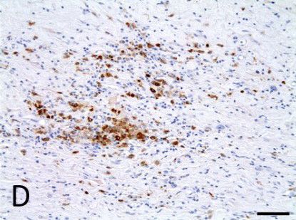
Case Report                                                        Veterinarni Medicina, 64, 2019 (02): 92–99

                                                                     https://doi.org/10.17221/70/2018-VETMED

(A)                             (B)                         (C)                          (D)

 (E)                            (F)                         (G)                          (H)

 (I)                                  (J)                                    (K)

Figure 1. Transverse (A–H), slightly right parasagittal (I and J) and midsagittal (K) images on magnetic resonance
imaging scan at initial examination. (A, E, J and K) T2-weighted images, (B and F) T1-weighted image, (C and G)
fluid-attenuated inversion recovery image, (D, H and I) contrast-enhanced T1-weighted image. (A–C, E–G and J)
A focal, ill-defined T2-, FLAIR hyperintense and T1- iso- to hypointense lesion (arrows) was evident in the right
thalamus. (D, H and I) Heterogeneous non-uniform enhancement was identified in the same region. (J) A broad, ill-
defined lesion (arrow heads) stretching from the pons to the medulla oblongata with an increased signal intensity was
observed. (K) Longitudinal syringomyelia (empty arrow) in the cervical spinal cord segment was identified
B = brainstem; C = cerebellum; F = forebrain; L = left

segment was no longer detected. The same therapy            clinical status without any neurological signs. At this
with imatinib mesylate plus hydroxyurea was main-           time, however, the lesions had generally progressed
tained, and the prednisolone dosage was tapered             to a more diffuse stage than at the third MRI check
slowly to 0.15 mg/kg once daily without any relapse         (Figure 2D). Further, a focal lesion within the left
of clinical signs. A third MRI scan was performed           thalamus, which was not identified previously, was
21 months after initial presentation. This MRI scan         detected. We again increased the imatinib mesylate
(Figure 2C) identified further ill-defined hyperin-         dosage to 10 mg/kg once daily. A fifth MRI scan was
tense lesions on both temporal lobes.                       performed 35 months after initial presentation for
  Approximately 23 months after initial presenta-           follow-up check. At that time the patient had not
tion, the patient was presented with an acute history       shown any clinical signs. Compared with the pre-
of vomiting. On complete blood count, leukopenia            ceding MRI (Figure 2E), the lesions involving the
(3.8 × 10⁹/l; reference range: 6 to 17 × 109/l) was         cerebrum improved again but the lesions within the
identified. We stopped prescribing hydroxyurea              two temporo-parietal lobes remained identical.
and decreased imatinib mesylate dosage to 8 mg/kg             Unfortunately, the patient did not recover from
per once daily. The vomiting stopped and the leu-           the anaesthesia and the owner requested eutha-
kopenia was gradually alleviated (after two weeks,          nasia. We surmised that multifocal lesions of the
8.8 × 10⁹/l; reference range: 6–17 × 109/l).                cerebrum and brainstem may have been associated
  A fourth MRI scan was performed 28 months after           with the failure to recover from anaesthesia. The
initial presentation. The patient maintained good           patient was donated by the owner, and we per-

94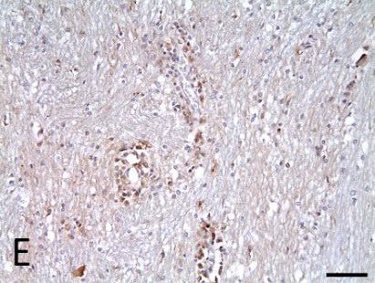
Veterinarni Medicina, 64, 2019 (02): 92–99                                                              Case Report

https://doi.org/10.17221/70/2018-VETMED

        T2-weigted                     T1-weigted            Fluid-attenuated inversion     Contrast-enhanced T1-
          images                         images                   recovery images             weighted images

(A)

(B)

(C)

(D)

(E)

Figure 2. Serial transverse magnetic resonance images at the level of the thalamus. Initial presentation (A), one month
(B), 21 months (C), 28 months (D) and 35 months (E) after initial presentation. After reduction of imatinib mesylate
dosage and discontinuation of the hydroxyurea, the lesions became markedly more diffuse (D). After the dosage of
imatinib mesylate was once again increased, the lesions improved again (E)

formed necropsy and histopathological examina-                 On histopathological examination (Figure 3A),
tion of the brain tissue.                                    multifocal perivascular cuffing lesions were distrib-
  At necropsy, gross findings from the brain showed          uted widely throughout the white matter of the cer-
generalised cerebrovascular congestion. On multi-            ebrum and brainstem. These inflammatory lesions
ple sections of the brain, multifocal discolourations        mainly consisted of macrophages and lymphocytes.
of the white matter in the cerebrum and brainstem            Based on these findings, the patient was definitively
were observed.                                               diagnosed with disseminated GME.

                                                                                                                    95
Case Report Veterinarni Medicina, 64, 2019 (02): 92–99
https://doi.org/10.17221/70/2018-VETMED
(A) (B) (A)
(C)
(D) (E) (F)
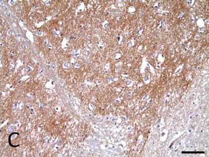
Figure 3. Histopathological and immunohistochemical findings. (A) Haematoxylin and eosin staining, × 100. Histo-
pathologically, multifocal perivascular cuffing lesions (arrows) were distributed widely throughout the white matter
of the cerebrum and brainstem. (B–F) × 200, scale bar size; 70 μm. Immunohistochemical analysis showing PDGFR-α
(B), PDGFR-ß (C), VEGFR-2 (D), c-Kit (E) and c-Abl (F) expression
Immunohistochemical staining was performed moderate), and 3+ (> 50%, widespread) for each
for five different TKs using antibodies specific for core. The immunohistochemical analysis revealed
the PDGFR-α, PDGFR-ß, VEGFR-2, c-Abl and c-Kit that the brain samples were positive for the expres-
proteins: a polyclonal rabbit PDGFR-α (Lifespan sion of all these TKs (Figures 3B–3F and Table 1).
BioSciences, Seattle, USA), a polyclonal rabbit Also, immunohistochemical staining of PDGFR-ß
PDGFR-ß (Abcam, Tokyo, Japan), a polyclonal rab- showed relatively strong intensity and widespread
bit VEGFR-2 (Abcam, Tokyo, Japan), a polyclonal distribution.
rabbit c-Abl (Santa Cruz Biotechnology, Santa
Cruz, USA) and a polyclonal affinity isolated rabbit
c-Kit (DAKO, Glostrup, Denmark). These proteins DISCUSSION AND CONCLUSIONS
are typical TKs that are inhibited by conventional
multi-targeted TKIs (e.g., imatinib, toceranib and The role of TKIs in autoimmune disorders, espe-
masitinib). The intensity and distribution of TK cially rheumatoid arthritis and multiple sclerosis
expression in each lesion were analysed by means (MS), has been suggested, and their efficacy in nu-
of a semi-quantitative scale. The intensity of TK merous animal models and clinical studies in human
expression was graded as follows: 0, negative; 1+, medicine has already been demonstrated (Eklund
mild; 2+, moderate and 3+, strong, and the distri- and Joensuu 2003; Akashi et al. 2011; Coffey et al.
bution of positively stained cells was graded as fol- 2012; Vermersch et al. 2012; Azizi et al. 2014). MS is
lows: 0 (0%, negative), 1+ (< 10%, scant), 2+ (< 50%, a human inflammatory disease characterised by the
T-cell mediated autoimmune response specific for
Table 1. The intensity and distribution of the expression myelin antigens of the CNS (Sospedra and Martin
of five different tyrosine kinases in immunohistochemi- 2016). The T-cell mediated immune response also
cal staining plays a key role in canine GME and necrotising
encephalitis, and it is also likely that these disor-
PDGFR-α PDGFR-ß VEGFR-2 c-Kit c-Abl ders are characterised by autoimmunity against the
Intensity 1+ 3+ 1+ 1+ 2+ CNS (Suzuki et al. 2003; Matsuki et al. 2004; Park
Distribution 2+ 3+ 1+ 2+ 2+ et al. 2012). According to a previous immunologi-
96Veterinarni Medicina, 64, 2019 (02): 92–99 Case Report
https://doi.org/10.17221/70/2018-VETMED
cal study, canine GME and necrotising encephalitis inhibitor which is used as an antineoplastic drug.
are considered similar to MS, which shares many Synergism of hydroxyurea with some TKIs has been
of the clinical, neuropathological and immuno- documented in certain neoplastic conditions via
logical characteristics of these conditions (Braund inhibition of the enzyme ribonucleotide reductase
1985; Greer et al. 2010). Multiple signal transduc- (Tipping et al. 2002; Shah et al. 2007; Reardon et al.
tion pathways involving TKs are implicated in the 2012). Hydroxyurea also has immunomodulatory
pathogenesis of MS. (Azizi et al. 2014; Mirshafiey effects which include reduction of T-cell prolifera-
et al. 2014). Uncontrolled signalling of these TKs tion and direct reduction of levels of inflammatory
results in aberrant inflammatory responses includ- cytokines (Lori 1999; Inayat et al. 2010). According
ing a T-cell mediated immune response. Although to these pharmacological studies, we surmised that
pathogenetic studies regarding role of TKs in ca- these properties of hydroxyurea might have ac-
nine GME and necrotising encephalitis have not counted for the good clinical status and long-term
yet been reported, we could diagnose GME and survival time with other drugs in the present case.
identify expression of five types of TK in the pre- However, since hydroxyurea is not a commonly
sent case. This result suggests that some proteins used drug in patients with immune-mediated in-
which act as ligand agonists for these TKs could be flammation, its efficacy has remained unclear, and
involved in key aspects of the pathogenesis of GME, a further well-designed placebo-controlled study
and multi-targeted therapy for over-expressed TKs will be needed.
may have potential as an effective treatment mo- The prognosis for GME is generally guarded to
dality for GME. poor but highly depends on the modality of the
Imatinib mesylate is a selective multi-targeted disease and on the treatment protocol followed.
TKI, which can inhibit BCR/Abl, PDGFR, c-KIT, According to previous studies, dogs with dis-
c-fms, TCR/Abl, Lck, FLT-3 and MAPK activities seminated GME (weeks to months) had signifi-
in various cell types (Azizi and Mirshafiey 2013). cantly shorter survival times than those with focal
The five types of TK which were expressed in this GME (weeks to years) (Munana and Luttgen 1998;
case are all included in the inhibition spectrum of O’Neill et al. 2005; Coates 2007). This patient’s
imatinib mesylate. Among these, PDGFR-ß, which survival time (survival for 1052 days with treat-
plays an important role in MS pathogenesis and ment) was relatively long compared those in previ-
is effectively inhibited by the imatinib mesylate, ously reported cases with disseminated GME, and
was relatively intensely and broadly expressed in improvement of clinical signs and maintenance
this case. Therefore, the clinically relevant effect were satisfactory. In other words, therapy using
of imatinib mesylate in the present case appears to imatinib mesylate might represent a good thera-
be at least partly based on inhibition of PDGFR-ß. peutic alternative to the other adjunctive immu-
Other TKs also appear to be highly associated with nosuppressive drugs due to its efficacy and lower
the disease process based on previous MS studies adverse effects.
(Mirshafiey et al. 2014) and the expression pat- In conclusion, to the authors’ knowledge, the
terns observed in this study. However, further present report describes the first case of the use
studies will be required to fully understand the of imatinib mesylate therapy for GME in the dog.
relationship between the expression of each TK The good response to the therapy and the results
and immune-mediated neuroinflammation in dogs. of the immunohistochemical staining suggest that
Furthermore, in our experience (unpublished data), these kinases may play a role in the pathogenesis
although immunohistochemical staining for TKs and development of GME. Therapy with multi-
was not investigated, some MUE patients were targeted TKIs plus glucocorticoids appears prom-
not responsive to multi-TK-targeted therapy using ising as a new therapeutic intervention in MUE.
imatinib mesylate. We thus surmised that expres- Furthermore, since multi-targeted TKIs inhibit
sion of specific TKs and expression patterns might diverse signal transduction pathways underlying
influence the efficacy of imatinib mesylate in dogs aberrant immune responses that cannot be con-
with MUE as in tumours. trolled by conventional immunosuppression, it may
The major limitation of this report is that the be useful to combine these drugs with conventional
therapy did not solely consist of imatinib me- therapy of MUE. However, further long-term pro-
sylate. Hydroxyurea is a ribonucleotide reductase spective studies in a larger population of dogs with
97Case Report Veterinarni Medicina, 64, 2019 (02): 92–99
https://doi.org/10.17221/70/2018-VETMED
GME and necrotising encephalitis are required for Inayat MS, El-Amouri IS, Bani-Ahmad M, Elford HL, Gal-
estimation of the clinical efficacy and survival times licchio VS, Oakley OR (2010): Inhibition of allogeneic
with imatinib mesylate treatment. inflammatory responses by the ribonucleotide reductase
inhibitors, didox and trimidox. Journal of Inflammation
7, doi: 10.1186/1476-9255-7-43.
REFERENCES Jung HW, Lee HC, Kim JH, Jang HM, Moon JH, Sur JH, Ha
J, Jung DI (2014): Imatinib mesylate plus hydroxyurea
Akashi N, Matsumoto I, Tanaka Y, Inoue A, Yamamoto K, chemotherapy for cerebellar meningioma in a Belgian
Umeda N, Tanaka Y, Hayashi T, Goto D, Ito S (2011): Malinois dog. The Journal of Veterinary Medical Science
Comparative suppressive effects of tyrosine kinase in- 76, 1545–1548.
hibitors imatinib and nilotinib in models of autoimmune Lori F (1999): Hydroxyurea and hiv: 5 years later-from an-
arthritis. Modern Rheumatology 21, 267–275. tiviral to immune-modulating effects. AIDS 13, 1433–
Azizi G, Mirshafiey A (2013): Imatinib mesylate: An in- 1442.
novation in treatment of autoimmune diseases. Recent Matsuki N, Fujiwara K, Tamahara S, Uchida K, Matsunaga
Patents on Inflammation & Allergy Drug Discovery 7, S, Nakayama H, Doi K, Ogawa H, Ono K (2004): Preva-
259–267. lence of autoantibody in cerebrospinal fluids from dogs
Azizi G, Haidari MR, Khorramizadeh M, Naddafi F, Sadria with various cns diseases. The Journal of Veterinary
R, Javanbakht MH, Sedaghat R, Zavareh FT, Mirshafiey Medical Science 66, 295–297.
A (2014): Effects of imatinib mesylate in mouse models Mirshafiey A, Ghalamfarsa G, Asghari B, Azizi G (2014):
of multiple sclerosis and in vitro determinants. Iranian Receptor tyrosine kinase and tyrosine kinase inhibitors:
Journal of Allergy, Asthma and Immunology 13, 198–206. New hope for success in multiple sclerosis therapy. In-
Bonkobara M (2015): Dysregulation of tyrosine kinases and novations in Clinical Neuroscience 11, 23–36.
use of imatinib in small animal practice. The Veterinary Munana KR, Luttgen PJ (1998): Prognostic factors for dogs
Journal 205, 180–188. with granulomatous meningoencephalomyelitis: 42 cases
Braund K (1985): Granulomatous meningoencephalomy- (1982–1996). Journal of the American Veterinary Medi-
elitis. Journal of the American Veterinary Medical As- cal Association 212, 1902–1906.
sociation 186, 138–141. O’Neill EJ, Merrett D, Jones B (2005): Granulomatous
Coates JR (2007): Emerging treatments for granulomatous meningoencephalomyelitis in dogs: A review. Irish Vet-
meningoencephalomyelitis (gme). Advances in Small erinary Journal 58, 86–92.
Animal Medicine and Surgery 20, 1–3. Park E, Uchida K, Nakayama H (2012): Comprehensive im-
Coffey G, Deguzman F, Inagaki M, Pak Y, Delaney SM, Ives munohistochemical studies on canine necrotizing menin-
D, Betz A, Jia ZJ, Pandey A, Baker D (2012): Specific in- goencephalitis (NME), necrotizing leukoencephalitis
hibition of spleen tyrosine kinase suppresses leukocyte (NLE), and granulomatous meningoencephalomyelitis
immune function and inflammation in animal models of (GME). Veterinary Pathology 49, 682–692.
rheumatoid arthritis. Journal of Pharmacology and Ex- Reardon DA, Norden AD, Desjardins A, Vredenburgh JJ,
perimental Therapeutics 340, 350–359. Herndon JE, Coan A, Sampson JH, Gururangan S, Peters
Eklund KK, Joensuu H (2003): Treatment of rheumatoid ar- KB, McLendon RE, Norfleet JA, Lipp ES, Drappatz J,
thritis with imatinib mesylate: Clinical improvement in Wen PY, Friedman HS (2012): Phase II study of Gleevec ®
three refractory cases. Annals of Medicine 35, 362–367. plus hydroxyurea (HU) in adults with progressive or re-
Fisher M (2002): Disseminated granulomatous meningoen- current meningioma. Journal of Neuro-Oncology 106,
cephalomyelitis in a dog. The Canadian Veterinary Jour- 409–415.
nal 43, 49–51. Shah GD, Silver JS, Rosenfeld SS, Gavrilovic IT, Abrey LE,
Granger N, Smith PM, Jeffery ND (2010): Clinical findings Lassman AB (2007): Myelosuppression in patients ben-
and treatment of non-infectious meningoencephalomy- efiting from imatinib with hydroxyurea for recurrent
elitis in dogs: A systematic review of 457 published cases malignant gliomas. Journal of Neuro-Oncology 85, 217–
from 1962 to 2008. The Veterinary Journal 184, 290–297. 222.
Greer K, Wong A, Liu H, Famula T, Pedersen N, Ruhe A, Sospedra M, Martin R (2016): Immunology of multiple scle-
Wallace M, Neff M (2010): Necrotizing meningoencepha- rosis. Seminars in Neurology 36, 115–127.
litis of pug dogs associates with dog leukocyte antigen Suzuki M, Uchida K, Morozumi M, Hasegawa T, Yanai T,
class ii and resembles acute variant forms of multiple Nakayama H, Tateyama S (2003): A comparative patho-
sclerosis. Tissue Antigens 76, 110–118. logical study on canine necrotizing meningoencephalitis
98Veterinarni Medicina, 64, 2019 (02): 92–99 Case Report
https://doi.org/10.17221/70/2018-VETMED
and granulomatous meningoencephalomyelitis. The Jour- Vermersch P, Benrabah R, Schmidt N, Zeehir H, Clavelou
nal of Veterinary Medical Science 65, 1233–1239. P, Vongsouthi C, Dubreuil P, Moussy A, Hermine O
Tipping AJ, Mahon FX, Zafirides G, Lagarde V, Goldman (2012): Masitinib treatment in patients with progressive
JM, Melo JV (2002): Drug responses of imatinib mesylate- multiple sclerosis: A randomized pilot study. BMC Neu-
resistant cells: Synergism of imatinib with other chemo- rology 12, doi: 10.1186/1471-2377-12-36.
therapeutic drugs. Leukemia 16, 2349–2357.
Received: April, 29, 2018
Accepted after corrections: January 4, 2019
99You can also read