Association between coenzyme Q10 and glucose transporter (GLUT1) deficiency
←
→
Page content transcription
If your browser does not render page correctly, please read the page content below
Yubero et al. BMC Pediatrics 2014, 14:284
http://www.biomedcentral.com/1471-2431/14/284

 CASE REPORT                                                                                                                                      Open Access

Association between coenzyme Q10 and glucose
transporter (GLUT1) deficiency
Delia Yubero1, Mar O’Callaghan1, Raquel Montero1, Aida Ormazabal1, Judith Armstrong1, Carmina Espinos2,
Maria A Rodríguez1, Cristina Jou1, Esperanza Castejon1, Maria A Aracil1, Maria V Cascajo3, Angela Gavilan3,
Paz Briones4, Cecilia Jimenez-Mallebrera1, Mercedes Pineda1, Plácido Navas3 and Rafael Artuch1*

  Abstract
  Background: It has been demonstrated that glucose transporter (GLUT1) deficiency in a mouse model causes a
  diminished cerebral lipid synthesis. This deficient lipid biosynthesis could contribute to secondary CoQ deficiency.
  We report here, for the first time an association between GLUT1 and coenzyme Q10 deficiency in a pediatric patient.
  Case presentation: We report a 15 year-old girl with truncal ataxia, nystagmus, dysarthria and myoclonic epilepsy
  as the main clinical features. Blood lactate and alanine values were increased, and coenzyme Q10 was deficient both
  in muscle and fibroblasts. Coenzyme Q10 supplementation was initiated, improving ataxia and nystagmus. Since
  dysarthria and myoclonic epilepsy persisted, a lumbar puncture was performed at 12 years of age disclosing
  diminished cerebrospinal glucose concentrations. Diagnosis of GLUT1 deficiency was confirmed by the presence
  of a de novo heterozygous variant (c.18+2T>G) in the SLC2A1 gene. No mutations were found in coenzyme Q10
  biosynthesis related genes. A ketogenic diet was initiated with an excellent clinical outcome. Functional studies in
  fibroblasts supported the potential pathogenicity of coenzyme Q10 deficiency in GLUT1 mutant cells when compared
  with controls.
  Conclusion: Our results suggest that coenzyme Q10 deficiency might be a new factor in the pathogenesis of G1D,
  although this deficiency needs to be confirmed in a larger group of G1D patients as well as in animal models.
  Although ketogenic diet seems to correct the clinical consequences of CoQ deficiency, adjuvant treatment with
  CoQ could be trialled in this condition if our findings are confirmed in further G1D patients.
  Keywords: Glucose transporter type I deficiency, SLC2A1 gene, Coenzyme Q10, Ataxia, Ketogenic diet

Background                                                                             it is unclear how decreased glucose flux leads to the mani-
GLUT1 deficiency syndrome (G1D) most often causes                                      festations of the disorder.
infantile-onset refractory epilepsy, cognitive impairment                                 G1D is a partially treatable condition with ketogenic
and motor abnormalities (ataxia, dystonia, chorea or dys-                              diet (KD), which can replace glucose for acetyl-CoA gen-
kinesia) [1-4]. The main pathophysiological mechanism of                               eration [7,8]. Energy failure has been proven in G1D
the disease is associated with impaired glucose transport                              astrocytes, while tricarboxilyc acid abundance in the brain
across the blood brain barrier and through astrocyte cell                              of G1D mouse model is normal. These findings support
membranes that are haploinsufficient in the GLUT1 glu-                                 the complementary or alternative view that additional
cose carrier encoded by the SLC2A1 gene [5,6]. However                                 mechanisms participate in disease pathogenesis, placing
                                                                                       new emphasis on G1D as a glial disease [9]. This conten-
                                                                                       tion is highlighted by the preliminary therapeutic efficacy
* Correspondence: rartuch@hsjdbcn.org
                                                                                       of triheptanoin, a dietary supplement with the potential to
1
 Clinical Biochemistry, Pediatric Neurology, Histopathology,                           stimulate cerebral anabolism and energy delivery [10].
Gastroenterology-Nutrition and Neuromuscular Unit Departments. Hospital                   Coenzyme Q10 (CoQ) is a lipidic electron carrier in
Sant Joan de Déu and Centre For research in rare diseases (CIBERER),
Instituto de Salud Carlos III, Passeig Sant Joan de Déu, 2, 08950 Esplugues,
                                                                                       the mitochondrial respiratory chain (MRC). One of the
Barcelona, Spain                                                                       essential substrates for CoQ generation is acetyl-CoA
Full list of author information is available at the end of the article

                                         © 2014 Yubero et al.; licensee BioMed Central Ltd. This is an Open Access article distributed under the terms of the Creative
                                         Commons Attribution License (http://creativecommons.org/licenses/by/4.0), which permits unrestricted use, distribution, and
                                         reproduction in any medium, provided the original work is properly credited. The Creative Commons Public Domain
                                         Dedication waiver (http://creativecommons.org/publicdomain/zero/1.0/) applies to the data made available in this article,
                                         unless otherwise stated.
Yubero et al. BMC Pediatrics 2014, 14:284 Page 2 of 5
http://www.biomedcentral.com/1471-2431/14/284
[11]. Mutations in genes involved in the CoQ biosynthesis disorders. CoQ deficiency was identified both in muscle
pathway are associated with different clinical phenotypes, and fibroblasts (see Results). Because of this finding, CoQ
being cerebellar ataxia the most common one [11]. How- supplementation (orally administered at 30 mg/Kg/day)
ever, “secondary” CoQ deficiency is also characteristic of was initiated. Ataxia improved dramatically after 6 months
other diseases, since CoQ biosynthesis involves an intri- of therapy, and, upon reassessment after 4 years of CoQ
cate, broad set of reactions that may be potentially im- treatment, ambulation remained essentially normal, with
pacted by “primary” disturbance of several biochemical a mild residual reduction in velocity. Her nystagmus
processes [12]. had also disappeared and her visual pursuit had normal-
Our aim is to describe, for the first time a relevant ized [13]. However, mild dysmetria, dysarthria, myoclonic
association between G1D and CoQ deficiency. Clinical, epilepsy and intellectual disability (perhaps refractory to
biochemical, molecular and therapeutic observations con- CoQ), were present. During CoQ therapy, concomitant
stitute the basis of this association. treatment with valproate and ethosuximide was given
to control myoclonic epilepsy. No noticeable side-effects
Case presentation were observed when antiepileptic doses were raised to
The proband is a 15-years old girl with an unremarkable maintain therapeutic levels along the evolution of the
family history. At 18 months old she manifested no ambu- disease due to the patient increasing weight. In order
lation, and by 3 years old she had developed ataxia and to further investigate these manifestations, a lumbar
epilepsy with normal neuroimaging. At 5 years old clinical puncture was performed at 12 years of age, revealing
examination showed axial hypotonia, truncal ataxia, limb diminished cerebrospinal glucose concentrations (Table 1).
dysmetria and hyperreflexia. Paroxysmal nystagmus, sac- Plasma glucose concentration was normal. Diagnosis of
cadization of visual pursuit and dysarthria were observed. GLUT1 deficiency (G1D) was established and a ketogenic
The patient also exhibited intellectual disability (IQ = 54). diet (4:1 ratio, containing medium chain triglyceride oil)
Biochemical analyses in blood disclosed elevated lactate was initiated (CoQ treatment was then discontinued).
in 3 separate occasions and elevated alanine in 12 de- Written informed consent was obtained from the parents.
terminations (Table 1). Muscle and skin biopsies were The study was approved by the Ethical Committee of our
analyzed at 8 years of age to search for mitochondrial Hospital.
Table 1 Main biochemical findings in a case with GLUT1 Laboratory investigations
and CoQ deficiency Muscle and skin biopsies were collected, stored and
Patient Reference values cultured following previously reported procedures [14].
Blood CoQ content was analyzed by HPLC with electrochemical
Lactate 1.8-2.6 (2.2) 0.5-1.7 mmol/L detection, and MRC enzyme activities by spectrophotom-
Alanine 300-691 (560) 150-270 μmol/L etry as reported [14].
Functional studies in fibroblasts: Human Dermal Fibro-
Cholesterol 4.0 < 5.2 mmol/L
blasts (HDF) were grown in Dulbecco’s modified essential
CoQ (baseline) 0.33 0.45-1.1 μmol/L
medium as previously reported [15]. Cells were grown
CoQ (after treatment) 3.9-7.8 (5.5) 0.45-1.1 μmol/L with 1 g/L of either glucose or galactose from plating and
Cerebrospinal fluid throughout the entire duration of the observation period.
Glucose 1.9 2.2-3.4 mmol/L Mycoplasma testing was negative. Also normal and G1D
Lactate 1.07 1.1-2.2 mmol/L fibroblasts were supplemented with 30 μmol/L CoQ.
Growth rate was analyzed as indicated [15].
Muscle biopsy
Molecular analysis of genomic DNA (gDNA) isolated
CoQ content 76 115-450 nmol/g protein
from blood included Sanger sequencing of 12 genes invol-
Complex I + III* 149 107-560 mU/citrate ved in CoQ biosynthesis (ADCK3, ADCK4, PDSS1, PDSS2,
synthase U
COQ2, COQ3, COQ4, COQ5, COQ6, COQ9, CQ10A and
Complex II + III* 82 75-149 mU/citrate COQ10B), in addition to the SLC2A1 gene, which encodes
synthase U
for GLUT1 transporter. In silico mutation analysis was
Fibroblast
done by the Mutation Taster software.
CoQ content 78 100-150 nmol/g protein Total RNA was isolated from control and patient’s fibro-
(incubated with glucose)
blast cultures with RNeasy Fibrous Tissue mini kit (Qiagen,
CoQ content (incubated 123 100-150 nmol/g protein Hilden, Germany). 1 μg of RNA was retro-transcribed with
with galactose)
GoTaq® Probe 2-Step for RT-PCR (Promega, Wisconsin,
Patient results from blood are expressed as range (median).
*Complex I + III: NADH:cytochrome C oxidoreductase. Complex II + III: succinate:
USA) to obtain cDNA. SLC2A1 mRNA transcripts were
cytochrome C oxidoreductase. studied on cDNA through PCR amplification with specificYubero et al. BMC Pediatrics 2014, 14:284 Page 3 of 5
http://www.biomedcentral.com/1471-2431/14/284
primers located in exon 1 (available on request), followed (data not shown). The cDNA region encompassing the
by direct sequencing. genomic DNA mutation was amplified to detect a possible
altered mRNA transcript in the patient. The PCR product
Results did not show alternate amplification bands other than the
Muscle and fibroblasts CoQ content was decreased control one (Figure 2B). Patient’s sample direct sequen-
(Table 1). Functional studies of cell viability in culture with cing showed only the wild type transcript, lacking any hint
either glucose or galactose are illustrated in Figure 1A. of an aberrant transcript consequence of the intronic mu-
The GLUT1 mutant cell line cultured in the presence of tation identified in gDNA. Concurrently, patient carried a
glucose displayed significantly decreased growth rate heterozygous SNP (rs1385129) located in exon 2, initially
when compared with cultures grown in medium with gal- identified in gDNA (both alleles (T/C) were represented).
actose. Furthermore, the control cell line growth rate was We analyzed this SNP on the cDNA sequence (Figure 2C),
significantly greater than those observed for the GLUT1 and only one allele was present (C), pointing out that half
mutant line when cultured in glucose-containing medium. doses of wild type mRNA was not present. Since the vari-
CoQ content was increased in GLUT1 mutant fibroblasts ant is located in the splice donor site and we only detected
after 10 days growth in galactose (Table 1) while it re- one allele of rs1385129 in the patient’s cDNA, down-
mained unchanged in fibroblasts incubated in glucose stream of c.18+2T>G, we hypothesize that splicing of the
media for the same length of time. CoQ supplementation SLC2A1 mRNA could be altered.
of patient’s fibroblasts for one week induced an increase
of 37% cell growth rate while the growth rate of control Therapeutic interventions
cells with the same treatment only increased by 15% After initiation of a ketogenic diet at 14 years old, dysarth-
(Figure 1B). ria was controlled. At this age and prior to KD, mioclonic
Sequencing of CoQ-related genes disclosed no known jerks with absences were present and EEG disclosed slow
pathogenic variants. G1D was established after the de- basal rhythm (alpha: 7–8 Hz) and generalized spike-wave
tection of a new heterozygous variant (c.18+2T>G) in discharges at 2.5 HZ, suggesting atypical absences. At that
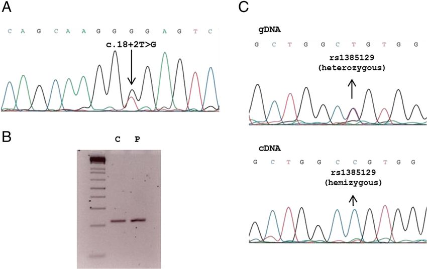
the first intron of the SLC2A1 gene (Figure 2A). The moment, patient was treated with valproate and ethosu-
variant was not present in 200 studied alleles of Spanish ximide. Epilepsy was totally controlled after 3 months of
ancestry. Conservation of the nucleotide position was KD, and EEG disclosed normal results, except for slow
high according to Phylophen scores and splice prediction basal activity. Antiepileptic treatment was progressively
software indicated a truncation of the splice site donor removed after 7 months of KD. At 15 years EEG showed a
A B
20000
18000
HDF
16000 450000
Total cells
14000
12000 400000
control medium
10000 GLUCOSE
350000
8000 GALACTOSE
6000 300000
nº cells
4000 medium supplemented with 30
250000 µM CoQ10
2000
0 10 13 14 17 19
200000
Days
150000
GLUT1 mutant
100000
12000
10000 50000
Total cells
HDF GLUT1
8000
GLUCOSE
6000
GALACTOSE
4000
2000
0 10 13 14 17 19
Days
Figure 1 Growth of GLUT1 fibroblast mutant cell line compared with control Human Dermal Fibroblasts (HDF). A). Both cells were
seeded (2.000 cells/plate) and incubated with either glucose or galactose. B). Both cells were seeded (50.000 cells/plate) and grown for one week
with or without 30 μM CoQ.Yubero et al. BMC Pediatrics 2014, 14:284 Page 4 of 5
http://www.biomedcentral.com/1471-2431/14/284
Figure 2 Genetic analysis. A- Electropherogram containing genomic SLC2A1 DNA mutation in a case with GLUT1 deficiency. B- 3% agarose gel
showing a 219 base pairs PCR amplification of SLC2A1 cDNA region enconmpassing the spling mutation. C (control) and P (patient). C- Patient’s
electropherograms showing a synonymous polymorphism (rs1385129, see dbSNP database) in heterozygous state in gDNA and in hemizygous
state in cDNA.
normal basal rhythm without paroxysms. Ataxia did not [15]. The human genome encodes for fourteen GLUT
appear during KD therapy. Plasma CoQ levels were nor- proteins that cooperate to transport different substrates
mal after initiation of a KD (0.58 μmol/L; reference values other than glucose [16].
shown in Table 1). The normalization of growth of G1D cells after the
incubation of fibroblasts with CoQ suggests the potential
Discussion benefit of this coadjuvant therapy for G1D patients,
We present for the first time, the identification of CoQ which deserves further investigations. Although the mu-
deficiency in a patient with G1D. Our clinical and bio- tation detected in SLC2A1 is compatible with the com-
chemical observations warrant further discussion be- mon G1D phenotype, we cannot rule out the existence
cause of their potential pathophysiological and therapeutic of mutations in other genes involved in CoQ metabolism
implications. given the fact that the CoQ metabolic pathway is not
Concerning the pathophysiology of G1D, Marin- well understood.
Valencia et al. [9], demonstrated in a G1D mouse model The observed clinical improvement after CoQ supple-
a diminished cerebral lipid synthesis. Importantly, this mentation in our patient supports these statements given
deficient lipid biosynthesis could contribute to secondary that the most severe cerebellar manifestations proved
CoQ deficiency. The muscle CoQ deficiency detected in amenable to CoQ therapy [13]. The simplest interpret-
our patient was mild, and this would explain that MRC ation of our clinical observations is provided by the notion
enzyme activities were low-normal. However, we interro- that the cerebellum is an extremely sensitive organ to oxi-
gated mitochondrial function due to the conjunction of dative stress and energy metabolic disorders, consistent
the clinical findings and the consistent hyperlactacide- with the near-universal demonstration of cerebellar mani-
mia and hyperalaninemia observed in our case. The ex- festations even in cases of mild CoQ deficiency [11,13].
periments conducted in fibroblasts confirmed the CoQ Once a ketogenic diet was initiated, the clinical outcome
deficiency, in agreement with its potential role in the was excellent, with a complete cessation of epilepsy
pathophysiology of G1D [11]. Especially relevant were and other signs. We did not simultaneously treat the
the differences of growth observed between fibroblasts patient with CoQ and the ketogenic diet in order to
cultured with either glucose or galactose and the recov- separately elucidate the individual effects of both in-
ery of CoQ in galactose growth. Glucose transport was terventions. It is possible that a ketogenic diet causes a
impaired in the mutant line, but not galactose influx, recovery in lipid biosynthesis that favorably impacts CoQ
which is able in turn to promote CoQ biosynthesis abundance.Yubero et al. BMC Pediatrics 2014, 14:284 Page 5 of 5
http://www.biomedcentral.com/1471-2431/14/284
Conclusions 3. Pons R, Collins A, Rotstein M, Engelstad K, De Vivo DC: The spectrum of
CoQ deficiency might be a new factor in the pathogenesis movement disorders in Glut-1 deficiency. Mov Disord 2010, 25:275–281.
4. Arsov T, Mullen SA, Rogers S, Phillips AM, Lawrence KM, Damiano JA,
of G1D, although CoQ deficiency needs to be confirmed Goldberg-Stern H, Afawi Z, Kivity S, Trager C, Petrou S, Berkovic SF,
in a larger group of G1D patients as well as in animal Scheffer IE: Glucose transporter 1 deficiency in the idiopathic generalized
models. Although KD seems to correct the clinical conse- epilepsies. Ann Neurol 2012, 72:807–815.
5. De Vivo DC, Trifiletti RR, Jacobson RI, Ronen GM, Behmand RA, Harik SI:
quences of CoQ deficiency, adjuvant treatment with CoQ Defective glucose transport across the blood–brain barrier as a cause of
could be trialled in this condition if our findings are con- persistent hypoglycorrahchia, seizures and developmental delay. N Engl J
firmed in further G1D patients. Furthermore, since mo- Med 1991, 325:703–709.
6. Seidner G, Álvarez MG, Yeh JL, O’Driscoll KR, Klepper J, Stump TS, Wang D,
lecular basis of CoQ deficiency syndrome remains elusive Spinner NB, Birnbaum MJ, De Vivo DC: GLUT-I deficiency syndrome
in most cases, the investigation of GLUT1 deficiency is caused by haploinsufficiency of the blood–brain barrier hexose carrier.
advisable in cases presenting ataxia and epilepsy. Nat Genet 1998, 18:188–191.
7. Klepper J, Scheffer H, Leiendecker B, Gertsen E, Binder S, Leferink M,
Hertzberg C, Näke A, Voit T, Willemsen MA: Seizure control and
Consent acceptance of the ketogenic diet in GLUT1 deficiency syndrome: a 2- to
5-year follow-up of 15 children enrolled prospectively. Neuropediatrics
Written informed consent was obtained from the patient’s 2005, 36:302–308.
parents for publication of this case report and any accom- 8. Wang D, Pascual JM, Yang H, Engelstad K, Jhung S, Sun RP, De Vivo DC:
panying images. A copy of written consent is available for Glut-1 deficiency syndrome: clinical, genetic, and therapeutic aspects.
Ann Neurol 2005, 57:111–118.
review by the Editor-in-Chief of this journal. 9. Marin-Valencia I, Good LB, Ma Q, Duarte J, Bottiglieri T, Sinton CM,
Heilig CW, Pascual JM: Glut1 deficiency (G1D): epilepsy and metabolic
Abbreviations dysfunction in a mouse model of the most common human phenotype.
G1D: GLUT1 deficiency syndrome; CoQ: Coenzyme Q10; MRC: Mitochondrial Neurobiol Dis 2012, 48:92–101.
respiratory chain; gDNA: Genomic DNA; HDF: Human Dermal Fibroblasts; 10. Marin-Valencia I, Good LB, Ma Q, Malloy CR, Pascual JM: Heptanoate as a
KD: Ketogenic diet. neural fuel: energetic and neurotransmitter precursors in normal and
glucose transporter I-deficient (G1D) brain. J Cereb Blood Flow Metab
Competing interests 2013, 33:175–182.
The authors declare that they have no competing interests. 11. Laredj LN, Licitra F, Puccio HM: The molecular genetics of coenzyme Q
biosynthesis in health and disease. Biochimie 2013, 100C:78–87.
Authors’ contributions 12. Emmanuele V, López LC, Berardo A, Naini A, Tadesse S, Wen B, D’Agostino E,
MP, PN and RA participated in the design of the study. DY, JA, CE, PB, MR, Solomon M, DiMauro S, Quinzii C, Hirano M: Heterogeneity of coenzyme Q10
MVC, AG, RM, CJM and AO provided all of the biochemical and molecular deficiency: patient study and literature review. Arch Neurol 2012, 69:978–983.
data. CJ did the hysthopathological investigations. MOC, MP, EC and AA 13. Pineda M, Montero R, Aracil A, O’Callaghan MM, Mas A, Espinos C,
were in charge of the neurological explorations and therapeutic Martinez-Rubio D, Palau F, Navas P, Briones P, Artuch R: Coenzyme
interventions. All authors helped in drafting the manuscript and read and Q(10)-responsive ataxia: 2-year-treatment follow-up. Mov Disord 2010,
approved the final manuscript. 25:1262–1268.
14. Montero R, Sánchez-Alcázar JA, Briones P, Hernández AR, Cordero MD,
Trevisson E, Salviati L, Pineda M, García-Cazorla A, Navas P, Artuch R:
Acknowledgments
Analysis of coenzyme Q10 in muscle and fibroblasts for the diagnosis of
This research was funded by grants PI11/02350, PI11/00078, PI1400028 and
CoQ10 deficiency syndromes. Clin Biochem 2008, 41:697–700.
PI14-01962 from the Spanish Ministry of Health (Fondo de Investigación
15. López-Martín JM, Salviati L, Trevisson E, Montini G, DiMauro S, Quinzii C,
Sanitaria, Instituto de Salud Carlos III). R. Artuch is supported by “programa
Hirano M, Rodriguez-Hernandez A, Cordero MD, Sánchez-Alcázar JA,
de intensificación de la actividad investigadora” and D. Yubero by “ayudas
Santos-Ocaña C, Navas P: Missense mutation of the COQ2 gene causes
predoctorales de formación en investigación” (Ref: 12/00580) from FIS.
defects of bioenergetics and de novo pyrimidine synthesis. Hum Mol
Genet 2007, 16:1091–1097.
Author details
1 16. Thorens B, Muexkler M: Glucose transporters in the 21st Century.
Clinical Biochemistry, Pediatric Neurology, Histopathology,
Am J Physiol Endocrinol Metab 2010, 298:E141–E145.
Gastroenterology-Nutrition and Neuromuscular Unit Departments. Hospital
Sant Joan de Déu and Centre For research in rare diseases (CIBERER),
Instituto de Salud Carlos III, Passeig Sant Joan de Déu, 2, 08950 Esplugues, doi:10.1186/s12887-014-0284-5
Barcelona, Spain. 2Insituto de Investigación Príncipe Felipe, CIBERER, Valencia, Cite this article as: Yubero et al.: Association between coenzyme Q10
and glucose transporter (GLUT1) deficiency. BMC Pediatrics 2014 14:284.
Spain. 3Centro Andaluz de Biología del Desarrollo, Universidad Pablo de
Olavide-CSIC-JA and CIBERER, Sevilla, Spain. 4Instituto de Bioquimica Clínica,
Hospital Clinic i provincial, CIBERER, Barcelona, Spain.
Received: 4 July 2014 Accepted: 21 October 2014 Submit your next manuscript to BioMed Central
and take full advantage of:
References
• Convenient online submission
1. Pearson TS, Akman C, Hinton VJ, Engelstad K, De Vivo DC: Phenotypic
spectrum of glucose transporter type 1 deficiency syndrome (Glut1 DS). • Thorough peer review
Curr Neurol Neurosci Rep 2013, 13:342–347. • No space constraints or color figure charges
2. Leen WG, Klepper J, Verbeek MM, Leferink M, Hofste T, van Engelen BG,
• Immediate publication on acceptance
Wevers RA, Arthur T, Bahi-Buisson N, Ballhausen D, Bekhof J, van Bogaert P,
Carrilho I, Chabrol B, Champion MP, Coldwell J, Clayton P, Donner E, • Inclusion in PubMed, CAS, Scopus and Google Scholar
Evangeliou A, Ebinger F, Farrell K, Forsyth RJ, de Goede CG, Gross S, • Research which is freely available for redistribution
Grunewald S, Holthausen H, Jayawant S, Lachlan K, Laugel V, Leppig K, et al:
Glucose transporter-1 deficiency syndrome: the expanding clinical and
genetic spectrum of a treatable disorder. Brain 2010, 133:655–670. Submit your manuscript at
www.biomedcentral.com/submitYou can also read