Water Clear Cell Parathyroid Adenoma: A Report of Two Cases - Arab Gulf Journal of ...
←
→
Page content transcription
If your browser does not render page correctly, please read the page content below
Water Clear Cell Parathyroid Adenoma: A
                 Report of Two Cases
                  Abdelrahman M. Radaideh1 MD, JBIM, JBED,
                Hisham Alkhalidi2 MD Mohamad Nusier3 MD. PhD,
                        and Mohammad Alqudah PhD3,4*
 1
     Endocrine division, Medical Department, Arrayan Hospital, Sulaiman Habib Medical
                                 Group, Riyadh, Saudi Arabia
             2
               Department of Pathology, College of Medicine, King Saud University,
                                     Riyadh, Saudi Arabia
        3
          Department of Physiology and Biochemistry. Jordan University of Science and
                     Technology, School of Medicine, Irbid, 22110 JORDAN
     4
       Department of Physiology, Arabian Gulf University, Manama, Kingdom of Bahrain
                              * E-mail: mohammada@agu.edu.bh

Abstract
Primary hyperparathyroidism is a disease characterized by hypercalcemia and               Received: 06/05/2021
                                                                                          Revised: 23/06/2021
excessive production of parathyroid hormone. It is the most common cause
                                                                                          Accepted: 27/06/2021
of hypercalcemia in outpatient practice and the third common endocrine
disorder. The incidence of primary hyperparathyroidism is increasing with age,
and it is more common in women than in men. 80-85% of cases of primary
hyperparathyroidism are due to single parathyroid adenoma. The majority of
parathyroid adenomas contain a mixed cell population with predominance of
chief cells. Primary hyperparathyroidism with clear cell adenoma is very rare.
Here we are sharing two cases of water clear cell parathyroid adenoma
associated with primary hyperparathyroidism presented with deferent clinical
manifestations.
Keywords: primary hyperparathyroidism, water clear cell, parathyroid adenoma,
osteoporosis, pancreatitis, hypertension diabetes mellitus, thalassemia.
Running title: Clear cell parathyroid adenoma associated with primary
hyperparathyroidism, report of two cases.

Introduction
Primary hyperparathyroidism (PHPT) is a disorder that mainly originates from
the parathyroid glands itself, wherein one or more of the glands become hyperactive
which results to over secretion of the parathyroid hormone (PTH). It is the third
most common endocrine disorder after Diabetes Mellitus (DM) and thyroid disease,
with a prevalence rate of 0.1 - 1% (Madkhali et al., 2016). It is characterized ideally
as hypercalcemia with elevated serum PTH concentration; it can now be recognized
extensively ranging from inappropriately high or even normal PTH in the setting of
high-normal or normal calcium (Madkhali et al., 2016) .
PHPT has an incidence of 28 cases per 100,000 individuals in general population
and is the highest between 50 and 60 years of age, affecting 2% of the population

AGJSR 37 (4) 2019: 33-42 Abdelrahman M. Radaideh et al                              33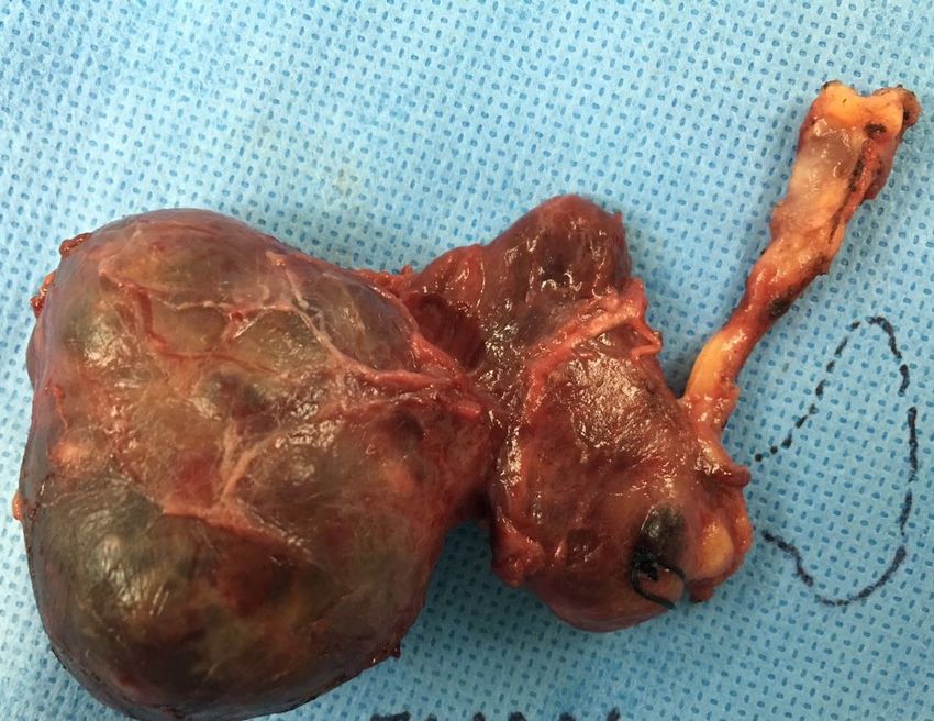
AGJSR   aged 55 years or older. It affects females 2 to 3 times more than males (Bilezikian et al., 2016;
        Madkhali et al., 2016). The vast majority of cases, 90- 95%, are sporadic, in which the PHPT
        is attributable to a solitary parathyroid adenoma in about 80% to 85% of the cases. A double
        adenoma is seen in up to 4% of cases and four-gland hyperplasia makes up the remaining
        10-15% of cases. While, parathyroid carcinoma is a very rare cause of PHPT, accounting for
        less than 1% of cases, familial parathyroid disorders are responsible for approximately 5% of
        the PHPT cases (Bilezikian et al., 2016; Madkhali et al., 2016).
        Although, adenomas usually arise as a single lesion, the majority of functional adenomas
        contain a mixed cell population with predominance of chief cells. There are also existing
        cases where clear cells and oxyphilic cells are mixed and rare cases where the majority of
        cells constituting the adenoma might be oxyphilic cells (Parelkar et al., 2012).
        Even though, hyperparathyroidism with clear cell adenoma is very rare (El Hussein & Poppiti,
        2017; Liang et al., 2010; Parelkar et al., 2012; Yazar et al., 2017), to date, around 40 cases
        have been reported in the literature with only four of them being intra-thyroid (Goodman et al.,
        2011; Gulati et al., 2012; Muthukrishnan et al., 2007; Parelkar et al., 2012).
        Water clear cell parathyroid adenoma (WCCA) is a rare cause of PHPT. Water-clear cells
        are not seen in normal human parathyroid glands (Ioannis et al., 2018). Usually, they are
        associated with hyperfunction of the parathyroid gland. They are characterized histologically
        based on large clear cells that might be transformed from parathyroid chief cells (ARık et al.,
        2017). On histopathological examinations; the cytoplasm is foamy and contains vacuoles
        of 0.2-2 microns in diameter. These cells represent end stage of hyperplastic chief cells (El
        Hussein & Poppiti, 2017).
        Here we present two cases of PHPT. The first case is a water clear cell intra-thyroid
        giant atypical parathyroid adenoma, and the second one is a small negative scan water clear
        cell parathyroid adenoma. Informed consent and approval from institutional review board
        were obtained for publication.

        Case 1:
        A fifty-year-old female patient, known to have β-thalassemia trait (minor thalassemia) and long-
        standing type 2 DM, treated with premixed insulin, Mixtard30, 60 units in the morning and 60
        units at night; presented for reevaluation on July 2016 because of uncontrolled blood sugar.
        One month before presentation, she experienced acute pancreatitis and was conservatively
        treated in a provincial hospital. No available data or medical reports were provided.
        Reviewing the history of the patient, she had a long-standing insulin treated type2 DM,
        hyperuricemia, diabetic peripheral neuropathy and chronic anemia. Additionally, she was
        diagnosed with β-thalassemia a year before presentation. Furthermore, a previous history
        of nonspecific abdominal pain and epigastric tenderness since 2012 that was associated
        with alternating diarrhea and constipation were reported. Patient was examined in a
        gastroenterology (GI) clinic without a definite diagnosis. Although panendoscopy was
        suggested but it was not performed at that time.
        On presentation, the patient complained of tiredness, easy fatigability, all over muscle and
        joints pain, numbness in both hands and lower limbs as well. On examination, the patient
        is overweight with a BMI of 40. Vital signs showed high systolic blood pressure 196/76,
        tachycardia, pulse was regular and high random blood sugar (319 mg/dL). Palpation of
        the neck revealed a right thyroid lobe enlargement. Chest auscultation: vesicular breathing

        34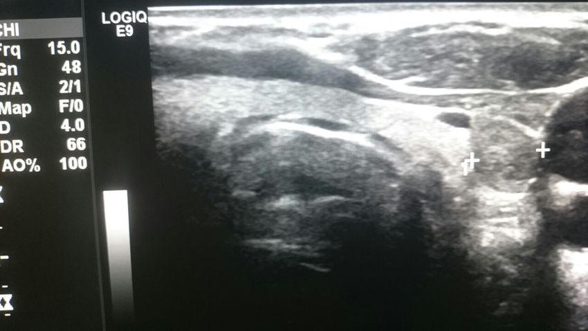
and normal heart sounds. Abdomen was soft, lax, with non-specific tenderness and no                  AGJSR
organomegaly. Lower limbs: pulses are present and no edema was detected, however a
decreased vibration sensation was noted.
Laboratory investigations showed uncontrolled blood sugar with high HbA1c, mixed
dyslipidemia, low vitamin D levels, hyperuricemia and high serum calcium. Once severe vitamin
D deficiency and high serum calcium with right thyroid lobe enlargement were observed,
further laboratory and radiological investigations were ordered. These investigations revealed
and confirmed a picture of hyperparathyroidism with severe vitamin D deficiency (Table 1).
Table 1: Laboratory Investigations Pre and Post-Surgery for case 1

                             Pre-Surgery              Post-Surgery              Normal range
Fasting Blood Sugar              327                      130                   80-100 mg/dl
HbA1c                           11.41                      7.5                    4.6-5.7%
Calcium                         12.07                      10                  8.5-10.5 mg/dl
TSH                               2.3                      2.4                  0.4-4.0 mU/l
Vitamin D                         17                       40                  50-150 nmol/L
ALP                              135                      149                   35-147 IU/L
PTH                              45.5                      6.6                 1.0-6.5 pmol/L

Additionally, neck ultrasound (U/S) revealed right thyroid lobe enlargement measuring
3.3x3.5x5 cm and a large cystic solid nodule was seen in the right lobe measuring 4.2x3.2x2.8
cm, showing surrounding vascularity and mild internal vascularity. The left lobe measured
1.2x1.7x3 cm and appeared normal in size, shape and texture. Multiple reactive lymph nodes
enlargement in the left parotid and submandibular region were observed (Figure 1).

               Figure 1: Neck ultrasound showing right thyroid lobe enlargement
             measuring 3.3x3.5x5 cm and a large cystic solid nodule in the right lobe.

                                                                                                35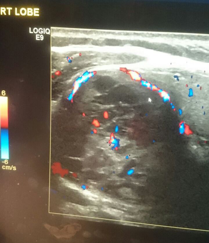
AGJSR   Abdominal U/S showed fatty liver, left renal simple cyst, tiny focus of cholesterolosis in the
        gall bladder and the visualized part of the pancreas appeared normal, but the tail of pancreas
        was not clearly visualized due to excessive bowel feces. Skeletal bone survey showed an
        overall decreased bone density, mild subperiosteal tunneling and absorption was seen in the
        proximal phalanges of both hands (Figure 3), small foci of lucency were observed along the
        skull bones, giving salt and pepper appearance (Figure 2).

                 Figure 2: Small foci of luceny                    Figure 3: Mild subperiosteal
               seen along the skull bones giving                 tunneling and absorption seen in
                  salt and pepper appearance                         the proximal phalanges

        Reviewing previous 2012 X-rays, bones appeared grossly osteopenic with vertebral endplate
        sclerosis. Vascular calcifications were observed and both sacroiliac joints were slightly
        sclerosed. Total bone mineral density (BMD) was 0.759/cm2 which is consistent with T-score
        of -3.2 and Z-score of -4.6 (Table 2).
        Table 2: Bone Mineral Density for case 1

                                                      T-score                        Z-score
                L1                                      -3.1                          -4.2
                Right femur                             -2.8                          -3.5
                Left femur                              -2.6                          -3.3
                Total body                              -3.2                          -4.6

        Parathyroid imaging with technetium-99m Sestamibi showed a large tracer-avid right thyroid
        nodule that could be an aggressive thyroid nodule or a large hyperfunctioning intra-thyroid
        parathyroid adenoma (Figure 4). Fine needle aspiration (FNA) revealed focally cellular foci
        where cohesive sheets that were formed by small and uniform cells, with rounded homogenous
        nuclei. Rare granulomas and focal Hurtle cell changes were noted in these samples and the
        pathology report was a follicular lesion (Figure 5).

        36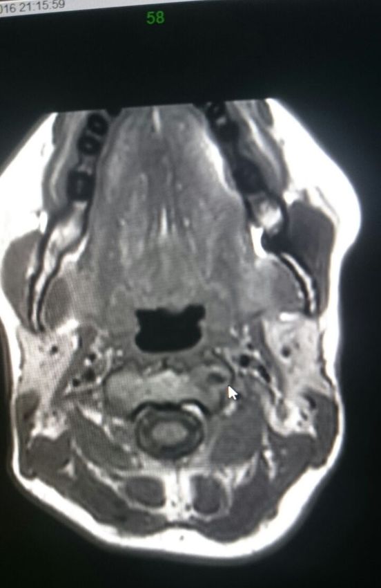
AGJSR

    Figure 4: Parathyroid technetium 99                  Figure 5: Fine need aspiration
     Sestamibi scan showing large tracer                 of patient number 1 neck mass
      avid right thyroid nodule that could               showing cellular clusters (white
   represent an aggressive thyroid nodule               arrow) of small uniform cells, with
   or a large hyperfunctioning intra-thyroid              rounded homogenous nuclei.
             parathyroid adenoma                                 DQx200arrows

The patient underwent hemithyroidectomy, parathyroidectomy and hospital course was
smooth without any complications. Even though, parathyroid hormone (PTH) dropped
from 51 to 0.8 pmol/L following surgery, she did not develop hypocalcemia, which is frequently
observed after a parathyroidectomy due to hungry bone syndrome. Laboratory investigations
before and after surgery are presented in (Table1).
Gross description of hemithyroidectomy and parathyroidectomy specimens (Figure 6)
revealed parathyroid adenoma of 5 cm in maximum dimension and weighing 36.3g.
Microscopic examination revealed predominantly water cell pattern with atypical features and
rim of benign thyroid tissue. The atypical features included fibrotic areas and fibrous septae
formation and clusters of tumor cells in the fibrous capsule (Figure 7). During follow up visits
postoperatively; the patient is much better clinically with no significant complaints and good
control of blood sugar, her most recent HbA1c was 7.5% (Table 1).

                   Figure 6: Hemi-thyroidectomy-parathyroidectomy specimen.

                                                                                              37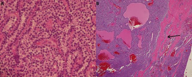
AGJSR

            Figure 7: A. High power view showing trabeculae of cellular parathyroid tissue with clear white
         cytoplasm and round monotonous nuclei (H&E, X 400). B. Low power microscopic view of the mass
        revealing trabeculae and sheets of cellular parathyroid tissue with microfollicles formation and fibrous
                                     septae in the stroma is seen (black arrow).

        Case 2:
        Thirty-three-year-old female patient, presented to endocrine clinic for reevaluation in 2015.
        She complained of tiredness, easy fatigability, bone and joint pain. For the last 2 years
        before presentation, she was labeled as a case of polycystic ovary syndrome (PCOS) and
        treated with metformin for short periods. She is clinically stable, asymptomatic, with normal
        vital signs and normal body mass index (BMI). No palpable goiter. On examination, neck,
        heart, abdomen and limbs were normal. Her laboratory investigations showed low vitamin D
        level 16 (15-150), high-normal calcium 2.56 (2.15-2.55 mmol/L), and high PTH 16.56 (1.6-6.9
        pmol/L). A diagnosis of PHPT was suggested.
        Neck U/S in 2014 revealed a small hypoechoic area in the left lobe of the thyroid measuring
        0.9x0.6 cm with increased peripheral vascularity. The possibility of parathyroid adenoma
        could not be entirely excluded and scintigraphy was warranted for further work up (Figure 8).

                                 Figure 8: Neck US showing left inferior intra-thyroid
                                    oval shaped hypoechoic lesion, 1.3x0.4 cm.

        Over the last 2 years, the patient continued to have high PTH, high calcium, and high alkaline
        phosphatase (ALP) with normal phosphorus levels (Table 3). Parathyroid adenoma was not
        considered at that time because BMD was normal and nuclear medicine investigations failed
        to detect parathyroid adenoma.

        38
Table 3: Laboratory Investigations for case 2 AGJSR
Pre-surgery Post-surgery Normal range
Albumin 45 44 40-49 g/l
Vitamin D 22 17 50-150 nmol/L
Calcium 10.7 10.4 8.5-10.5 mg/dl
Corrected Ca 10.5 8 8.5-10.5 mg/dl
Urine, Calcium 7.22 8.5-10.5 mg/dl
PTH 16.56 2.4 1.0-6.5 pmol/L
ALP 186 105 44-147 U/l
Phosphorus 3.63 3.66 3.4 to 4.5 mg/dl
HbA1c 5.1 4.5 4.6-5.7%
FBS 94 88 80-100 mg/dl
PTH 16.56 2.4 1.6-6.9 pmol/L
TSH 1.6
Upon reviewing medical records, the patient had nonspecific abdominal pain that was
investigated in the GI clinic. No significant abnormalities were detected and therefore, no
definite diagnosis was established. Meanwhile, at that time, in 2012, when she was diagnosed
with PCOS, her laboratory investigations failed to show high normal corrected calcium of 2.55
(2.2-2.60) mmol/L with severe vitamin D deficiency of 12 (50-150 nmol/L).
Repeated neck U/S reported left inferior intra-thyroid oval shaped hypoechoic lesion of
1.3x0.4 cm (Figure 8). However, a year later, when the patient was reevaluated, biochemical
picture was consistent with and confirmed the diagnosis of PHPT.
Furthermore, repeated neck Computed Tomography scan (CT scan) (Figure 9) and Magnetic
Resonance Imaging (MRI) were suggestive of left lower parathyroid adenoma with similar
picture as was observed earlier by neck U/S. In spite of the negative Sestamibi scan, minimal
neck invasive surgery was performed and 0.3 gm left lower parathyroid adenoma measuring
1.2x0.7x0.6 cm was resected. Post-operative course was uneventful.
Figure 9: Neck computed tomography scan (CT scan).
39AGJSR Histopathological examination of the mass demonstrated water clear cell parathyroid
adenoma with a rim of non-neoplastic parathyroid tissue (Figure 10). Patient did not develop
hypocalcemia, her ionized calcium was normal during hospitalization and PTH dropped
significantly, from 11.1 to 2.4 (1.6-6.9 pmol/L). The patient became asymptomatic with no
further complaints.
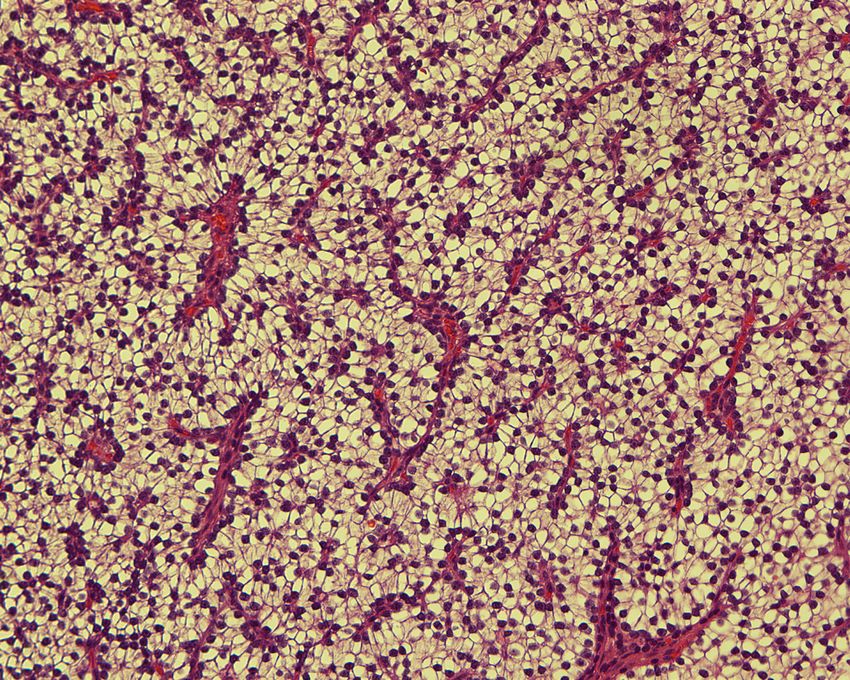
Figure 10: The mass similarly consists of trabeculae of cellular parathyroid tissue with
clear white pale cytoplasm. Delicate capillary network can be seen in the background
Discussion
The diagnosis of PHPT is usually established biochemically, based on serum calcium, PTH,
and phosphate levels (Bilezikian et al., 2016; Madkhali et al., 2016). While elevated levels
of serum calcium and PTH are associated with 95% of classical cases of PHPT, patients
however may present with normal levels of phosphate, calcium or PTH (Bilezikian et al.,
2016; Madkhali et al., 2016). Twenty-four-hour urine collection for calcium measurement is
therefore necessary to rule out familial hypocalciuric-hypercalcemia. Even though, 24-hour-
urine collection was not performed for our patients, random calcium in urine was high in
the first case. While parathyroidectomy success rate is high, persistent and recurrence still
represent clinical challenge (Bilezikian et al., 2016; Madkhali et al., 2016). Therefore, pre-
operative localization techniques should be considered, not only to confirm the diagnosis of
PHP, but also to guide the endocrine surgeons intraoperatively, as well as to evaluate for any
ectopic glands, and to assist in planning of minimally invasive parathyroidectomy (Agarwal &
Pradhan, 2012; Bilezikian et al., 2016; Madkhali et al., 2016).
Usually ultrasonography, CT scan and MRI are common techniques used for parathyroid
adenoma workup. Nevertheless, Sestamibi scan with single photon emission computerized
tomography (SPECT) is superior due to the high sensitivity (Bilezikian et al., 2016; Madkhali
et al., 2016).
Sestamibi scan was performed for both patients. In the first case, it was localized as a
persistent increased area of activity in the right thyroid lobe that was seen on the neck U/S. In
the second patient, ultrasonography and CT scan were suggestive of localized adenoma with
negative Sestamibi scan. However, negative scan could be due to several factors including
40small size of adenomas, thyroid nodules, superior position and the paucity of oxyphillic cells AGJSR
in the parathyroid adenoma (Bilezikian et al., 2016; Grenko et al., 1995; Kojidi et al., 2016;
Madkhali et al., 2016). Water-clear cell adenoma (WCCA) is extremely rare condition, only
40 cases has been reported in the literature with variant clinical presentations (Bai et al.,
2012; Kojidi et al., 2016).
While water-clear cell hyperplasia (WCCH) is another rare condition, it should be distinguished
from WCCA, where in the case of WCCH, all four parathyroid glands have water clear cells
on their histological examination (El Hussein & Poppiti, 2017; Ezzat et al., 2013; Liang et al.,
2010; Parelkar et al., 2012; Yadav et al., 2017; Yazar et al., 2017). In fact, recent studies
reported 0.3% incidence of water clear cell parathyroid disease, which supports the decline in
the incidence of WCCH from 13% in 1930 to less than 1% (El Hussein & Poppiti, 2017; Ezzat
et al., 2013; Parelkar et al., 2012). On the other hand another study showed the prevalence of
WCCA of 46% in 66 patients with PHPT with an overt clinical picture (Varshney et al., 2013).
Parathyroid clear cell carcinoma is very rare and only few cases were reported recently (El
Hussein & Poppiti, 2017; Naganuma et al., 2013; Parelkar et al., 2012).
In the two cases reported here, the first one was an ectopic intra-thyroid adenoma, which
is still uncommon, even though few cases have been previously reported (Abboud et al.,
2007; Mondal, 2013; Parelkar et al., 2012; Prasad et al., 2004; Shi et al., 2016). Parathyroid
adenoma in the first case weighted 36,5 gm which is considered a giant adenoma.Currently,
fine needle aspiration (FNA) is usually performed prior to surgery for thyroid nodules as well
as for suspicious parathyroid adenomas. Previously, it has been reported that FNA might be
misleading and/or challenging for the diagnosis of parathyroid adenoma and therefore, other
neoplasms should be considered in the differential diagnosis (Abboud et al., 2007; Heo et al.,
2013; Papanicolau‐Sengos et al., 2013; Parelkar et al., 2012)Interestingly, FNA biopsy for the
first patient was reported as thyroid follicular lesion.
Histopathological examination of the specimen of the first case, revealed atypical features
of clear cell adenoma, which should be differentiated from parathyroid carcinoma clinically
and histopathologically (Ioannis et al., 2018; LiVolsi et al., 2014). In our case, the diagnosis
of WCCA was confirmed by both clinical and histopathological examinations. Even though,
atypical adenomas are defined as adenomas that exhibit some features of malignancy,
such as broad fibrous bands crossing the tumor or pseudocapsular invasion (clusters of
parathyroid cells trapped within the capsule), they do not show vascular invasion, metastases
or apparently increased mitotic activity. To best of our knowledge, this observation has not
been reported previously for a water-clear clear cell adenoma.
The first case, parathyroid adenoma was located ectopically intra-thyroid which accounts for
1.4-6% of parathyroid adenomas of ectopic location. Parathyroid ectopia occurs in 4-20%
of patients because of abnormal migration during embryogenesis or secondary to acquired
migration (Issoufou et al., 2015; Parelkar et al., 2012; Roy et al., 2013).
Currently, clinical presentation of PHPT is either asymptomatic, with minor symptoms or it
could be with an overt clinical picture and radiological manifestations (Bilezikian et al., 2016;
Madkhali et al., 2016; Parelkar et al., 2012). Nonetheless, our first patient developed acute
pancreatitis a month prior to presentation and hyperparathyroidism could be the cause of
pancreatitis excluding other causes (Bilezikian et al., 2016; Madkhali et al., 2016; Parelkar
et al., 2012). However acute pancreatitis was reported as initial manifestation of PHPT that
was due to WCCA10 and the first case reported here should be the second in literature. Bone
radiological manifestations in our case is a well-documented clinical picture reported in other
41AGJSR cases of PHPT (Varshney et al., 2013; Zanocco & Yeh, 2017).
Furthermore, our first case of WCCA has β-Thalassemia trait. This might be the first case of
hyperparathyroidism associated with β-Thalassemia, as it is well known for β-Thalassemia to
be associated with hypoparathyroidism (Shetty & Shenoy, 2014). Additionally, the first patient
has type 2 DM and hypertension, however blood sugar control became possible following
parathyroidectomy, which has been reported previously (Jena et al., 2016; Kumar & Singh,
2015; Reddy et al., 2009)..
The second case underwent minimal invasive surgery and parathyroidectomy and the
adenoma was small weighing 0.3 gm, a clear cell parathyroid adenoma that might explain the
negative Sestamibi scan as well as the low functional activity.
The differential diagnosis of WCCA includes, WCCH, clear cell changes in a thyroid tumor,
paraganglioma, parathyroid chief cell adenoma with focal clear cell changes, clear cell lung
tumors, clear cell sarcoma, Primary clear cell carcinoma of the thymus, parathyroid carcinoma,
and metastatic renal cell carcinoma (Zinovkin et al., 2020). The distinctive histopathologic
features of WCCA include optical clear cytoplasm, large clear cells with fine cytoplasmic
vacuolization, high levels of cytoplasmic glycogen and fat, and absence of vascular invasion,
metastases or apparently increased mitotic activity.
Conclusion
PHPT is a common endocrine disorder. Clinical presentation of PHPT varies from asymptomatic
to insignificant, it might present with other complications such as pancreatitis. The most
common cause of PHPT is parathyroid adenoma. Therefore, any suspected clinical condition
associated with calcium abnormalities should be further investigated with biochemical and
imaging workup to confirm the diagnosis of PHPT. The aim of this report is to highlight this
rare variant of parathyroid adenomas, especially with WCCA could be the underlying cause
of the clinical picture.
Conflict of Interest
All authors declare that they do not have any conflict of interest of any type.
Informed consent
Informed consent and approval from institutional review board were obtained for publication
from Arrayan Hospital, Sulaiman Habib Medical Group, Riyadh, Saudi Arabia.
Funding information
This wok was not financially supported from any institution.
Ethical approval
Informed consent and approval from institutional review board were obtained for publication
from Arrayan Hospital, Sulaiman Habib Medical Group, Riyadh, Saudi Arabia.
42References AGJSR
Abboud, B., Sleilaty, G., Ayoub, S., Hachem, K., Smayra, T., Ghorra, C., & Abadjian, G.
(2007). Intrathyroid parathyroid adenoma in primary hyperparathyroidism: can it be
predicted preoperatively? World journal of surgery, 31(4), 817-823.
Agarwal, A., & Pradhan, R. (2012). Failed parathyroidectomy: the road ahead. Indian journal
of endocrinology and metabolism, 16(Suppl 2), S221.
ARık, D., DünDAR, E., YılMAz, E., & SİvRİkoz, C. (2017). Water-clear cell adenoma of the
mediastinal parathyroid gland.
Bai, S., LiVolsi, V. A., Fraker, D. L., & Bing, Z. (2012). Water-clear parathyroid adenoma:
report of two cases and literature review. Endocrine pathology, 23(3), 196-200.
Bilezikian, J. P., Cusano, N. E., Khan, A. A., Liu, J.-M., Marcocci, C., & Bandeira, F. (2016).
Primary hyperparathyroidism. Nature Reviews Disease Primers, 2(1), 1-16.
El Hussein, S., & Poppiti, R. (2017). Water clear cell adenoma of the parathyroid gland: a
forgotten cause of primary hyperparathyroidism. International journal of surgical pathology,
25(5), 384-388.
Ezzat, T., Maclean, G., Parameswaran, R., Phillips, B., Komar, V., Mihai, R., Sadler, G., &
Courtney, S. (2013). Primary hyperparathyroidism with water clear cell content: the impact
of histological diagnosis on clinical management and outcome. The Annals of The Royal
College of Surgeons of England, 95(3), e6-e8.
Goodman, A., Politz, D., Lopez, J., & Norman, J. (2011). Intrathyroid parathyroid adenoma:
incidence and location—the case against thyroid lobectomy. Otolaryngology--Head and
Neck Surgery, 144(6), 867-871.
Grenko, R. T., Anderson, K. M., Kauffman, G., & Abt, A. B. (1995). Water-clear cell adenoma
of the parathyroid. A case report with immunohistochemistry and electron microscopy.
Archives of pathology & laboratory medicine, 119(11), 1072-1074.
Gulati, H., Anand, M., & Deshmukh, S. (2012). Asymptomatic intrathyroid” Water Clear Cell”
parathyroid adenoma: A rare entity. Thyroid Research and Practice, 9(2), 72-72.
Heo, I., Park, S., Jung, C. W., Koh, J. S., Lee, S.-S., Seol, H., Choi, H. S., & Cho, S. Y. (2013).
Fine needle aspiration cytology of parathyroid lesions. Korean journal of pathology, 47(5),
466.
Ioannis, P., Stavros, P., Nektarios, K., Chrysa, S., & Kalliopi, P. (2018). Water-clear cell
adenoma of parathyroid gland: a case report and concerns on differential diagnosis. J
Endocrinol Diabetes Obes, 6(1), 1117.
Issoufou, I., Belliraj, L., Rabiou, S., Ghalimi, J., Rchachi, M., Lakranbi, M., Ajdi, F., Ouadnouni,
Y., & Smahi, M. (2015). Intrathymic Parathyroid Adenoma. Case Reports in Clinical
Medicine, 4(08), 297.
Jena, A., Patnayak, R., Suresh, V., Kalawat, T. C., Phaneendra, B. V., Lakshmi, A. Y., &
Sachan, A. (2016). Parathyroid adenomas: A case series and clinicopathological study
from a tertiary care center in South India. Medical Journal of Dr. DY Patil University, 9(4),
495.
Kojidi, H. T., Vagharimehr, N., Mohseni, S., Pajouhi, M., & Mohajeri-Tehrani, M. R. (2016).
Unusual ectopic parathyroid adenoma: a case report. Acta Medica Iranica, 547-550.
43AGJSR Kumar, A., & Singh, S. (2015). Parathyroidectomy Ameliorates Glucose and Blood Pressure
Control in a Patient with Primary Hyperparathyroidism, Type 2 Diabetes, and Hypertension.
Clinical Medicine Insights: Endocrinology and Diabetes, 8, CMED. S31292.
Liang, Y., Mojica, W., & Chen, F. (2010). Water-clear cell adenoma of parathyroid gland: a
case report and literature review. North American Journal of Medicine and Science, 3(4).
LiVolsi, V. A., Montone, K. T., & Baloch, Z. N. (2014). Parathyroid: the pathology of
hyperparathyroidism. Surgical pathology clinics, 7(4), 515-531.
Madkhali, T., Alhefdhi, A., Chen, H., & Elfenbein, D. (2016). Primary hyperparathyroidism.
Turkish Journal of Surgery/Ulusal cerrahi dergisi, 32(1), 58.
Mondal, S. K. (2013). Histopathologic Analysis of Female Genital Tuberculosis: A Fifteen-\
Year Retrospective Study of 110 Cases in Eastern India/Kadın Genital Sistem Tüberkülozu:
Hindistan’da 110 Olgunun Retrospektif Histopatolojik Analizi. Turkish journal of pathology,
29(1), 41-45.
Muthukrishnan, J., Verma, A., Modi, K., Kumaresan, K., & Jha, S. (2007). Ectopic parathyroid
adenoma-the hidden culprit. JAPI, 55.
Naganuma, H., Shibuya, R., Takaya, K., Asakura, T., Mori, Y., & Kameyama, K. (2013).
Water-clear Cell Carcinoma of Parathyroid Gland with Primary Hyperparathyroidism: First
Case Report with Review of the Literature. Journal of Basic and Clinical Medicine, 2(2).
Papanicolau‐Sengos, A., Brumund, K., Lin, G., & Hasteh, F. (2013). Cytologic findings of a
clear cell parathyroid lesion. Diagnostic cytopathology, 41(8), 725-728.
Parelkar, S. V., Oak, S. N., Patel, J. L., Sanghvi, B. V., Joshi, P. B., Sahoo, S. K., & Sampat,
N. (2012). 首页» 文章» 文章详细信息. Journal of Indian Association of Pediatric Surgeons,
17(4), 180-183.
Prasad, K. K., Agarwal, G., & Krishnani, N. (2004). Water-clear cell adenoma of the parathyroid
gland: a rare entity. Indian journal of pathology & microbiology, 47(1), 39-40.
Reddy, P., Harinarayan, C., Suresh, V., Jena, A., Reddy, M., Kalawat, T., Moorthy, M., Vittal,
S., & Sachan, A. (2009). Can diabetes associated with hyperparathyroidism be an
additional indication for parathyroidectomy? A case report.
Roy, M., Mazeh, H., Chen, H., & Sippel, R. S. (2013). Incidence and localization of ectopic
parathyroid adenomas in previously unexplored patients. World journal of surgery, 37(1),
102-106.
Shetty, B., & Shenoy, U. (2014). Prevalence of hypoparathyroidism (HPT) in beta thalassemia
major. Journal of clinical and diagnostic research: JCDR, 8(2), 24.
Shi, C., Guan, H., Qi, W., Ji, J., Wu, J., Yan, F., & Wang, H. (2016). Intrathyroidal parathyroid
adenoma: diagnostic pitfalls on fine‐needle aspiration: two case reports and literature
review. Diagnostic cytopathology, 44(11), 921-925.
Varshney, S., Bhadada, S. K., Nahar, U., Shah, V. N., Bhansali, A., & Behera, A. (2013).
Chief cell and clear cell parathyroid adenoma do not influence clinical and biochemical
expression of the sporadic primary hyperparathyroidism. Endocrine, 43(2), 440-443.
Yadav, S., Singh, K., & Sathe, P. (2017). Water Clear Parathyroid Adenoma: A report of 2
cases. Annals of Pathology and Laboratory Medicine, 4(3), C66-68.
Yazar, F. M., Karaağaç, M., İşler, A., Bülbüloğlu, E., & Ezberci, F. (2017). An unusual cause
44of hypercalcemic crisis: Water-clear cell double parathyroid adenoma. Turkish journal of AGJSR
surgery, 33(4), 243.
Zanocco, K. A., & Yeh, M. W. (2017). Primary hyperparathyroidism: effects on bone health.
Endocrinology and Metabolism Clinics, 46(1), 87-104.
Zinovkin, D., Zhandarov, M. Y., & Pranjol, M. (2020). Water clear cell adenoma of parathyroid
gland: a rare lesion.
45ورم الغدة الجار درقية ذو الخاليا الصافية :تقرير عن حالتين
*4-3
عبد الرحمن محمد ردايدة ،1هشام الخالدي ،2محمد نصير 3ومحمد القضاة
1قسم الغدد الصماء ،القسم الطبي ،مستشفى العريان ،مجموعة سليمان حبيب الطبية
الرياض ،المملكة العربية السعودية
2قسم الباثولوجيا ،كلية الطب ،جامعة الملك سعود ،الرياض ،المملكة العربية السعودية
3قسم الفسيولوجيا والكيمياء الحيوية ،جامعة العلوم والتكنولوجيا األردنية ،كلية الطب ،إربد 22110 ،األردن
4قسم علم وظائف األعضاء ،جامعة الخليج العربي ،مملكة البحرين
*بريد الكترونيmohammada@agu.edu :
ال ُمـسـتَخـلَص
تاريخ استالم البحث2021/05/06 : فــرط نشــاط جــارات الدرقيــة األولــي هــو مــرض يتميــز بفــرط كالســيوم الــدم واإلفــراط فــي
تاريخ تعديل البحث2021/06/23 :
تاريخ قبول البحث2021/06/27 : إنتــاج هرمــون الغــدة الجــار درقيــة .وهــو الســبب األكثــر شــيو ًعا لفــرط كالســيوم الــدم فــي
العيــادات الخارجيــة وثالــث االمــراض االكثــر شــيوعما مــن اضطرابــات الغــدد الصمــاء.
يــزداد معــدل اإلصابــة بفــرط نشــاط جــارات الدرقيــة األولــي مــع تقــدم العمــر وهــو أكثــر
شــيو ًعا عنــد النســاء منــه عنــد الرجــال 85٪-80 .مــن حــاالت فــرط نشــاط جــارات الــدرق
األولــي ناتجــة عــن ورم الفــدة الجــار درقيــة .تحتــوي غالبيــة األورام الغديــة جــارات الدرقيــة
علــى مجموعــة خاليــا مختلطــة مــع غلبــة الخاليــا الرئيســية .فــرط نشــاط جــارات الدرقيــة
األولــي مــع الــورم الحميــد الخلــوي الصافــي نــادر جـدًا.
نحــن هنــا نشــارك حالتيــن مــن الــورم الحميــد جــارات الدرقيــة ذو الخاليــا الصافيــة المرتبــط
بفــرط نشــاط جــارات الدرقيــة األولــي مــع عــرض المظاهــر الســريرية مختلفــة.
الكلمــات الدالــة :فــرط نشــاط جــارات الــدرق األولــي ،خليــة صافيــة للمــاء ،ورم الغــدة الجار
درقيــة ،هشاشــة العظــام ،التهــاب البنكريــاس ،ارتفــاع ضغــط الدم ،داء الســكري ،الثالســيميا.
46 AGJSR 37 (4) 2019: 33-46 Abdelrahman M. Radaideh et alYou can also read